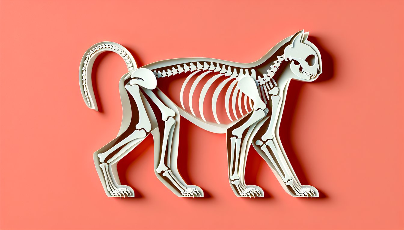
Calling all cat lovers and aspiring veterinarians! Are you ready to master the cat appendicular skeleton and prove your understanding of feline limb bones? Dive into our interactive feline limb bones quiz and test your cat skeletal anatomy know-how as you identify each bone in your furry friend's limbs. From the scapula to the phalanges, this feline appendicular skeleton guide will challenge you like never before. Think you have what it takes to ace this free appendicular skeleton quiz and sharpen your cat bone identification test skills? Take the plunge, tackle the parts of the bones quiz , and start learning today!
Which bone forms the shoulder blade in cats?
Coracoid
Scapula
Clavicle
Humerus
The scapula, or shoulder blade, is the flat bone that connects the humerus with the clavicle. In cats, it provides attachment for several shoulder muscles. It is a major bone of the forelimb's appendicular skeleton. See more on scapula anatomy.
What is the largest bone in the feline forelimb?
Scapula
Humerus
Ulna
Radius
The humerus is the longest and largest bone of the cat's forelimb, spanning from the shoulder to the elbow. It articulates proximally with the scapula and distally with the radius and ulna. It supports major muscle attachments for locomotion. More on humerus anatomy.
Which two bones form the antebrachium in a cat's forelimb?
Radius and Ulna
Carpals and Metacarpals
Tibia and Fibula
Humerus and Scapula
The antebrachium refers to the forearm region, which is composed of the radius and ulna. These two bones run parallel between the elbow and the carpus. They allow flexion and extension at the elbow and wrist joints. Review radius anatomy.
Where are the carpal bones located in a cat?
Ankle region of the hindlimb
Within the pelvic girdle
Between the shoulder and elbow
Wrist region of the forelimb
Carpal bones are the small bones that make up the wrist (carpus) in cats. They lie distal to the radius and ulna and proximal to the metacarpals. They allow complex movements of the forepaw. Details on carpal bones.
Which bones support the paw between the wrist and the digits?
Carpals
Phalanges
Tarsals
Metacarpals
Metacarpals are the long bones that form the palm of the forepaw, located between the carpals and the phalanges. Cats have five metacarpals in each forelimb. They support weight and allow digit articulation. Learn about metacarpus.
Where are the proximal phalanges found in a cat?
Between the tibia and tarsals
At the base of each digit
Between the scapula and humerus
At the hip joint
Proximal phalanges are the first set of small bones in each digit, immediately distal to the metacarpals or metatarsals. They serve as the base segment of each toe or finger. They facilitate flexion and extension of the claws. More on phalanges.
Which bone is part of the pelvic girdle that connects to the femur?
Pubis
Sacrum
Ischium
Ilium
The ilium is the largest bone of the pelvic girdle and articulates with the femur at the acetabulum. It provides attachment for muscles of the hindlimb and supports abdominal organs. Cats' iliac wings are slender, aiding agility. Ilium details.
Into which socket does the femur fit proximally in the pelvis?
Olecranon fossa
Trochlea
Acetabulum
Glenoid cavity
The acetabulum is the concave cavity of the pelvis where the head of the femur articulates. It forms the hip joint, allowing range of motion in the hindlimb. This ball-and-socket joint is critical for feline jumping and running. More on the acetabulum.
On which side of the feline hindlimb is the tibia located?
Medial side
Anterior side
Lateral side
Posterior side
The tibia is situated on the medial (inner) side of the hindlimb between the stifle (knee) and tarsus (hock). It bears the majority of weight transmitted from the femur. The fibula is lateral and slender in cats. Tibia anatomy overview.
Which bone is the smaller, lateral bone of the feline lower hindlimb?
Calcaneus
Femur
Fibula
Tibia
The fibula is the slender, lateral bone of the lower hindlimb, running parallel to the tibia. In cats, it is relatively thin and provides muscle attachment sites. It does not bear as much weight as the tibia. Fibula details.
Where are the tarsal bones located in a cat?
Hock (ankle) region of the hindlimb
Wrist region of the forelimb
Between femur and pelvis
Within the skull
Tarsal bones make up the hock, or ankle, in the hindlimb. They lie distal to the tibia and fibula and proximal to the metatarsals. These small bones allow articulation and shock absorption. Learn more about tarsals.
Which bones form the main support of the hind paw between the tarsals and digits?
Metatarsals
Phalanges
Patella
Ischium
Metatarsals are the long bones in the hind paw located between the tarsal bones and the phalanges. In cats, there are typically five metatarsals per hindlimb. They help distribute weight and facilitate toe movement. Metatarsus information.
How many metacarpal bones does each cat forelimb have?
Seven
Six
Five
Four
Cats normally have five metacarpal bones in each forelimb, corresponding to each digit. The first metacarpal is often reduced in size and is analogous to a dewclaw. These bones support the paw structure. Read more on metacarpals.
The olecranon process is a prominence of which feline bone?
Radius
Scapula
Ulna
Humerus
The olecranon is the prominent, proximal end of the ulna forming the point of the elbow. It serves as the attachment for the triceps brachii muscle. In cats, it is well-developed to support jumping. More about the ulna.
Which structure on the femur serves as the major lateral attachment for gluteal muscles?
Greater trochanter
Patellar groove
Medial condyle
Lesser trochanter
The greater trochanter is the large, lateral projection on the proximal femur that provides attachment for gluteal and other hip muscles. It is key for hindlimb propulsion in cats. The lesser trochanter is more medial and smaller. Femur anatomy details.
Which sesamoid bone is found near the caudal aspect of the carpus in cats?
Pisiform
Accessory carpal bone
Fabella
Patella
The accessory carpal bone is a sesamoid bone located on the caudal side of the wrist (carpus), serving as a lever for flexor tendons. It is distinct from the small pisiform which lies medially. This bone improves tendon leverage during paw placement. Carpal bones overview.
Which process of the scapula serves as the attachment for the acromiodeltoid muscle in cats?
Glenoid tubercle
Spine of scapula
Coracoid process
Acromion
The acromion is a bony projection on the scapula that articulates with the clavicle in species that have one. In cats, it provides attachment for the deltoid and acromiodeltoid muscles. It is distinct from the scapular spine which runs medially. Feline scapula anatomy.
Which carpal bone is considered a sesamoid within the flexor carpi ulnaris tendon in cats?
Accessory carpal
Lunar
Pisiform
Scaphoid
The pisiform is a sesamoid bone embedded in the tendon of the flexor carpi ulnaris muscle. It sits medial to the other carpal bones and increases tendon leverage. The accessory carpal is a separate sesamoid on the caudal side. Pisiform bone info.
Which bone feature of the humerus articulates with the ulna at the elbow joint?
Trochlea
Capitulum
Greater tubercle
Head
The trochlea is a spool-shaped structure on the distal humerus that articulates medially with the ulna's trochlear notch. The capitulum, on the lateral side, articulates with the radial head. These structures form the elbow hinge. Distal humerus anatomy.
Which region of the feline ulna forms the point of the elbow?
Trochlear notch
Styloid process
Coronoid process
Olecranon tuberosity
The olecranon tuberosity is the prominent proximal projection of the ulna that forms the elbow's point. It serves as the insertion for the triceps brachii. The trochlear notch is the articulation surface, not the external prominence. Olecranon details.
Which bone of the feline hindlimb contributes to the patellar mechanism?
Fabella
Sesamoid of gastrocnemius
Patella
Acetabular sesamoid
The patella is the largest sesamoid bone, located within the quadriceps tendon at the stifle joint. It protects the tendon and improves leverage during extension of the stifle. Cats also have smaller fabellae behind the femoral condyles. Patella anatomy.
Which structure on the cat's femur separates the lateral and medial condyles?
Trochanteric fossa
Patellar groove
Intercondylar fossa
Linea aspera
The intercondylar fossa is the deep notch between the medial and lateral condyles of the distal femur. It accommodates cruciate ligaments of the stifle. The patellar groove lies anteriorly between the condyles. Femur distal features.
Which bone in the cat hindlimb bears most weight and articulates with the femur at the stifle?
Femur
Patella
Fibula
Tibia
The tibia is the primary weight-bearing bone of the lower hindlimb, articulating with the femoral condyles at the stifle (knee). It transmits forces from the femur to the tarsus. The fibula in cats is slender and less load-bearing. Tibia function.
Which tarsal bone articulates with the calcaneus to form the calcaneal tuber prominence?
Fourth tarsal
Central tarsal
Talus
Medial tarsal
The talus articulates proximally with the tibia and fibula and posteriorly with the calcaneus, forming the calcaneal tuber (heel) prominence. It supports the hock's lever mechanism. Other tarsals lie more distally. Talus bone info.
Which small bone forms part of the medial carpal row in cats?
Accessory carpal
Lunar (lunate)
Scaphoid
Pisiform
The lunar (lunate) is one of the central bones in the proximal carpal row on the medial side. It articulates with the radius and contributes to the carpal joint. The scaphoid lies lateral, while the pisiform is a sesamoid. Carpal bone rows.
What is the primary function of sesamoid bones in the feline appendicular skeleton?
Improve tendon leverage and reduce wear
Produce red blood cells
Store fat for energy
Connect two long bones directly
Sesamoid bones, such as the patella or accessory carpal bone, develop in tendons to alter the direction of muscle pull and reduce friction. They improve mechanical advantage in joints. They do not store fat or directly connect long bones. Sesamoid bone function.
Which limb bone in cats contains the nutrient foramen near its midshaft?
Scapula
Femur
Calcaneus
Patella
Long bones like the femur typically have a nutrient foramen on their diaphysis to allow entry of vessels that nourish bone. While other long bones also have foramina, the femur's is prominent. Flat bones like the scapula do not have a similar feature. Nutrient foramen details.
Which bone landmark is found on the medial side of the feline tibia?
Lateral malleolus
Medial malleolus
Intercondylar eminence
Lateral condyle
The medial malleolus is the bony prominence on the distal medial side of the tibia, forming part of the hock joint. The lateral malleolus is part of the fibula. The intercondylar eminence is on the proximal tibia. Tibia features.
Which bone of the feline forelimb contributes to the structure of the elbow along with the humerus?
Ulna
Carpal bones
Scapula
Radius
The ulna, particularly its trochlear notch, articulates with the humerus at the elbow joint. The radius also contributes but the ulna forms the main hinge. The scapula is proximal, and carpal bones lie distal. Elbow joint anatomy.
Which structure distinguishes the proximal femur from the distal in cats?
Adductor tubercle
Lateral condyle
Femoral head
Linea aspera
The femoral head is the spherical proximal extension of the femur that fits into the acetabulum. Condyles and the adductor tubercle lie distally. The linea aspera runs along the shaft. Proximal femur features.
Which specific carpal bone articulates with the radius and supports the majority of weight in the feline wrist?
Central carpal
Lunate (lunar)
Pisiform
Scaphoid (radial carpal)
The scaphoid, or radial carpal bone, lies directly distal to the radius and bears most of the load transmitted through the forelimb. The lunate also articulates but shares load. The pisiform is a sesamoid and central carpal lies distal. Radial carpal detail.
Which anatomical feature of the scapula is reduced or absent in most domestic cats compared to other mammals?
Infraspinous fossa
Acromion process
Supraspinous fossa
Coracoid process
Domestic cats often have a small or absent acromion, reducing shoulder blade bulk and increasing flexibility. The coracoid process is also reduced but present. The fossae remain well-developed for muscle attachment. Compare feline scapula.
In cats, which tarsal bone lies directly distal to the tibia and articulates with the central tarsal bone?
Fourth tarsal
Talus
Calcaneus
Medial tarsal
The talus articulates proximally with the tibia and distally with the central tarsal bone. It forms the roof of the tarsal canal. The calcaneus lies posterior, forming the heel. Talus articulation.
Which bone in the feline pelvic girdle forms the dorsal roof of the pelvic canal?
Sacrum
Ilium
Pubis
Ischium
The sacrum, composed of fused sacral vertebrae, forms the dorsal roof of the pelvis and articulates with the ilia. It transmits forces from the hindlimb to the vertebral column. The ilium forms the lateral walls. Sacrum anatomy.
Which femoral ligament attaches to the intertrochanteric crest in cats?
Ligament of head of femur
Iliofemoral ligament
Pubofemoral ligament
Ischiofemoral ligament
The iliofemoral ligament reinforces the capsule of the hip joint anteriorly, attaching between the ilium and the intertrochanteric line/crest of the femur. It limits overextension. Other ligaments attach elsewhere. Hip joint ligaments.
Which bone feature serves as the insertion for the gluteus superficialis muscle in cats?
Lesser trochanter
Third trochanter of femur
Linea aspera
Greater trochanter
Cats have a third trochanter on the proximal lateral femur where the gluteus superficialis (gluteus superficialis) attaches. This is an adaptation for powerful hindlimb abduction. Many species lack this distinct prominence. Feline femur features.
Which carpal bone articulates distally with the first metacarpal in cats?
Trapezium
Hamate
Lunate
Capitate
The trapezium is one of the distal carpal bones that articulates with the first metacarpal, forming the first carpometacarpal joint. This joint allows limited opposition in cats. The capitate and hamate articulate with other metacarpals. Distal carpal bones.
Which structure on the distal tibia forms part of the medial support for the tarsal joint?
Lateral malleolus
Tibial crest
Medial malleolus
Anterior tubercle
The medial malleolus is the distal projection of the tibia that provides medial stability to the tarsal joint. The lateral malleolus is part of the fibula. The tibial crest is on the shaft. Tibia landmarks.
Which sesamoid bone lies within the tendon of the gastrocnemius muscle in the feline hindlimb?
Accessory tarsal
Paired fabellae
Patella
Calcaneus
Fabellae are small sesamoid bones located within the heads of the gastrocnemius muscle behind the femoral condyles. They protect tendon fibers and improve leverage. They are separate from the patella. Sesamoid overview.
Which feature distinguishes the third metacarpal from the second and fourth in cats?
It is the largest and bears the most weight
It is absent in some cats
It has an accessory sesamoid
It is fused to the fourth metacarpal
The third metacarpal is typically the longest and strongest, bearing the majority of weight during locomotion. The second and fourth are slightly shorter. No routine fusion or absence occurs. Metacarpal differences.
Which ligamentous structure spans between the calcaneus and the proximal phalanges in the hindfoot?
Fibular collateral ligament
Long plantar ligament
Calcaneocuboid ligament
Plantar aponeurosis
The long plantar ligament runs along the plantar aspect of the hindpaw from the calcaneus to the proximal phalanges, supporting the plantar arch. Cats rely on it for paw stability. Other ligaments have different attachments. Ligament anatomy.
Which accessory bone is sometimes present on the medial side of the distal tibia in kittens and ossifies later?
Tibial tuberosity ossification center
Medial malleolus sesamoid
Lateral malleolar sesamoid
Accessory navicular
In growing kittens, the tibial tuberosity has a separate ossification center that fuses over time. It can appear as an accessory bone on radiographs. It is distinct from sesamoids. Ossification centers.
Which microscopic feature of feline long bone growth plates allows for endochondral ossification?
Chondrocyte column organization
Volkmann's canals
Periosteal ridges
Haversian lamellae
In growth plates, chondrocytes are organized in columns that proliferate and then hypertrophy, which allows cartilage to be replaced by bone (endochondral ossification). Haversian and Volkmann's canals are features of mature bone. Learn about ossification.
Which variation in the feline scapula can predispose to supraspinatus tendinopathy?
Narrowing of the scapular spine
Thickening of the coracoid process
Deepening of the supraspinous fossa
Shortening of the acromion
A deeper supraspinous fossa can cause impingement of the supraspinatus tendon, leading to tendinopathy. Other scapular variations are less commonly implicated. Veterinary orthopedic resources.
Which genetic mutation affects the development of appendicular bone epiphyses in certain cat breeds?
COL1A1 frameshift
FGFR3 gain-of-function
SOX9 promoter deletion
PAX3 missense
A gain-of-function mutation in FGFR3 can lead to abnormal endochondral ossification and epiphyseal dysplasia, as seen in some feline chondrodysplastic breeds. COL1A1 relates to collagen and osteogenesis imperfecta. FGFR3 gene info.
Which rare sesamoid is sometimes found within the tendon of the tensor fasciae latae on the feline femur?
Patellar fabella
Calcaneal fabella
Third trochanter sesamoid
Lesser trochanter sesamoid
An uncommon sesamoid within the tensor fasciae latae tendon has been reported near the lesser trochanter. It is not routinely present. Other fabellae exist behind the femoral condyles. Veterinary case reports.
How does the arrangement of the coxofemoral joint capsule in cats differ from that of dogs, contributing to greater hip mobility?
Fusion with the sacrial ligament
Looser capsular fibers and less thickening medially
Thicker medial ligament and tighter capsule
Additional sesamoid reinforcement
Cats have a looser hip joint capsule with fewer thickened medial fibers, allowing greater range of abduction and rotation. Dogs have tighter medial capsular thickenings (pubofemoral ligament). This difference aids feline agility. Hip joint comparative anatomy.
0
{"name":"Which bone forms the shoulder blade in cats?", "url":"https://www.quiz-maker.com/QPREVIEW","txt":"Which bone forms the shoulder blade in cats?, What is the largest bone in the feline forelimb?, Which two bones form the antebrachium in a cat's forelimb?","img":"https://www.quiz-maker.com/3012/images/ogquiz.png"}
Score17/47
Easy5/16
Medium4/14
Hard5/12
Expert3/5
AI Study Notes
Email these to me
You can bookmark this page to review your notes in future, or fill out the email box below to email them to yourself.
Study Outcomes
Identify Major Limb Bones -
Learn to name and locate key bones in the cat appendicular skeleton, including the humerus and femur.
Differentiate Forelimb and Hindlimb Structures -
Distinguish between forelimb and hindlimb bones by their anatomical positions and functions.
Apply Anatomical Terminology -
Use correct cat skeletal anatomy terms to accurately describe bone features and landmarks.
Analyze Bone Morphology -
Recognize specific shapes and surface landmarks of feline limb bones for precise identification.
Assess Knowledge with Feline Limb Bones Quiz -
Test and reinforce your understanding of feline limb bones through the interactive cat bone identification test.
Relate Anatomy to Veterinary Practice -
Connect your cat bone identification skills to practical scenarios in veterinary and clinical settings.
Cheat Sheet
Scapula and Clavicle: Foundation of the Forelimb -
Despite cats lacking a bony clavicle, their floating clavicular remnants allow the scapula to slide for agile pounces (Textbook of Veterinary Anatomy). Look for the distinct spine and acromion on the scapula; a handy mnemonic is "SCAle: Scapula Cranks Agile Leaps Easily" to recall its role in the cat appendicular skeleton. Review these landmarks to excel in the feline limb bones quiz.
Humerus, Radius & Ulna: Lever Arms for Leaping -
The humerus features the greater and lesser tubercles, which serve as muscle attachment sites, while the radius and ulna form the elbow joint crucial for swift extension (American Association of Veterinary Anatomists). Use the mnemonic "HurRay!" to remember Humerus→Radius→Ulna along the proximal-to-distal axis. Identifying these bones accurately boosts your score on the cat bone identification test.
Carpus & Metacarpus: The Cat's Wrist and Hand -
The feline carpus comprises seven small bones - scaphoid, lunar, intermedium, pisiform, and distal carpals I - III - that allow flexible paw movement (University of Edinburgh Vet School). Remember "Some Lovers Try Positions That They Can't Handle" to sequence carpal bones from proximal to distal row. Knowing these tiny structures is key in the feline appendicular skeleton guide and will sharpen your cat skeletal anatomy knowledge.
Pelvic Girdle & Femur: Powerhouse of the Hindlimb -
The pelvis, made up of ilium, ischium, and pubis, connects to the femur at the acetabulum for powerful kicks and leaps (Evans & de Lahunta's Veterinary Neuroanatomy). Identify the femoral head, neck, and greater trochanter; a simple rhyme "Hip hop on the trochanter's top" helps commit these landmarks to memory. Mastery of the pelvic limb bones is essential for any cat appendicular skeleton study.
Tibia, Fibula & Tarsus: Stability and Spring -
The tibia bears most weight while the slender fibula provides lateral support, and the tarsal bones (talus, calcaneus, central, and fourth tarsal) act like springs during leaps (Journal of Feline Medicine & Surgery). Use "Tall Centers Never Take Shots" to recall tarsal sequence from proximal to distal on the medial side. This knowledge cements your understanding of feline limb bones for quiz success.