Unlock hundreds more features
Save your Quiz to the Dashboard
View and Export Results
Use AI to Create Quizzes and Analyse Results

Sign inSign in with Facebook
Sign inSign in with Google

Identify the Pelvic Appendicular Muscles of a Cat - Take the Quiz!

Think you can ace cat leg muscles labeled? Start this appendicular muscles quiz now!
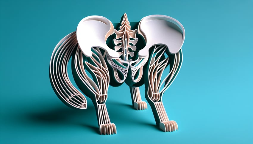
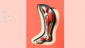
Difficulty: Moderate
2-5mins
Learning OutcomesCheat Sheet
Paper art cat hind leg muscles labeled on teal background for pelvic appendicular muscle quiz.

Ready to flex your feline anatomy skills? This appendicular muscles labeled quiz welcomes vet students and cat enthusiasts alike to test knowledge of pelvic appendicular muscles in a cat. Dive into our cat muscle labeling challenge and explore the cat appendicular skeleton . You'll identify the rectus femoris cat, adductor femoris cat, and master cat leg muscles labeled in our appendicular muscles quiz. Enjoy instant feedback and detailed explanations to reinforce your learning. Perfect for anyone craving a muscles of the leg quiz or leg muscle label practice. Tap "Start" to prove your prowess and pounce on your new skills!

Which muscle is the primary extensor of the stifle joint in the cat hindlimb?
Quadriceps femoris
Biceps femoris
Gastrocnemius
Sartorius
The quadriceps femoris is a group of four muscles responsible for extending the stifle (knee) joint in mammals, including cats. It originates from the femur and pelvis and inserts on the patella. Contraction of this muscle group straightens the stifle. Learn more about the quadriceps femoris
Which muscle originates from the ischial tuberosity and flexes the stifle in cats?
Semitendinosus
Rectus femoris
Tensor fascia latae
Tibialis cranialis
The semitendinosus arises from the ischial tuberosity and inserts on the medial tibia, functioning to flex the stifle and extend the hip. It is one of the hamstring muscles in cats. This muscle also helps adduct the limb. More on the semitendinosus muscle
Which muscle forms the prominent ‘calf’ on the caudal aspect of a cat’s hindlimb?
Gastrocnemius
Gluteus medius
Pectineus
Vastus lateralis
The gastrocnemius is the main superficial muscle of the calf, originating on the femoral condyles and inserting on the calcaneus via the common calcaneal (Achilles) tendon. It plantarflexes the tarsus (hock) and flexes the stifle. This muscle is easily visible in dorsal view. Details on the gastrocnemius muscle
Which muscle abducts the thigh and lies on the lateral aspect of the cat’s pelvis?
Tensor fascia latae
Gracilis
Semimembranosus
Iliopsoas
The tensor fascia latae originates on the ilium and inserts into the fascia lata, assisting in abducting and flexing the hip. It is located on the lateral aspect of the pelvis and helps stabilize the limb during locomotion. Learn more about the tensor fascia latae
Which muscle acts as a major adductor on the medial side of the cat’s thigh?
Gracilis
Sartorius
Gluteus medius
Tibialis cranialis
The gracilis spans from the pubic symphysis to the medial tibia, functioning primarily as an adductor of the thigh. It also assists in stifle flexion and hock extension. Its superficial position makes it easy to identify in dissection. More on the gracilis muscle
Which strap-like muscle runs superficially along the medial thigh of the cat?
Sartorius
Semitendinosus
Vastus medialis
Pectineus
The sartorius is a long, thin muscle that runs from the ilium to the medial tibia. It helps flex the hip and extend the stifle. In cats, it is easily visible as a superficial structure on the medial thigh. Read more about the sartorius
Which muscle has two heads and functions in abducting the thigh and flexing the stifle in cats?
Biceps femoris
Rectus femoris
Gracilis
Pectineus
The biceps femoris has a long and short head originating on the ischial tuberosity and inserting on the patella, tibia, and fascia lata. It abducts the thigh, extends the hip, and flexes the stifle. As part of the hamstrings, it is important for major hindlimb movements. Details on biceps femoris
Which deeper hamstring muscle lies beneath the semitendinosus in cats?
Semimembranosus
Gluteus maximus
Iliopsoas
Vastus intermedius
The semimembranosus lies deep to the semitendinosus, originating on the ischial tuberosity and inserting on the medial condyle of the tibia. It extends the hip and flexes the stifle. Its deep location distinguishes it from its more superficial neighbor. Explore the semimembranosus muscle
From which bone does the rectus femoris originate in the cat hindlimb?
Ilium
Ischium
Pubis
Femur
The rectus femoris, one of the quadriceps muscles, originates from the ilium at the ventral border of the acetabulum. It crosses both the hip and stifle joints, contributing to hip flexion and stifle extension. See rectus femoris details
Which muscle’s tendon contributes to the common calcaneal tendon in cats?
Gastrocnemius
Semimembranosus
Vastus lateralis
Pectineus
The gastrocnemius tendon, along with those of the superficial digital flexor and other small muscles, forms the common calcaneal tendon inserting on the calcaneus. This tendon is crucial for plantarflexion of the hock. More on the common calcaneal tendon
Which muscle acts as a dorsiflexor of the hock (ankle) in the cat?
Tibialis cranialis
Gracilis
Gluteus medius
Biceps femoris
The tibialis cranialis (tibialis anterior in some texts) originates on the tibia and inserts on the first and second metatarsals, pulling the hock into dorsiflexion. It counteracts the plantarflexion caused by the gastrocnemius. Learn about tibialis cranialis
Which small muscle lies deep between the gracilis and sartorius in the cat’s medial thigh?
Pectineus
Vastus medialis
Semitendinosus
Tensor fascia latae
The pectineus is a small adductor arising from the pubis and inserting on the proximal femur. It sits deep between the gracilis and sartorius, assisting in thigh adduction. Details on the pectineus muscle
The iliopsoas in cats is formed by which two muscles?
Psoas major and iliacus
Psoas minor and sartorius
Iliacus and gracilis
Psoas major and gluteus medius
The iliopsoas group consists of the psoas major and the iliacus, which fuse near their insertion on the lesser trochanter of the femur. They are the primary flexors of the hip in cats. Read about the iliopsoas complex
Which head of the quadriceps femoris is located most medially in cats?
Vastus medialis
Vastus lateralis
Rectus femoris
Vastus intermedius
The vastus medialis is the medial portion of the quadriceps group, situated along the inner thigh. It assists in stifle extension and stabilizes the patella medially. More about vastus medialis
What is the primary action of the semimembranosus muscle in cats?
Extends the hip and flexes the stifle
Abducts the thigh
Plantarflexes the hock
Dorsiflexes the hock
The semimembranosus originates on the ischial tuberosity and inserts on the medial tibia, acting to extend the hip and flex the stifle. It also contributes to medial stabilization of the stifle. Details on semimembranosus
Which nerve innervates the quadriceps femoris muscle in the cat?
Femoral nerve
Sciatic nerve
Obturator nerve
Tibial nerve
The femoral nerve provides motor innervation to the quadriceps femoris group, enabling stifle extension. Injury to this nerve impairs the ability to bear weight on the limb. Learn about the femoral nerve
Which artery supplies the adductor muscle group in the cat’s hindlimb?
Obturator artery
Femoral artery
Popliteal artery
Deep femoral artery
The obturator artery, a branch of the internal iliac artery, supplies blood to the medial adductor group in the hindlimb. It travels through the obturator foramen to reach these muscles. More on obturator artery
From which bony landmark does the middle gluteal muscle originate in cats?
Iliac crest and dorsal ilium
Ischial tuberosity
Greater trochanter
Pubic symphysis
The middle gluteal muscle arises from the gluteal surface of the ilium and the iliac crest, inserting on the greater trochanter of the femur to abduct and extend the hip. Learn about middle gluteal anatomy
Which muscle originates on the transverse processes of lumbar vertebrae and inserts on the lesser trochanter of the femur?
Psoas major
Iliacus
Tensor fascia latae
Gluteus maximus
The psoas major originates on the lumbar vertebrae and joins the iliacus to form the iliopsoas, inserting on the lesser trochanter of the femur. It is a powerful hip flexor. View iliopsoas details
Which muscle’s tendon wraps around the lateral malleolus in cats?
Fibularis longus
Gastrocnemius
Tibialis cranialis
Semimembranosus
The fibularis longus tendon passes behind the lateral malleolus before inserting on the plantar metatarsals, aiding in plantarflexion and eversion of the foot. This wrapping mechanism provides mechanical advantage. Learn about fibularis longus
Which of the following is NOT part of the pes anserinus in the cat?
Vastus lateralis
Gracilis
Sartorius
Semitendinosus
The pes anserinus is formed by the tendons of gracilis, sartorius, and semitendinosus inserting on the medial tibia. Vastus lateralis is part of the quadriceps femoris group and does not contribute to this structure. More on pes anserinus
Which muscle is considered part of the hamstring group in cats?
Semimembranosus
Pectineus
Tibialis cranialis
Tensor fascia latae
The semimembranosus is one of the three hamstring muscles (with semitendinosus and biceps femoris) that extend the hip and flex the stifle. It lies deep to the semitendinosus. Learn about the hamstring group
During a biopsy of the iliopsoas muscle in cats, which nerve is at greatest risk of injury?
Femoral nerve
Obturator nerve
Sciatic nerve
Tibial nerve
The iliopsoas is innervated by branches of the femoral nerve. A biopsy in the iliopsoas region risks direct injury to this nerve, which would impair hip flexion and stifle extension. Femoral nerve anatomy
Rupture of the common calcaneal tendon in cats would affect all of the following muscles EXCEPT:
Quadriceps femoris
Gastrocnemius
Superficial digital flexor
Semitendinosus
The common calcaneal tendon is formed by the gastrocnemius, superficial digital flexor, biceps femoris, semitendinosus, and gracilis. The quadriceps femoris does not contribute to this tendon and thus is not affected by its rupture. Common calcaneal (Achilles) tendon
Which muscle originates on the gluteal surface of the ilium and inserts on the greater trochanter in cats?
Middle gluteal
Gluteus maximus
Piriformis
Tensor fascia latae
The middle gluteal muscle arises from the gluteal surface of the ilium and inserts on the greater trochanter of the femur, acting as a powerful hip extensor and abductor. It is the primary gluteal muscle in cats. Details on middle gluteal
0
{"name":"Which muscle is the primary extensor of the stifle joint in the cat hindlimb?", "url":"https://www.quiz-maker.com/QPREVIEW","txt":"Which muscle is the primary extensor of the stifle joint in the cat hindlimb?, Which muscle originates from the ischial tuberosity and flexes the stifle in cats?, Which muscle forms the prominent ‘calf’ on the caudal aspect of a cat’s hindlimb?","img":"https://www.quiz-maker.com/3012/images/ogquiz.png"}

Study Outcomes

  1. Identify Pelvic Appendicular Muscles -

    Recognize the major pelvic appendicular muscles on a labeled cat diagram, reinforcing your knowledge of appendicular muscles labeled in the quiz.

  2. Recall Muscle Origins and Insertions -

    Memorize each muscle's name, origin, and insertion to deepen your understanding of cat leg muscles labeled and their structural relationships.

  3. Differentiate Cat Leg Muscle Groups -

    Distinguish between the various muscles of the pelvis and hindlimb to compare appendicular structures and highlight key anatomical differences.

  4. Explain Function of Rectus Femoris and Adductor Femoris -

    Describe how the rectus femoris cat and adductor femoris cat contribute to limb movement and stability, emphasizing their specific roles in locomotion.

  5. Apply Labeling Skills -

    Use your anatomy knowledge to accurately label muscles on the interactive quiz, reinforcing appendicular muscles quiz skills through hands-on practice.

  6. Assess Your Appendicular Muscle Knowledge -

    Receive instant feedback on your performance to pinpoint areas for improvement and measure your progress in mastering pelvic appendicular muscles.

Cheat Sheet

  1. Rectus Femoris (Rectus Femoris Cat) -

    The rectus femoris is a powerful extensor of the stifle and flexor of the hip in feline appendicular muscles labeled studies (University of Edinburgh Vet School). It originates from the iliac spine and inserts on the patella, creating a straight line over the femur. Use the mnemonic "ReFINE: Rectus Femoris INExtends" to recall its dual joint action.

  2. Adductor Femoris (Adductor Femoris Cat) -

    Adductor femoris cat is key for medial thigh stabilization and leg adduction in cats (Cornell University College of Veterinary Medicine). It arises from the pelvic symphysis and fans out to the femoral shaft. Remember "Add to the middle" to link its adduction function and central origin.

  3. Superficial Gluteal Muscle -

    In appendicular muscles labeled diagrams, the superficial gluteal sits caudal to the ilium, promoting hip extension and abduction (Michigan State University). Its broad origin on the gluteal surface and insertion on the third trochanter resemble the human gluteus maximus. Think "Glutes Go Up" to reinforce its upward pull on the femur.

  4. Tensor Fasciae Latae -

    The tensor fasciae latae in cat leg muscles labeled resources tenses the fascia lata to stabilize the hip and stifle (University of California Davis Vet Anatomy). It springs from the ilium's crest and attaches to the iliotibial band. Use the phrase "TFL Tightens Fascia" to recall its key role in lateral stability.

  5. Iliopsoas Group -

    Iliopsoas comprises psoas major and iliacus, crucial flexors of the hip joint in appendicular muscles quizzes (Texas A&M College of Vet Medicine). Each muscle originates from lumbar vertebrae or ilium and inserts on the lesser trochanter. Picture "Psoas Pulls Up" to remember its essential role in hip flexion.

Powered by: Quiz Maker