Thigh Muscles Practice Quiz
Boost your anatomy skills with guided practice
Editorial: Review CompletedUpdated Aug 26, 2025
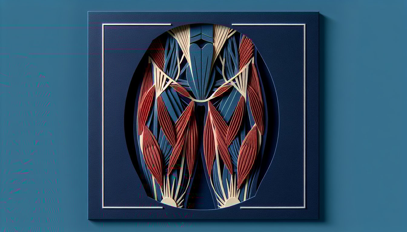
This Thigh Muscles quiz helps you review the main muscles in your thigh, including their names, locations, and actions. Work through 20 quick questions to spot gaps before a test or lab and lock in key facts. Great for Grade 10 anatomy, and useful for anyone who wants a fast practice round.
Study Outcomes
- Identify the major muscles of the thigh, including the quadriceps, hamstrings, and adductors.
- Describe the anatomical locations and functions of each thigh muscle group.
- Differentiate between superficial and deep layers of the thigh muscles.
- Analyze how individual thigh muscles contribute to movement and stability.
- Evaluate potential injury mechanisms associated with thigh muscle strains.
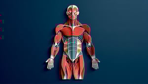
Thigh Muscles Cheat Sheet
- Quadriceps Femoris - The quadriceps femoris sits front and center on your thigh, powering knee extension and hip flexion every time you kick a ball or stand up from a chair. Made up of the rectus femoris, vastus lateralis, vastus medialis, and vastus intermedius, it's basically your thigh's superhero squad. Remember "rectus" means straight, hinting at its direct line down the front.
- Hamstrings - Running down the back of your thigh, the hamstrings handle hip extension and knee flexion, giving you that powerful push-off when you sprint or jump. This trio - biceps femoris, semitendinosus, and semimembranosus - can be remembered by the mnemonic "Boys To Men." Stretch and strengthen them to avoid those dreaded pulls!
- Adductor Group - Hiding on the inner thigh, the adductor group squeezes your legs together for stability and balance. With the adductor longus, brevis, magnus, gracilis, and pectineus, you can recall their names by thinking "Long, Brief, Magnificent Gracious Pectineus." These muscles keep you grounded and graceful.
- Sartorius - The sartorius is the longest muscle in the body, draping diagonally across your thigh like a tailor's ribbon. It helps you flex, abduct, and laterally rotate your hip - and even flex your knee. Named after the Latin word for "tailor," it's your go-to for cross-legged lounging.
- Femoral Triangle - This anatomical "triangle of power" in your upper thigh is bordered by the inguinal ligament, sartorius, and adductor longus. It houses the femoral nerve, artery, and vein - so keep it protected! Remember "SAIL" for Sartorius, Adductor longus, Inguinal Ligament.
- Iliopsoas - Comprising the iliacus and psoas major, the iliopsoas is your prime hip flexor, lifting the thigh toward your torso when you march or climb stairs. Think "Iliopsoas lifts the leg" to lock in that function. Neglect it, and you'll feel the difference in every step!
- Tensor Fasciae Latae - On the outer thigh, the tensor fasciae latae assists with hip abduction and medial rotation, while also stabilizing your pelvis as you walk or run. Its name literally means "tensor of the wide band," referencing its job of tightening the iliotibial band.
- Gracilis - Part of the adductor crew, the gracilis is slender yet mighty, crossing both hip and knee joints to help with hip adduction and knee flexion. Its Latin name means "slender," and it's key for smooth, coordinated leg movements.
- Pectineus - Nestled at the upper, medial thigh, the pectineus flexes and adducts the hip, and even helps rotate the thigh inward. Its name comes from the Latin for "comb," thanks to its comb-like shape. It might be small, but it's crucial for thigh control.
- Nerve Supply - The femoral nerve powers your anterior thigh, the obturator nerve serves the medial compartment, and the sciatic nerve commands the posterior thigh. To keep nerves happy, use the mnemonic "FOSe" for Femoral, Obturator, Sciatic and avoid nerve impingements!