Unlock hundreds more features
Save your Quiz to the Dashboard
View and Export Results
Use AI to Create Quizzes and Analyse Results

Sign inSign in with Facebook
Sign inSign in with Google

Muscle Labeling Practice Test

Master muscle diagrams with our engaging review

Difficulty: Moderate
Grade: Grade 10
Study OutcomesCheat Sheet
Colorful paper art promoting the interactive Muscle Mapping Challenge trivia quiz.

Which muscle is primarily responsible for flexing the forearm at the elbow?
Biceps brachii
Triceps brachii
Deltoid
Pectoralis major
The biceps brachii is the main muscle involved in flexing the forearm at the elbow, clearly distinguishing its role from the muscles located at the back of the arm. Its anatomical position on the anterior aspect of the upper arm makes it immediately identifiable in muscle mapping.
Which muscle is located on the posterior aspect of the upper arm and is key for extending the forearm?
Triceps brachii
Biceps brachii
Deltoid
Latissimus dorsi
The triceps brachii is found on the posterior part of the upper arm and is pivotal for extending the forearm. Its distinct location helps differentiate its function from that of the biceps brachii.
Which muscle is mainly responsible for abducting the arm at the shoulder?
Deltoid
Supraspinatus
Pectoralis major
Latissimus dorsi
The deltoid muscle is the prime mover in abducting the arm at the shoulder due to its broad, rounded structure. Although the supraspinatus initiates the first portion of abduction, the deltoid takes over to complete the movement.
Which major muscle located on the chest is involved in pushing movements?
Pectoralis major
Rectus abdominis
Latissimus dorsi
Serratus anterior
The pectoralis major is a large, fan-shaped muscle on the chest that plays a critical role in pushing movements. Its size and location make it a prominent feature in muscle mapping exercises.
Which muscle group is primarily responsible for extending the spine and maintaining upright posture?
Erector spinae
Rectus abdominis
Latissimus dorsi
Gluteus maximus
The erector spinae is a group of muscles that extends the spine and is fundamental for maintaining proper posture. Its long, columnar arrangement along the back makes it a key focus in muscle mapping tasks.
Which muscle, part of the rotator cuff, is most commonly injured in shoulder impingement syndrome?
Supraspinatus
Infraspinatus
Teres minor
Subscapularis
The supraspinatus muscle plays a key role in the rotator cuff and is especially prone to injury during shoulder impingement. Its anatomical position beneath the acromion contributes to its susceptibility during repetitive overhead movements.
Which large back muscle, often dubbed the 'swimmer's muscle,' has a wide attachment across the back?
Latissimus dorsi
Trapezius
Rhomboids
Deltoid
The latissimus dorsi is a broad, flat muscle that covers a large portion of the back and is pivotal in swimming and pulling movements. Its extensive attachment makes it a central muscle in back mapping exercises.
Which muscle forms the primary barrier between the thoracic and abdominal cavities and is essential for respiration?
Diaphragm
Intercostal muscles
Abdominals
Obliques
The diaphragm is a dome-shaped muscle that plays the most crucial role in breathing by separating the thoracic and abdominal cavities. Its contraction increases the volume of the chest cavity, making it indispensable for respiration.
Which muscle in the calf is primarily responsible for plantarflexion and is often prominently labeled in anatomical diagrams?
Gastrocnemius
Soleus
Tibialis anterior
Peroneus longus
The gastrocnemius is the most visible muscle in the calf and is a key driver in plantarflexion, such as when jumping or running. Its size and superficial location make it a frequent focus in muscle mapping of the lower leg.
Which muscle is the primary hip extensor and is critical for activities such as climbing stairs and standing up?
Gluteus maximus
Gluteus medius
Hamstrings
Adductor magnus
The gluteus maximus is the largest of the gluteal muscles and is primarily responsible for extending the hip during movements like climbing and rising. Its significant size and power are key identifiers in muscle mapping exercises of the lower body.
Which muscle is primarily involved in flexing the vertebral column, commonly resulting in the 'six-pack' appearance?
Rectus abdominis
External oblique
Internal oblique
Transversus abdominis
The rectus abdominis is the primary muscle responsible for flexing the vertebral column and is visibly recognized as the 'six-pack' muscle. Its linear, segmented appearance makes it a distinct landmark in muscle mapping of the torso.
Which muscle is primarily responsible for dorsiflexion of the foot?
Tibialis anterior
Gastrocnemius
Soleus
Peroneus longus
The tibialis anterior, located on the front of the lower leg, is essential for dorsiflexing the foot during walking. Its distinct anatomical location makes it an important muscle to identify in lower limb mapping.
Which muscle is primarily involved in controlling scapular elevation and retraction in the upper back?
Trapezius
Serratus anterior
Latissimus dorsi
Deltoid
The trapezius muscle is key to scapular stabilization, controlling both elevation and retraction of the shoulder blade. Its large size and expansive coverage of the upper back are clearly evident during muscle mapping.
Which muscle is chiefly responsible for lateral rotation of the shoulder joint?
Infraspinatus
Subscapularis
Deltoid
Biceps brachii
The infraspinatus, a part of the rotator cuff, primarily facilitates lateral rotation of the shoulder joint. Its specific attachment to the scapula helps distinguish its role from other muscles around the shoulder.
Which small muscle aids in stabilizing the knee joint by controlling tibial rotation?
Popliteus
Quadriceps femoris
Hamstrings
Gastrocnemius
The popliteus is essential for initiating the unlocking of the knee by controlling the rotation of the tibia. Its unique role in knee stabilization makes it a notable muscle in detailed mapping of the lower extremity.
The biceps brachii has two heads; which option correctly identifies the origin of its long head?
Supraglenoid tubercle of the scapula
Infraglenoid tubercle of the scapula
Coracoid process of the scapula
Acromion of the scapula
The long head of the biceps brachii originates from the supraglenoid tubercle of the scapula, which is a crucial anatomical landmark. Recognizing this origin is important for understanding the muscle's trajectory and function.
Which lower leg muscle exhibits a pennate fiber arrangement that boosts its force production during plantarflexion?
Soleus
Tibialis anterior
Gastrocnemius
Peroneus longus
The soleus muscle has a pennate fiber arrangement, meaning its fibers are oriented at an angle relative to its tendon. This structure allows more fibers to be packed into the muscle, thereby increasing its force output during plantarflexion.
When mapping the gluteal muscles, which function is primarily attributed to the gluteus medius?
Stabilizes the pelvis during walking
Extends the hip
Flexes the hip
Adducts the hip
The gluteus medius is critical for maintaining pelvic stability during walking by preventing excessive tilting of the pelvis. Its function in abduction and stabilization distinguishes it from other gluteal muscles primarily involved in hip movement.
Which of the following muscle and nerve pairings is correct?
Biceps brachii - radial nerve
Deltoid - axillary nerve
Triceps brachii - median nerve
Pectoralis major - long thoracic nerve
The deltoid muscle is correctly paired with the axillary nerve, which is responsible for its motor function. The other pairings are incorrect as they misidentify the primary nerve supply for the associated muscles.
In muscle mapping and coordinated movement, what is the primary role of synergistic muscles?
They work antagonistically to oppose the prime mover.
They provide additional force and stabilize joints during movement.
They are solely responsible for movement independent of prime movers.
They only function during static postures.
Synergistic muscles assist the primary movers by adding extra force and stabilizing the joints during movement. This cooperative action ensures smooth and coordinated motion, reducing the risk of injury.
0
{"name":"Which muscle is primarily responsible for flexing the forearm at the elbow?", "url":"https://www.quiz-maker.com/QPREVIEW","txt":"Which muscle is primarily responsible for flexing the forearm at the elbow?, Which muscle is located on the posterior aspect of the upper arm and is key for extending the forearm?, Which muscle is mainly responsible for abducting the arm at the shoulder?","img":"https://www.quiz-maker.com/3012/images/ogquiz.png"}

Study Outcomes

  1. Identify key human muscles and their anatomical locations accurately.
  2. Map muscle structures on diagrams to reinforce spatial relationships.
  3. Analyze the functional associations between different muscle groups.
  4. Apply anatomical knowledge to effectively prepare for labeling tests and exams.

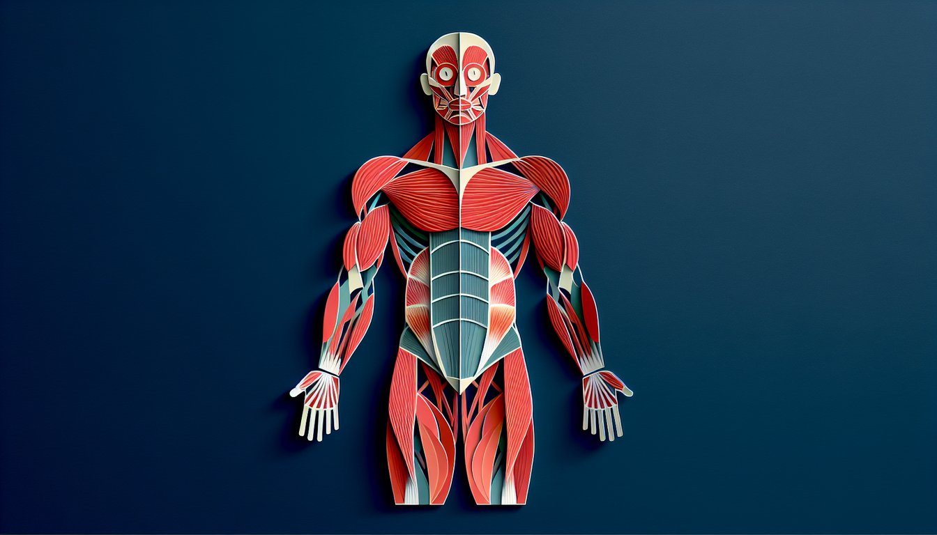
Muscle Labeling Test Cheat Sheet

  1. Identify Major Muscle Groups - Dive into the world of muscle anatomy by exploring key players like the deltoids, pectorals, quadriceps, and hamstrings. Building a solid foundation here will make every workout and movement click into place. Muscle Anatomy Materials
  2. Know Muscle Functions - Understanding what each muscle does (for instance, the biceps brachii flexes the elbow while the triceps brachii extends it) turns confusing terminology into real-life actions. This clarity makes it easier to remember and describe muscle movements. Anatomy Flashcards
  3. Practice Labeling Diagrams - Hands-on practice with muscle diagrams supercharges your visual memory and cements names in your brain. Labeling over and over turns rote memorization into an interactive game you'll actually enjoy. Labeling Exercises
  4. Learn Origins & Insertions - Origins and insertions tell the story of how muscles attach and pull to create movement. Grasping these attachment points deepens your understanding of biomechanics and bodily motion. Origins & Insertions Guide
  5. Use Quizzes & Flashcards - Interactive quizzes and digital flashcards turn studying into a friendly challenge, pinpointing what you know and highlighting what needs more work. Instant feedback is the key to steady improvement. Muscle Quizzes
  6. Master Movement Terminology - Terms like flexion, extension, abduction, and adduction are the vocabulary of muscle action. Once you speak this language, you'll describe exercises and injuries like a pro. Muscle Movements Terminology
  7. Compare Muscle Tissue Types - Skeletal, cardiac, and smooth muscle each have a unique look and job. Spotting their differences reveals why your heart never tires and why your biceps flex on command. Types of Muscle Tissue
  8. Study Neuromuscular Basics - The neuromuscular junction and excitation-contraction coupling are where nerves meet muscles to spark movement. Grasp this connection, and you'll see how brain signals translate into every twitch and lift. Neuromuscular Junction Info
  9. Explore Energy Pathways - ATP and creatine phosphate are the fuels that power your muscle contractions. Knowing how these energy sources work together helps you optimize performance and recovery. Muscle Energy Pathways
  10. Apply Mnemonics - Memory tricks like "SITS" for the rotator cuff - Supraspinatus, Infraspinatus, Teres minor, Subscapularis - turn long lists into short, catchy phrases. These handy shortcuts stick in your mind long after the exam. Rotator Cuff Mnemonics
Powered by: Quiz Maker