Unlock hundreds more features
Save your Quiz to the Dashboard
View and Export Results
Use AI to Create Quizzes and Analyse Results

Sign inSign in with Facebook
Sign inSign in with Google

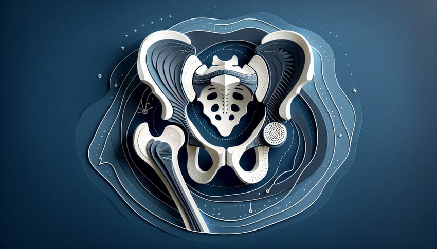
Coxal Bone Practice Quiz Challenge

Enhance bone anatomy mastery with engaging practice.

Difficulty: Moderate
Grade: Grade 10
Study OutcomesCheat Sheet
Paper art representing a trivia quiz on Coxal Bone anatomy for students.

Which three bones fuse to form the coxal bone?
Ilium, Ischium, Pubis
Ilium, Sacrum, Pubis
Ischium, Pubis, Femur
Ilium, Ischium, Sacrum
The coxal bone is formed by the fusion of the ilium, ischium, and pubis bones. This process occurs during adolescence and is fundamental to the structure of the pelvis.
What is the socket of the hip joint called?
Acetabulum
Iliac fossa
Pubic symphysis
Ischial tuberosity
The acetabulum is the concave surface on the coxal bone that articulates with the femoral head, forming the hip joint. Its structure is essential for providing both mobility and stability.
Which portion of the coxal bone forms the broad, upper part of the pelvis?
Ilium
Pubis
Ischium
Acetabulum
The ilium is the large, fan-shaped section of the hip bone that forms the upper part of the pelvis. It provides attachment for numerous muscles and is central to pelvic structure.
What is the joint between the two pubic bones called?
Pubic symphysis
Acetabulum
Iliac crest
Sacroiliac joint
The pubic symphysis is the cartilaginous joint where the two pubic bones meet anteriorly. It allows limited movement and helps absorb shock during movement.
Which part of the coxal bone is most involved in weight-bearing when sitting?
Ischium
Ilium
Pubis
Acetabulum
The ischium is the lower and posterior part of the coxal bone, and its tuberosity bears most of the weight when sitting. Its robust structure is adapted to withstand compressive forces.
What is the name of the large concave surface on the ilium that serves as a major muscle attachment site?
Iliac fossa
True pelvis
Obturator foramen
Acetabulum
The iliac fossa is a broad, concave inner surface of the ilium that provides a large area for muscle attachment. Its curvature is important for the leverage and function of muscles such as the iliacus.
Which bony prominence on the ilium serves as an important landmark for the attachment of the sartorius muscle?
Anterior Superior Iliac Spine (ASIS)
Posterior Superior Iliac Spine (PSIS)
Ischial spine
Iliac crest
The ASIS is a prominent projection at the anterior edge of the ilium and is a key landmark for muscle attachment, including the sartorius. It also is used clinically for measurement and orientation of the pelvis.
Which of the following best describes the primary function of the acetabulum?
It forms a socket for the femoral head, enabling hip joint movement.
It serves as a surface for muscle attachment.
It protects the pelvic organs.
It connects the coxal bones with the sacrum.
The acetabulum is designed as a deep socket that articulates with the head of the femur, forming a stable hip joint. Its shape ensures that the joint has both range of motion and stability during activities.
Which landmark on the coxal bone is a key attachment point for the inguinal ligament?
Anterior Inferior Iliac Spine (AIIS)
Posterior Superior Iliac Spine (PSIS)
Pubic tubercle
Iliac crest
The AIIS is located on the anterior border of the ilium and is an essential landmark where the inguinal ligament attaches. This attachment helps maintain the integrity of the anterior pelvic structure.
Which part of the coxal bone contributes most significantly to the formation of the lateral wall of the pelvic cavity?
Ilium
Pubis
Ischium
Acetabulum
The ilium forms the majority of the lateral wall of the pelvic cavity, providing structural support and anchoring various muscles. Its broad, curved anatomy is essential for the stability and support of the pelvis.
The ischial spine is an important landmark used to assess which of these features?
Pelvic shape and birthing canal dimensions.
Muscle attachment areas for the hamstrings.
Origin of the iliopsoas muscle.
The articulation of the hip joint.
The ischial spine serves as a critical anatomical landmark in evaluating pelvic morphology, important in obstetrics. Its position helps in assessing the dimensions of the pelvic canal which is crucial during childbirth.
What does the pectineal line, found on the superior pubic ramus, provide?
Attachment for the pectineus muscle.
A site for the insertion of the gluteus medius muscle.
A pathway for the sciatic nerve.
Stability to the acetabulum.
The pectineal line is a ridge along the superior pubic ramus that serves as the attachment site for the pectineus muscle. This anatomical feature is important for understanding muscle dynamics in the pelvic region.
Which structure of the coxal bone forms a significant part of the acetabular rim?
The iliopectineal eminence
The ischial tuberosity
The iliac fossa
The pubic symphysis
The iliopectineal eminence contributes to the formation of the acetabular rim, offering structural reinforcement to the socket. This landmark is important both for anatomical orientation and surgical reference.
The ischial tuberosity is primarily known as the attachment site for which muscle group?
Hamstrings
Quadriceps
Abdominals
Adductors
The ischial tuberosity serves as the origin for the hamstring muscles. Its robust, weight-bearing design makes it an essential landmark for lower limb movement during actions such as running and jumping.
Which part of the coxal bone articulates with the sacrum to form the sacroiliac joint?
Ilium
Ischium
Pubis
Acetabulum
The ilium is the part of the coxal bone that joins with the sacrum to form the sacroiliac joint. This joint is crucial for transferring load and maintaining the stability between the spine and the lower limbs.
How does the acetabulum's design enhance the hip joint's stability during dynamic movements?
Its deep, hemispherical shape securely houses the femoral head and includes a labrum for added stability.
It is shallow, allowing a wide range of uncontrolled movement.
It provides minimal contact, relying primarily on ligaments for support.
It is supported mainly by the pelvic floor muscles.
The acetabulum's deep and hemispherical design, combined with the presence of a fibrocartilaginous labrum, helps keep the femoral head tightly in place. This architectural configuration offers both enhanced stability and flexibility during dynamic movements.
What clinical complication could arise from a fracture involving the ilium portion of the coxal bone near the acetabulum?
Instability of the hip joint leading to impaired mobility.
Weakening of the lumbar spine without affecting the hip joint.
Isolated damage to the pubic symphysis.
Purely cosmetic deformity without functional loss.
A fracture near the acetabulum can destabilize the hip joint because it compromises the bony support needed for proper articulation with the femoral head. This instability can lead to significant mobility issues and often requires surgical intervention.
In terms of biomechanical function, how do the ilium and ischium differ in supporting body weight during locomotion?
The ilium mainly supports weight by forming the lateral pelvic wall while the ischium is critical during seated positions.
The ischium supports weight in standing while the ilium is important only in hip rotation.
Both bones contribute equally without specialization.
The ilium supports less weight compared to the ischium in all activities.
The ilium plays a major role in forming the lateral wall of the pelvic cavity and thus supports body weight during standing and walking. In contrast, the ischium, particularly its tuberosity, is adapted to handle load during sitting.
How can variations in the morphology of the coxal bone affect pelvic dimensions relevant to childbirth?
Differences in the shape and size of the coxal bone can alter the pelvic inlet and outlet, impacting labor progress.
Morphological variations only affect aesthetic appearance without functional consequences.
Only abnormalities in the ilium influence childbirth, with no effect from the ischium or pubis.
Pelvic dimensions are exclusively determined by the sacrum, not the coxal bone.
Variations in the morphology of the ilium, ischium, and pubis can modify the dimensions of the pelvic inlet and outlet. Such changes are clinically significant in obstetrics as they can impact the ease or difficulty of childbirth.
How might developmental disorders impact the formation and structural integrity of the coxal bone?
They can result in irregular fusion patterns of the ilium, ischium, and pubis, leading to deformity and reduced joint function.
They only affect the soft tissue structures without altering bone morphology.
They enhance the strength of the coxal bone by promoting additional bone growth.
They solely impair the acetabulum, leaving the rest of the coxal bone unaffected.
Developmental disorders can disrupt the normal fusion process of the ilium, ischium, and pubis, potentially leading to structural deformities. These irregularities compromise the integrity and function of the hip joint, often resulting in clinical problems.
0
{"name":"Which three bones fuse to form the coxal bone?", "url":"https://www.quiz-maker.com/QPREVIEW","txt":"Which three bones fuse to form the coxal bone?, What is the socket of the hip joint called?, Which portion of the coxal bone forms the broad, upper part of the pelvis?","img":"https://www.quiz-maker.com/3012/images/ogquiz.png"}

Study Outcomes

  1. Identify key anatomical landmarks of the coxal bone.
  2. Label major regions of the hip bone on diagrams.
  3. Understand the structural relationships between coxal bone features.
  4. Analyze the functional significance of coxal bone landmarks in hip movement.
  5. Apply anatomical knowledge to answer quiz questions effectively.

Coxal Bone Quiz - Anatomy Review Cheat Sheet

  1. Coxal Bone Fusion - The coxal bone, or hip bone, is actually three bones (ilium, ischium, pubis) elegantly fused together at the acetabulum. This fusion creates a sturdy basin for the hip joint, letting you kick, run, and dance. kenhub hip bone
  2. Kenhub Article
  3. Ilium & Iliac Crest - The ilium is the largest part of the hip bone, crowned by the iliac crest, which you can feel just under your hands when you place them on your waist. It serves as an anchor for core and thigh muscles, making it a hotspot for strength and stability. getbodysmart hip bone intro
  4. GetBodySmart Overview
  5. Ischium & Ischial Tuberosity - The ischium makes up the lower back portion of the hip bone, featuring the ischial tuberosity ("sit bone") that bears your weight when you park yourself on a chair. It also anchors your powerful hamstring muscles for sprinting and leaping. kenhub hip bone
  6. Kenhub Article
  7. Pubis & Pubic Symphysis - The pubis is the front piece of the hip bone, meeting its partner at the pubic symphysis - a slightly movable joint cushioned by cartilage. This gives just enough flexibility to help you walk smoothly and absorb shocks. getbodysmart hip bone intro
  8. GetBodySmart Overview
  9. Acetabulum Socket - The acetabulum is the deep, cup-shaped socket where the head of the femur nests, forming the classic ball-and-socket hip joint. Its snug fit provides both stability for weight-bearing and freedom for a wide range of motion. en.wikipedia acetabulum
  10. Wikipedia: Acetabulum
  11. Obturator Foramen - The obturator foramen is a large opening forged by the ischium and pubis, creating a gateway for nerves and vessels to travel into your legs. A thin membrane covers most of it, making this passage super-efficient yet protected. kenhub hip bone
  12. Kenhub Article
  13. Greater Sciatic Notch - Nestled between the ilium and ischium, the greater sciatic notch transforms into a foramen by ligaments, letting the sciatic nerve - the body's longest nerve - glide down to your thigh. It's a key corridor for nerve traffic control. getbodysmart hip bone intro
  14. GetBodySmart Overview
  15. Ischiofemoral Ligament - This strong band wraps around the back of the hip joint, limiting excessive internal rotation and adduction when your hip is flexed. It's one of the superheroes keeping your hip from over-rotating. en.wikipedia ischiofemoral ligament
  16. Wikipedia: Ischiofemoral Ligament
  17. Iliac Crest Landmark - Stretching from the anterior to posterior superior iliac spines, the iliac crest is a prime real estate for muscle attachments - from your abs to lower back. It's also a go-to surface landmark for clinicians and bodybuilders alike. getbodysmart hip bone intro
  18. GetBodySmart Overview
  19. Ischial Spine - The ischial spine is a sharp projection from the ischium, acting as a pulley point for the sacrospinous ligament. You'll find it sandwiched between the greater and lesser sciatic notches. kenhub hip bone
  20. Kenhub Article
Powered by: Quiz Maker