Unlock hundreds more features
Save your Quiz to the Dashboard
View and Export Results
Use AI to Create Quizzes and Analyse Results

Sign inSign in with Facebook
Sign inSign in with Google

Free High School Anatomy Quiz: Semester 1 Practice Final

Ready for an anatomy practice test? Tackle these anatomy quiz questions now!

Difficulty: Moderate
2-5mins
Learning OutcomesCheat Sheet
Paper art layers of human organs, bones, and muscles for a high school anatomy quiz on dark blue background

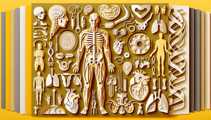
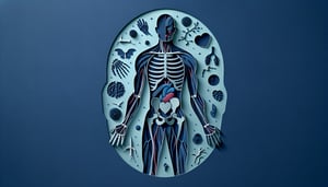
Calling all future biologists and medical whizzes! Ready to test your smarts with a high school anatomy quiz built to help you master your semester 1 anatomy test? This free anatomy quiz challenges you with varied anatomy quiz questions, letting you hone your recall and build confidence. From bones and muscles to vital organs, you'll get hands-on practice for an anatomy practice final. Dive into detailed sections inspired by the anatomy physiology final exam and kickstart your learning with our engaging human anatomy quiz . Jump in now and ace your next test!

What is the basic functional unit of the human body?
Tissue
Cell
Organ
Organ system
The cell is the fundamental unit of structure and function in all living organisms, including humans. Similar cells group together to form tissues, which then organize into organs and organ systems. Cells carry out essential processes such as metabolism, energy production, and reproduction. Mastery of cellular anatomy lays the groundwork for understanding more complex physiological systems. Britannica: Cell Biology
Which tissue type covers body surfaces and lines cavities?
Connective tissue
Nervous tissue
Muscle tissue
Epithelial tissue
Epithelial tissue forms continuous sheets that cover external surfaces and line internal cavities and organs. These cells serve as protective barriers, regulate permeability, and facilitate secretion in glands. The close packing of epithelial cells and presence of tight junctions help maintain their barrier function. This tissue type is essential for protection, absorption, and secretion throughout the body. Innerbody: Epithelial Tissue
What bone is commonly referred to as the collarbone?
Humerus
Scapula
Clavicle
Sternum
The clavicle, or collarbone, is a long, slender bone that connects the sternum to the scapula. It serves as a strut to support the shoulder and allows for a wide range of arm movements. The clavicle also helps protect underlying neurovascular structures. Fractures of the clavicle are common due to its subcutaneous position. Britannica: Clavicle
Which organ is responsible for pumping blood throughout the body?
Brain
Liver
Heart
Lungs
The heart is a muscular organ that contracts rhythmically to pump blood through the blood vessels. It is divided into four chambers: two atria and two ventricles. The coordinated contraction of cardiac muscle ensures blood circulates to the lungs for oxygenation and then to the rest of the body. Proper heart function is vital for delivering oxygen and nutrients to tissues. American Heart Association: How the Heart Works
What is the largest organ of the human body?
Lungs
Skin
Liver
Large intestine
The skin is the largest organ of the human body by surface area and weight. It serves as a protective barrier against pathogens, regulates body temperature, and enables sensory perception. The skin consists of three layers: epidermis, dermis, and hypodermis. Studying skin anatomy is essential for understanding integumentary system health and diseases. Britannica: Integumentary System
What is the name of the tube that connects the throat to the stomach?
Trachea
Larynx
Pharynx
Esophagus
The esophagus is a muscular tube that transports food and liquids from the pharynx (throat) to the stomach. It moves contents via coordinated muscular contractions known as peristalsis. The esophageal lining protects against friction and mechanical damage. Disorders of the esophagus can affect swallowing and digestion. Britannica: Esophagus
Which part of a neuron receives signals from other neurons?
Axon
Cell body
Myelin sheath
Dendrites
Dendrites are branched extensions of a neuron that receive chemical signals from the synaptic terminals of other neurons. These signals are converted into electrical impulses that travel toward the neuron's cell body. The number and complexity of dendrites increase the synaptic connections a neuron can make. Understanding dendrite function is key to neuroanatomy and neural communication. Britannica: Dendrite
What is the name of the process by which a single cell divides to form two identical daughter cells?
Apoptosis
Binary fission
Mitosis
Meiosis
Mitosis is the process of nuclear division that produces two genetically identical daughter cells from a single parent cell. It is essential for growth, tissue repair, and asexual reproduction in multicellular organisms. The process includes phases such as prophase, metaphase, anaphase, and telophase. Understanding mitosis is crucial for cell biology and anatomy studies. Khan Academy: Mitosis
Which mineral is most abundant in the human bone matrix?
Calcium
Potassium
Magnesium
Iron
Calcium phosphate in the form of hydroxyapatite crystals provides strength and rigidity to the bone matrix. Calcium is the most abundant mineral in bones, accounting for about 99% of the body's total calcium. Bones also serve as a reservoir to maintain blood calcium levels. Disorders in calcium metabolism can lead to conditions like osteoporosis. Britannica: Bone Anatomy
What is the primary muscle used for breathing?
Intercostal muscles
Sternocleidomastoid
Diaphragm
Pectoralis major
The diaphragm is a dome-shaped muscle that separates the thoracic cavity from the abdominal cavity. During inhalation, it contracts and flattens, increasing thoracic volume and drawing air into the lungs. It is the most critical muscle for normal respiration. Dysfunction of the diaphragm can lead to breathing difficulties. Britannica: Diaphragm Muscle
Which component of blood is primarily responsible for transporting oxygen?
Red blood cells
White blood cells
Plasma
Platelets
Red blood cells contain hemoglobin, a protein that binds and transports oxygen from the lungs to tissues throughout the body. They have a biconcave shape that maximizes surface area for gas exchange. Red blood cells also help transport a small amount of carbon dioxide back to the lungs. Adequate red blood cell production is vital for maintaining oxygen delivery. Britannica: Red Blood Cell
Which layer of the skin contains hair follicles and sweat glands?
Hypodermis
Epidermis
Stratum corneum
Dermis
The dermis is the middle layer of the skin containing connective tissue, blood vessels, nerves, hair follicles, and sweat glands. It provides structural strength and elasticity due to collagen and elastin fibers. The dermis supports thermoregulation and sensation. Damage to the dermis can affect wound healing and skin function. Britannica: Dermis
What structure in the kidney filters blood to begin urine formation?
Loop of Henle
Collecting duct
Glomerulus
Proximal tubule
The glomerulus is a network of capillaries located in the renal corpuscle of the nephron. It filters blood plasma, allowing water and small solutes to pass into Bowman's capsule while retaining larger molecules and blood cells. This filtration is driven by hydrostatic pressure. Glomerular function is critical for kidney health and waste removal. Khan Academy: Glomerular Filtration
Which blood vessel carries oxygenated blood from the lungs to the heart?
Superior vena cava
Pulmonary vein
Aorta
Pulmonary artery
The pulmonary veins carry oxygen-rich blood from the lungs back to the left atrium of the heart. In humans, there are typically four pulmonary veins, two from each lung. Unlike most veins, pulmonary veins transport oxygenated blood. Proper function is essential for systemic oxygen delivery. Britannica: Pulmonary Vein
Which part of the brain coordinates voluntary muscle movements and balance?
Hypothalamus
Thalamus
Cerebellum
Medulla oblongata
The cerebellum, located at the back of the brain beneath the occipital lobes, fine-tunes voluntary movements and maintains posture and balance. It integrates sensory input with motor commands to ensure smooth, coordinated actions. Damage to the cerebellum can lead to ataxia and coordination difficulties. Understanding cerebellar function is crucial in neuroanatomy and clinical neurology. Britannica: Cerebellum
What term describes programmed cell death essential for development and homeostasis?
Autophagy
Necrosis
Endocytosis
Apoptosis
Apoptosis is a controlled process of cellular self-destruction that removes damaged or unneeded cells without causing inflammation. It plays a key role in development, immune function, and tissue homeostasis. Dysregulation of apoptosis can lead to diseases such as cancer or degenerative disorders. Cells undergoing apoptosis exhibit characteristic morphological changes, including membrane blebbing and DNA fragmentation. Britannica: Apoptosis
Which organ produces the hormone insulin?
Liver
Pancreas
Thyroid gland
Adrenal gland
The pancreas contains clusters of cells called islets of Langerhans, where beta cells secrete insulin. Insulin is essential for regulating blood glucose levels by facilitating cellular glucose uptake. Defects in insulin production or action can lead to diabetes mellitus. Understanding pancreatic anatomy is critical for endocrinology studies. Britannica: Pancreas
Where in the digestive system does most nutrient absorption occur?
Large intestine
Small intestine
Esophagus
Stomach
The small intestine, consisting of the duodenum, jejunum, and ileum, is the primary site for digestion and absorption of nutrients. Its lining has villi and microvilli, which increase surface area to maximize nutrient uptake. Enzymes and bile released into the duodenum aid in macronutrient breakdown. Disorders of the small intestine can impair nutrient absorption and lead to malnutrition. Britannica: Small Intestine
Which valve prevents backflow of blood from the left ventricle to the left atrium?
Tricuspid valve
Aortic valve
Mitral valve
Pulmonary valve
The mitral valve, also known as the bicuspid valve, is located between the left atrium and left ventricle. It opens during atrial systole to allow blood flow into the ventricle and closes during ventricular systole to prevent backflow. Its two leaflets ensure a tight seal under high pressure. Mitral valve dysfunction can lead to regurgitation or stenosis, affecting cardiac output. Britannica: Mitral Valve
What is the functional unit of the kidney?
Lobule
Neuron
Nephron
Alveolus
The nephron is the microscopic functional unit of the kidney responsible for filtering blood, reabsorbing needed substances, and excreting waste as urine. Each human kidney contains approximately one million nephrons. A nephron consists of a glomerulus, renal tubule, and collecting duct. Nephron health is vital for maintaining fluid and electrolyte balance. Britannica: Nephron
Which part of the eye controls the amount of light entering the pupil?
Cornea
Iris
Lens
Retina
The iris is the colored, muscular ring around the pupil that adjusts its diameter to control light entry. In bright conditions, the iris constricts the pupil; in low light, it dilates the pupil. This regulation helps protect the retina and optimize visual clarity. Iris anatomy is important in ophthalmology and clinical examinations. Britannica: Iris
Which hormone regulates the body's sleep-wake cycle?
Thyroxine
Growth hormone
Melatonin
Cortisol
Melatonin is secreted by the pineal gland in response to darkness and helps regulate circadian rhythms, including the sleep-wake cycle. Its production decreases with exposure to light, signaling wakefulness. Disruption in melatonin levels can affect sleep patterns and mood. Melatonin supplements are sometimes used to treat sleep disorders. Britannica: Melatonin
Which type of muscle tissue is involuntary and found in the walls of hollow organs?
Cardiac muscle
Smooth muscle
Striated muscle
Skeletal muscle
Smooth muscle is non-striated and involuntary, found in the walls of hollow organs such as blood vessels, the gastrointestinal tract, and the bladder. It contracts slowly and can maintain tone over extended periods. Smooth muscle cells are spindle-shaped and controlled by the autonomic nervous system. Disorders of smooth muscle function can affect digestion and vascular tone. Britannica: Smooth Muscle
What is the primary role of the sodium-potassium pump in neurons?
Transport neurotransmitters
Catalyze protein synthesis
Generate ATP from ADP
Maintain resting membrane potential
The sodium-potassium pump actively transports three sodium ions out of and two potassium ions into the neuron, using ATP. This activity maintains the negative resting membrane potential essential for excitability. By preserving ionic gradients, the pump enables action potentials to propagate. Dysfunction of this pump can impair neuronal signaling. Khan Academy: Sodium-Potassium Pump
Which type of joint is formed by the shoulder socket?
Hinge joint
Saddle joint
Ball-and-socket joint
Pivot joint
The shoulder (glenohumeral) joint is a ball-and-socket synovial joint where the spherical head of the humerus fits into the glenoid cavity of the scapula. This structure allows the greatest range of motion of any joint in the body, including flexion, extension, abduction, adduction, rotation, and circumduction. Stability is provided by the rotator cuff muscles and ligaments. Due to its mobility, the shoulder joint is prone to dislocation. Britannica: Ball-and-Socket Joint
What is the function of the myelin sheath in neurons?
Produce neurotransmitters
Provide metabolic support
Protect neurons from pathogens
Increase the speed of nerve impulse conduction
The myelin sheath is a lipid-rich wrapping around axons formed by Schwann cells in the peripheral nervous system and oligodendrocytes in the central nervous system. It insulates the axon and allows rapid, saltatory conduction of action potentials between nodes of Ranvier. Efficient impulse transmission is essential for coordinated motor and sensory functions. Demyelinating diseases like multiple sclerosis disrupt this process. Britannica: Myelin
Which artery is most commonly used to measure blood pressure in the arm?
Brachial artery
Femoral artery
Radial artery
Carotid artery
Blood pressure is typically measured at the brachial artery located in the medial aspect of the upper arm. This artery is easily occluded by a sphygmomanometer cuff and provides accurate readings of systemic arterial pressure. The brachial pulse can also be palpated for clinical assessments. Proper cuff placement and technique are crucial for reliable measurements. American Heart Association: Blood Pressure Measurement
What is the primary functional subunit of a muscle fiber called?
Sarcolemma
Myofibril
Sarcomere
Sarcoplasm
The sarcomere is the smallest contractile unit of a muscle fiber, composed of overlapping thick (myosin) and thin (actin) filaments. During muscle contraction, sarcomeres shorten via the sliding filament mechanism. Z-discs define the boundaries of each sarcomere. Coordination of sarcomere shortening across myofibrils produces tension and movement. Khan Academy: Sarcomere Structure
Which neurotransmitter is primarily excitatory in the central nervous system?
GABA
Glutamate
Serotonin
Dopamine
Glutamate is the most abundant excitatory neurotransmitter in the central nervous system, involved in synaptic plasticity, learning, and memory. It activates various receptor types that allow ion flux, depolarizing the postsynaptic membrane. Excessive glutamate release can lead to excitotoxicity and neuronal damage. Balanced glutamate signaling is essential for healthy brain function. Britannica: Glutamate
Which part of the nephron is responsible for concentrating urine?
Loop of Henle
Proximal tubule
Bowman's capsule
Distal tubule
The loop of Henle, particularly its descending and ascending limbs, creates an osmotic gradient in the renal medulla that concentrates urine. Water is reabsorbed in the descending limb, while ions are reabsorbed in the ascending limb. This countercurrent mechanism allows the kidney to conserve water and produce concentrated urine. Disruption of loop function can lead to fluid imbalances. Britannica: Loop of Henle
Which lobe of the brain is primarily responsible for processing visual information?
Parietal lobe
Temporal lobe
Frontal lobe
Occipital lobe
The occipital lobe, located at the back of the cerebral cortex, is the main center for visual processing. It receives and interprets information from the retina via the optic radiations. Damage to this lobe can result in visual field defects and impairments in perception. Studies of the occipital lobe provide insight into how the brain interprets sight. Britannica: Occipital Lobe
Which hormone increases blood calcium levels?
Parathyroid hormone
Aldosterone
Calcitonin
Insulin
Parathyroid hormone (PTH) is secreted by the parathyroid glands and acts to raise blood calcium levels by stimulating bone resorption, increasing renal calcium reabsorption, and activating vitamin D. PTH plays a critical role in calcium homeostasis. Imbalances in PTH can lead to hypercalcemia or hypocalcemia. Understanding PTH is essential in endocrinology and clinical medicine. Britannica: Parathyroid Hormone
What is the innermost layer of the heart wall called?
Endocardium
Epicardium
Myocardium
Pericardium
The endocardium is the thin, smooth inner lining of the heart chambers and valves. It is composed of endothelial cells that reduce friction as blood flows through the heart. Beneath the endocardium lies the myocardium, the muscular layer responsible for contraction. The pericardium, including the epicardium, encloses the heart externally. Britannica: Endocardium
What mechanism allows the thin and thick filaments in muscle fibers to slide past each other during contraction?
Sodium-potassium pump
Calcium-induced calcium release
Sliding filament theory
Allosteric modulation
The sliding filament theory describes how muscle contraction occurs when actin (thin) and myosin (thick) filaments slide past each other, shortening the sarcomere without changing filament length. Cross-bridge cycling powered by ATP enables this sliding motion. Calcium ions trigger the interaction by exposing binding sites on actin. This mechanism is fundamental in muscle physiology. NCBI: Sliding Filament Theory
Which reflex arc is monosynaptic, involving only one synapse between sensory and motor neurons?
Golgi tendon reflex
Withdrawal reflex
Pupillary light reflex
Patellar (knee-jerk) reflex
The patellar reflex, also known as the knee-jerk reflex, is a monosynaptic stretch reflex. When the patellar tendon is tapped, sensory neurons synapse directly on motor neurons in the spinal cord, causing quadriceps contraction. This simple circuit has only one synapse and no interneurons. It is commonly tested clinically to assess nervous system integrity. Britannica: Monosynaptic Reflex
What primary structure constitutes the blood-brain barrier?
Astrocyte end-feet
Tight junctions between endothelial cells
Pericyte layers
Oligodendrocyte myelin
The blood-brain barrier is formed by tight junctions between endothelial cells lining cerebral capillaries. These junctions restrict the passage of most substances from the bloodstream into the brain extracellular fluid. Astrocyte end-feet support barrier function but do not form the junctions themselves. The BBB protects neural tissue and maintains a stable environment. Britannica: Blood-Brain Barrier
How many pairs of cranial nerves are present in the human nervous system?
14
10
8
12
Humans have 12 pairs of cranial nerves that arise directly from the brain and brainstem. These nerves serve sensory, motor, or mixed functions, including smell (I), vision (II), facial movement (VII), and more. Each nerve is numbered based on its rostral-caudal position. Knowledge of cranial nerves is essential in neurology. Britannica: Cranial Nerve
0
{"name":"What is the basic functional unit of the human body?", "url":"https://www.quiz-maker.com/QPREVIEW","txt":"What is the basic functional unit of the human body?, Which tissue type covers body surfaces and lines cavities?, What bone is commonly referred to as the collarbone?","img":"https://www.quiz-maker.com/3012/images/ogquiz.png"}

Study Outcomes

  1. Identify Cellular Structures -

    Recognize major cell organelles and their functions as covered in semester 1 anatomy test, enabling accurate responses to quiz questions.

  2. Differentiate Tissue Types -

    Distinguish between epithelial, connective, muscle, and nervous tissues and describe their key characteristics for high school anatomy quiz scenarios.

  3. Explain Bone Physiology -

    Articulate the structure, growth, and remodeling processes of bones as tested in anatomy practice final questions.

  4. Apply Anatomical Terminology -

    Use correct anatomical terms for body regions, cavities, and directional planes when answering free anatomy quiz items.

  5. Assess Knowledge Gaps -

    Analyze instant feedback to identify strengths and areas for improvement, guiding your study plan for semester 1 anatomy test success.

  6. Reinforce Key Concepts -

    Review correct and incorrect responses to reinforce core concepts from cellular structure to bone physiology and boost your confidence.

Cheat Sheet

  1. Cell Anatomy Essentials -

    Understanding organelles like the nucleus, mitochondria, and ribosomes is crucial for your high school anatomy quiz. Use the mnemonic "MR. NERd" (Mitochondria, Ribosomes; Nucleus, Endoplasmic reticulum) to recall their primary functions: energy production, protein synthesis, and genetic control. Supplement your review with diagrams from reliable sources like Khan Academy or university biology departments to reinforce semester 1 anatomy test topics.

  2. Tissue Type Classification -

    Memorize the four primary tissue types - epithelial, connective, muscle, and nervous - and their key characteristics. For example, simple squamous epithelium (one cell layer) facilitates diffusion in alveoli, while connective tissue's extracellular matrix provides support. A handy acronym "ECMN" streamlines recall for your anatomy practice final.

  3. Integumentary System Layers -

    Recall the three skin layers - epidermis, dermis, and hypodermis - and their distinct roles in protection, sensation, and thermoregulation. The phrase "Every Day Heals" can help you remember Epidermis, Dermis, Hypodermis in order. Cross-check free anatomy quiz resources from medical school websites for detailed histology images.

  4. Bone Structure & Types -

    Classify bones into long, short, flat, and irregular categories: long bones (femur) for leverage, short bones (carpals) for stability, flat bones (skull) for protection, and irregular bones (vertebrae) for complex shapes. Remember that bone matrix is roughly 70% mineral (hydroxyapatite) and 30% collagen, which ensures strength and flexibility. Use official NIH diagrams to solidify your knowledge before tackling anatomy quiz questions.

  5. Sliding Filament Muscle Theory -

    Understand how an action potential triggers Ca²❺ release, exposing actin binding sites so myosin heads initiate the power stroke using ATP. The mnemonic "Cool Athletes Can Move Rapidly" stands for Ca²❺ release, Actin exposure, Cross-bridge formation, Myosin stroke, Release for quick recall. Flashcards from university physiology courses can boost retention for your free anatomy quiz.

Powered by: Quiz Maker