Unlock hundreds more features
Save your Quiz to the Dashboard
View and Export Results
Use AI to Create Quizzes and Analyse Results

Sign inSign in with Facebook
Sign inSign in with Google

Free Wrist Bones Quiz: How Well Do You Know Your Wrist Anatomy?

Ready for a hand anatomy quiz? Test your carpal bones knowledge now!

Editorial: Review CompletedCreated By: Saleh El SaidaweyUpdated Aug 28, 2025
Difficulty: Moderate
2-5mins
Learning OutcomesCheat Sheet
Paper art illustration for carpal bones quiz on dark blue background

This wrist bones quiz helps you practice naming the carpals and spot each bone on sight. Use it to find gaps before an exam and feel solid on the basics. For more practice, try the forearm and wrist quiz or the hand bones quiz .

How many carpal bones are in the human wrist?
9
6
8
7
The human wrist contains eight carpal bones arranged in two rows of four bones each, allowing complex movements and articulations with the forearm bones. These small bones provide the stable yet flexible architecture required for wrist function. Identifying the total number of carpal bones is fundamental for understanding hand anatomy.
How many rows of carpal bones are there in the wrist?
2
3
4
1
The carpal bones in the wrist are organized into two distinct rows: the proximal and distal rows. This arrangement helps distribute forces and allows for the wrist's range of motion. Each row consists of four bones that articulate with each other and with the radius or metacarpals.
Which carpal bone has a hook-like process on its palmar surface?
Pisiform
Hamate
Scaphoid
Lunate
The hamate bone features a prominent hook, known as the hamulus, on its palmar surface, which serves as an attachment site for ligaments and the flexor retinaculum. This distinctive feature aids in identifying the hamate in palpation and imaging. Fractures of the hook of the hamate are clinically significant injuries in athletes using bats or rackets.
Which carpal bone is considered a sesamoid bone embedded in a tendon?
Pisiform
Trapezoid
Capitate
Lunate
The pisiform is a small sesamoid bone embedded within the tendon of the flexor carpi ulnaris muscle. It acts to improve the mechanical advantage of this tendon and serves as an attachment point for ligaments. The pisiform is easily palpated on the ulnar side of the wrist.
Which carpal bone articulates with the first metacarpal?
Capitate
Hamate
Trapezium
Trapezoid
The trapezium sits at the base of the thumb and articulates directly with the first metacarpal, forming the carpometacarpal joint of the thumb. This saddle joint allows for the thumb's wide range of motion. The trapezium's unique shape facilitates opposition and precision grip.
Which carpal bone is most commonly palpable at the base of the anatomical snuffbox on the radial side?
Triquetrum
Pisiform
Scaphoid
Lunate
The scaphoid forms the floor of the anatomical snuffbox and is palpable in this region. Clinically, tenderness in the snuffbox after trauma raises suspicion for a scaphoid fracture. Early diagnosis is crucial to prevent complications like avascular necrosis.
What type of synovial joint is the radiocarpal (wrist) joint?
Hinge joint
Saddle joint
Pivot joint
Condyloid joint
The radiocarpal joint is classified as a condyloid (ellipsoid) synovial joint, allowing flexion, extension, abduction, and adduction of the wrist. This joint involves the distal radius and the proximal row of carpal bones (scaphoid, lunate, triquetrum). Its structure provides stability while permitting a wide range of motion.
Which carpal bone articulates with the base of the second metacarpal?
Trapezoid
Trapezium
Lunate
Capitate
The trapezoid is the smallest bone in the distal carpal row and articulates with the base of the second metacarpal. Its wedge shape provides stability to the index finger's carpometacarpal joint. Understanding these articulations is important for diagnosing wrist injuries.
Which artery primarily supplies the scaphoid bone?
Interosseous artery
Radial artery
Median artery
Ulnar artery
The scaphoid's blood supply is predominantly from branches of the radial artery entering distally. Proximal pole fractures can disrupt this supply and lead to avascular necrosis. Recognizing the vascular pattern is crucial in managing scaphoid injuries.
What is the most common site of fracture in the scaphoid?
Proximal pole
Waist
Distal pole
Tubercle
The waist of the scaphoid, located between its proximal and distal poles, is the most frequent fracture site. Fractures here account for the majority of scaphoid injuries due to the bone's narrow waist. Prompt diagnosis and immobilization are essential to prevent nonunion.
Which ligament connects the scaphoid and lunate bones?
Ulnocarpal ligament
Radiocarpal ligament
Lunotriquetral ligament
Scapholunate ligament
The scapholunate ligament stabilizes the scaphoid and lunate, maintaining carpal alignment. Injury to this ligament can cause wrist instability and degenerative arthritis if left untreated. Clinical tests like the Watson shift assess its integrity.
In which row of carpal bones is the capitate located?
Distal row
Neither row
Proximal row
Both rows
The capitate is the largest carpal bone and sits in the center of the distal row. It articulates proximally with the lunate and scaphoid, and distally with the third metacarpal. Its position is key to central wrist stability.
Which carpal bone is most commonly dislocated?
Hamate
Capitate
Scaphoid
Lunate
The lunate is the most frequently dislocated carpal bone, often in a volar direction following trauma. Lunate dislocations can compress the median nerve and disrupt carpal stability. Emergency reduction is critical to prevent complications.
The pisiform articulates on the palmar surface of which carpal bone?
Trapezium
Lunate
Scaphoid
Triquetrum
The pisiform sits on the palmar surface of the triquetrum and increases the leverage of the flexor carpi ulnaris tendon. Its sesamoid nature provides mechanical advantage for wrist flexion. Palpation of the pisiform helps assess ulnar-sided wrist pain.
Which nerve passes through the carpal tunnel?
Musculocutaneous nerve
Ulnar nerve
Median nerve
Radial nerve
The median nerve travels through the carpal tunnel alongside the flexor tendons, supplying motor and sensory innervation to the hand. Compression of this nerve causes carpal tunnel syndrome, characterized by numbness and tingling in the lateral three and a half digits. Early intervention can prevent permanent nerve damage.
The hamate bone articulates with which metacarpal?
Fifth metacarpal
Fourth metacarpal
Second metacarpal
Third metacarpal
The hamate's proximal articulation includes the triangular facet for the fourth metacarpal and a separate facet for the fifth metacarpal. The hook of the hamate serves as an attachment for the flexor retinaculum and ulnar collateral ligament. Recognizing its articulations helps diagnose ulnar-sided wrist injuries.
Which ligament forms the roof of the carpal tunnel?
Transverse carpal ligament (flexor retinaculum)
Palmar radiocarpal ligament
Dorsal radiocarpal ligament
Ulnar collateral ligament
The transverse carpal ligament, also known as the flexor retinaculum, forms the roof of the carpal tunnel. It spans from the pisiform and hook of the hamate to the scaphoid and trapezium. This ligament maintains the carpal arch and confines the flexor tendons and median nerve.
Which two carpal bones articulate directly with the distal end of the radius?
Capitate and Hamate
Scaphoid and Triquetrum
Lunate and Triquetrum
Scaphoid and Lunate
The scaphoid and lunate bones articulate with the distal radius at the radiocarpal joint. The triquetrum contacts the articular disc of the distal radioulnar joint rather than directly with the radius. Understanding these articulations is essential for interpreting wrist biomechanics.
Which carpal bones ossify first during human development?
Trapezium and Trapezoid
Scaphoid and Lunate
Pisiform and Triquetrum
Capitate and Hamate
The capitate and hamate are the first carpal bones to ossify, typically appearing at birth or shortly thereafter. Other carpal bones ossify at various ages during childhood. Knowledge of ossification timelines assists in pediatric radiographic assessment.
The distal radioulnar joint is classified as which type of synovial joint?
Hinge joint
Pivot joint
Ball and Socket joint
Saddle joint
The distal radioulnar joint is a pivot synovial joint allowing pronation and supination of the forearm. It is formed by the head of the ulna and the ulnar notch of the radius, stabilized by the triangular fibrocartilage complex. Its design permits rotation while maintaining stability.
Which carpal bone does NOT articulate with the third metacarpal?
Trapezium
Hamate
Capitate
Trapezoid
The third metacarpal articulates primarily with the capitate. The trapezoid articulates with the second metacarpal, while the trapezium articulates with the first metacarpal. The hamate has facets for the fourth and fifth metacarpals.
In a carpal tunnel (tangential) radiographic view, which bone's hook is best visualized?
Pisiform
Trapezium
Hamate
Scaphoid
The tangential, or carpal tunnel, view is designed to visualize the hook of the hamate and the pisiform. This specialized projection helps detect hamate hook fractures. Proper positioning is critical for accurate imaging.
Which nerve is most at risk with a fracture of the hook of the hamate?
Radial nerve
Musculocutaneous nerve
Ulnar nerve
Median nerve
The ulnar nerve passes adjacent to the hook of the hamate within Guyon's canal. A fracture of the hook can damage or compress the nerve, leading to sensory and motor deficits in the ulnar distribution of the hand. Early recognition and management are essential.
Which tendon lies superficial to the flexor retinaculum of the wrist?
Flexor carpi ulnaris tendon
Extensor carpi radialis longus tendon
Palmaris longus tendon
Flexor digitorum superficialis tendon
The palmaris longus tendon crosses the wrist superficial to the flexor retinaculum in most individuals. It is often used as a surgical tendon graft due to its expendability. Not all people have a palmaris longus, but when present it is visible with wrist flexion.
Which carpal bone directly articulates with the triangular fibrocartilage complex (TFCC)?
Lunate
Hamate
Pisiform
Triquetrum
The TFCC sits between the distal ulna and the proximal carpal row, articulating directly with the ulnar side of the triquetrum. It stabilizes the distal radioulnar joint and transmits load from the hand to the forearm. Injuries to the TFCC often cause ulnar-sided wrist pain.
0
{"name":"How many carpal bones are in the human wrist?", "url":"https://www.quiz-maker.com/QPREVIEW","txt":"How many carpal bones are in the human wrist?, How many rows of carpal bones are there in the wrist?, Which carpal bone has a hook-like process on its palmar surface?","img":"https://www.quiz-maker.com/3012/images/ogquiz.png"}

Study Outcomes

  1. Identify Carpal Bones -

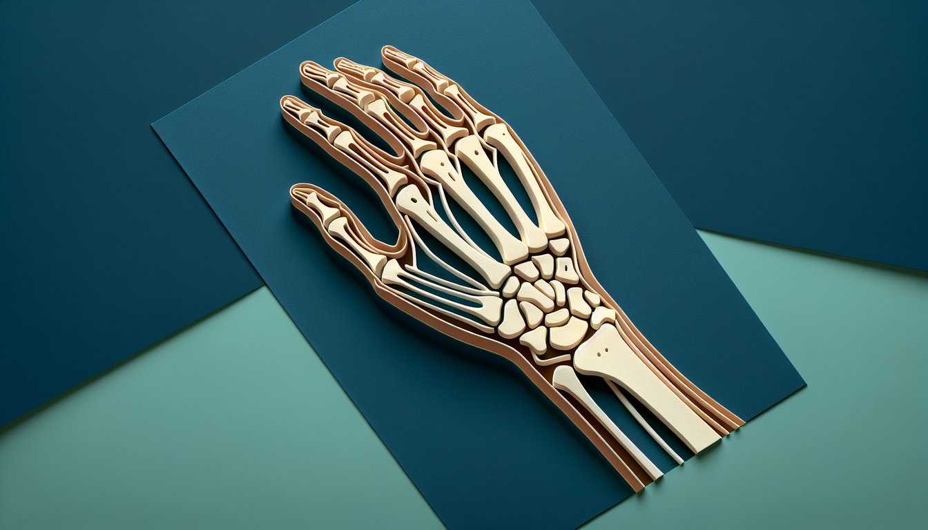
    Visually pinpoint and name each of the eight carpal bones featured in the wrist bones quiz, strengthening your grasp of hand anatomy.

  2. Understand Joint Articulations -

    Explain the primary articulations and movements of the wrist joints, preparing you for the carpal bones quiz challenges.

  3. Analyze Ligament and Muscle Roles -

    Examine how ligaments and muscle attachments support wrist stability and facilitate motion.

  4. Apply Knowledge to Injury Prevention -

    Utilize your understanding of wrist anatomy to recommend strategies for avoiding common wrist injuries and enhancing performance.

  5. Evaluate Carpal Alignment -

    Assess how carpal bone alignment shifts during various hand movements, improving your interpretation skills for the wrist anatomy challenge.

  6. Recall Clinical Relevance -

    Describe the clinical importance of wrist anatomy in diagnosing and treating fractures and disorders of the carpal bones.

Cheat Sheet

  1. Carpal Bone Rows & Mnemonic -

    Memorize the proximal and distal carpal bones in order using "Some Lovers Try Positions That They Can't Handle," which stands for Scaphoid, Lunate, Triquetrum, Pisiform (proximal) and Trapezium, Trapezoid, Capitate, Hamate (distal). This classic mnemonic, endorsed by leading anatomy courses, makes recalling all eight bones in our wrist bones quiz a breeze.

  2. Scaphoid Anatomy & Clinical Significance -

    The scaphoid lies in the anatomical snuffbox and relies on a retrograde blood supply from the radial artery, making fractures prone to avascular necrosis. Recognizing its shape and location is critical in any carpal bones quiz and guides prompt diagnosis in wrist trauma.

  3. Radiocarpal & Midcarpal Joint Movements -

    The wrist comprises the radiocarpal and midcarpal joints, allowing approximately 80° of flexion, 70° of extension, and 20° of ulnar deviation. Understanding these articulations is key in our hand anatomy quiz for grasping how carpal bones coordinate during motion.

  4. Key Wrist Ligaments & Stability -

    The scapholunate and lunotriquetral ligaments maintain carpal alignment and prevent dorsal intercalated segment instability (DISI). Familiarity with dorsal and palmar wrist ligaments boosts your wrist anatomy challenge performance and supports effective injury prevention.

  5. Carpal Tunnel & Flexor Retinaculum -

    The transverse carpal ligament attaches to the tubercles of the trapezium, hook of the hamate, and pisiform, forming the roof of the carpal tunnel. Knowing these attachment points and the tunnel's contents - median nerve and flexor tendons - is essential for acing our wrist bones quiz and understanding carpal tunnel syndrome.

Powered by: Quiz Maker