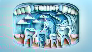
Are you ready to master teeth labeling and deepen your grasp of dental anatomy? Our Master Teeth Labeling Quiz is perfect for students, budding hygienists, educators, and trivia buffs ready to test every crown, root, and canal. In this tooth identification quiz, you'll learn to name the parts of a tooth - from enamel and dentin to pulp chamber - while sharpening your precision. Need a quick review on teeth label positions? Explore our labelling of teeth guide, then dive into morphology of the teeth insights to boost your accuracy. Ready to put your skills to the test on our interactive teeth labeling diagram? Let's begin the challenge now!
What is the hard, outermost layer covering the anatomical crown of a tooth?
Enamel
Dentin
Cementum
Pulp
Enamel is the hardest, most mineralized tissue in the human body and covers the anatomical crown of the tooth. It protects underlying dentin and pulp from physical and chemical damage. Enamel is composed primarily of hydroxyapatite crystals. Learn more about enamel composition.
Which part of the tooth contains nerves and blood vessels?
Pulp chamber
Enamel
Crown
Cementum
The pulp chamber houses the dental pulp, which includes nerves, blood vessels, and connective tissues. It is located centrally in the tooth under enamel and dentin. The pulp supplies nutrients and sensory function to the tooth. More on pulp anatomy.
What structure covers the root of a tooth and helps anchor it within the alveolus?
Cementum
Enamel
Dentin
Periodontal ligament
Cementum is a calcified connective tissue covering the tooth root, providing attachment for periodontal ligament fibers. It helps seal the dentin tubules in the root area. Cementum is less mineralized than enamel but essential for tooth stability. Read about cementum function.
Which tissue lies between the cementum of the root and the alveolar bone?
Periodontal ligament
Gingiva
Enamel
Dentin
The periodontal ligament is a fibrous connective tissue that attaches cementum to alveolar bone. It absorbs occlusal forces and helps maintain tooth position. The PDL also contains blood vessels and nerves for support and proprioception. More on the periodontal ligament.
What is the term for the biting edge of anterior teeth?
Incisal edge
Cusp tip
Occlusal surface
Root surface
The incisal edge is the cutting or biting surface of incisors and canines. It is formed by the junction of the labial and lingual surfaces. This edge assists in shearing food during biting. Details on tooth surfaces.
What is the name of the junction between the enamel and cementum?
Cementoenamel junction
Dentinoenamel junction
Enamel-dentin junction
Cephalometric plane
The cementoenamel junction (CEJ) is the anatomical boundary where enamel of the crown meets cementum on the root. It is also known as the cervical line. The CEJ marks the division between crown and root and is clinically important in periodontal assessments. Read more about the CEJ.
Which of the following surfaces faces the cheeks on posterior teeth?
Buccal surface
Lingual surface
Mesial surface
Distal surface
The buccal surface of posterior teeth faces the cheeks. In anterior teeth, this same surface is termed the labial surface. It plays a key role in mastication and oral hygiene access. More on tooth surfaces.
What anatomical feature is the raised part on the chewing surface of premolars and molars?
Cusp
Fissure
Pit
Groove
A cusp is a pointed or rounded elevation on the chewing surface of premolars and molars. Cusps help in grinding and tearing food. Molars typically have multiple cusps, while premolars have two cusps each. Details on cusps and their functions.
Which part of the pulp extends into the roots of a tooth?
Root canal
Pulp horn
Crown chamber
Cementoenamel junction
The root canal is the portion of the pulp cavity that extends from the pulp chamber down through the root. It contains nerves and blood vessels that supply the tooth. Root canals are treated during endodontic therapy to remove infected tissue. Learn about root canal anatomy.
What is the anatomical depression found near the tip of a cusp called?
Pit
Fossa
Groove
Ridge
A pit is a small pinpoint depression on the occlusal surface, often at the junction of developmental grooves. Pits can trap plaque and are common sites for dental caries. They differ from fossae, which are broader depressions. More on occlusal anatomy.
What specialized cells line the pulp chamber and are responsible for forming dentin?
Odontoblasts
Ameloblasts
Osteoblasts
Cementoblasts
Odontoblasts are columnar cells responsible for the formation of dentin throughout life. They line the periphery of the pulp chamber and extend processes into dentinal tubules. Ameloblasts, by contrast, form enamel and are lost after crown formation. Read on odontoblast function.
What is the term for the space on a radiograph where the periodontal ligament appears as a thin radiolucent line?
PDL space
Lamina dura
Alveolar crest
Cortical bone
The periodontal ligament space appears as a thin radiolucent line between the cementum and lamina dura on radiographs. It represents the soft tissue ligament fibers. The lamina dura is the radiopaque line adjacent to it, representing bone. Assessment of PDL space is important in diagnosing trauma or infection. See radiographic features of PDL.
Which anatomical landmark on a molar separates the chewing surface into mesial and distal fossae?
Central groove
Buccal pit
Transverse ridge
Oblique ridge
The central groove runs mesiodistally across the occlusal surface, dividing it into mesial and distal fossae. It is a developmental groove formed during tooth morphogenesis. The oblique and transverse ridges are raised enamel lines on molars but do not bisect the surface similarly. Glossary of endodontic terms.
What name is given to the tiny channels within dentin that extend from the pulp to the enamel or cementum?
Dentinal tubules
Canaliculi
Haversian canals
Lacunae
Dentinal tubules are microscopic channels running through dentin, housing odontoblastic processes. They transmit sensory stimuli and fluid between the pulp and enamel or cementum. Canaliculi and Haversian canals are found in bone tissue, not dentin. More on dentin structure.
Which developmental cell type secretes enamel matrix and is lost after tooth eruption?
Ameloblasts
Odontoblasts
Cementoblasts
Fibroblasts
Ameloblasts are specialized epithelial cells that form enamel by secreting enamel proteins during tooth development. After enamel formation is complete and the tooth erupts, ameloblasts degenerate and are not present in mature teeth. This is why enamel cannot regenerate. Details on amelogenesis.
0
{"name":"What is the hard, outermost layer covering the anatomical crown of a tooth?", "url":"https://www.quiz-maker.com/QPREVIEW","txt":"What is the hard, outermost layer covering the anatomical crown of a tooth?, Which part of the tooth contains nerves and blood vessels?, What structure covers the root of a tooth and helps anchor it within the alveolus?","img":"https://www.quiz-maker.com/3012/images/ogquiz.png"}
Study Outcomes
Understand Tooth Anatomy -
Describe the main components of a tooth - from enamel to pulp - and their functions within overall tooth structure.
Identify Tooth Parts -
Recognize and name key areas such as cusps, root canal, and periodontal ligament using a teeth labeling diagram.
Label Teeth Accurately -
Complete a tooth identification quiz by correctly matching labels to corresponding tooth parts.
Differentiate Tooth Layers -
Distinguish between enamel, dentin, cementum, and pulp based on composition and protective roles.
Apply Dental Terminology -
Use precise anatomical terms to enhance your communication and knowledge in tooth labeling.
Cheat Sheet
Enamel: The Super Shield -
Enamel is the hardest substance in the human body, composed of about 96% hydroxyapatite crystals, making it your tooth's first line of defense (American Dental Association). When you tackle a teeth labeling diagram, look for this glossy, white outer layer covering the crown. A handy mnemonic is "E-N-A-M-E-L: Every Nice Athlete Makes Every Lap" to recall it's the topmost layer.
Dentin: The Sensitive Support -
Directly beneath enamel lies dentin, a porous tissue with microscopic tubules that transmit sensations, sourced from odontoblasts (Journal of Dental Research). In a tooth identification quiz, dentin appears slightly darker and forms the bulk of the tooth structure. Remember "Denti-NET" to link dentin's network of nerve pathways for sensitivity.
Pulp Chamber: The Living Core -
The pulp contains nerves, blood vessels, and connective tissue, keeping the tooth alive and responsive (University of Michigan School of Dentistry). When you name the parts of a tooth, the pulp chamber is the central space extending into root canals. Think "POP" (Pulp = Organizing Power) to emphasize its vital role.
Cementum & Periodontal Ligament: Root Anchors -
Cementum is a calcified layer covering the root, working with the periodontal ligament fibers to secure teeth in the jawbone (Journal of Periodontology). In your tooth identification quiz, these parts sit below the cementoenamel junction (CEJ) and attach to alveolar bone. Use "C & P Lock" (Cementum and Periodontal ligament locking teeth in place) to nail this pair.
Tooth Numbering Systems: ID Made Easy -
Familiarize yourself with the Universal and FDI systems to ace any teeth label challenge - Universal uses numbers 1 - 32, while FDI uses quadrant+tooth codes (WHO guidelines). Practicing on a teeth labeling diagram helps cement which code matches each tooth. A quick mnemonic is "F-DI goes First-Distrib, U-NI goes Universal-Numerical" to sort them out.