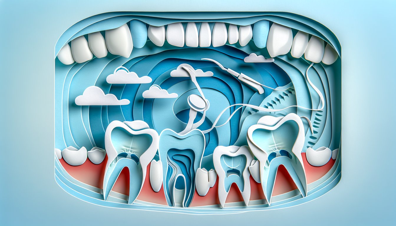
Ready to sharpen your knowledge with our free dental anatomy molars quiz? Whether you're a dental student studying upper molars anatomy or a keen learner exploring lower molars cusps and chewing with premolars train maxilla, this molars identification quiz puts your skills to the test. Dive into interactive teeth labeling challenges and explore the intricate morphology of the teeth . You'll uncover the secrets of cusps, ridges, and chewing pathways while building confidence in molar structure. Ready to prove your expertise? Start now and conquer every cusp!
Which tooth is designated as the adult maxillary right first molar in the Universal Numbering System?
#3
#2
#14
#19
In the Universal Numbering System, tooth #3 corresponds to the maxillary right first molar. This system is widely used in North America to identify permanent teeth. It numbers the upper right quadrant from 1 to 8, then continues around the arch. More on tooth numbering.
How many major cusps does a typical maxillary first molar have?
2
3
4
5
A typical maxillary first molar has four major cusps: mesiobuccal, distobuccal, mesiolingual, and distolingual. Occasionally a fifth cusp (Cusp of Carabelli) may be present, but it's not considered a primary cusp. The four major cusps form the primary occlusal table. Read about molar cusp anatomy.
The largest cusp on a mandibular first molar is:
Mesiobuccal cusp
Mesiolingual cusp
Distobuccal cusp
Distolingual cusp
On the mandibular first molar, the mesiolingual cusp is the largest and most prominent cusp. It plays a major role in grinding food along with the other buccal and distal cusps. Its size helps maintain proper occlusion with maxillary molars. Lower molar cusp details.
The Cusp of Carabelli is most frequently found on which molar?
Maxillary first molar
Maxillary second molar
Mandibular first molar
Mandibular second molar
The Cusp of Carabelli is an additional morphological feature found on the mesiolingual surface of maxillary first molars. It is a variable trait, present in many individuals as a small tubercle or groove. This cusp is absent on mandibular molars and maxillary second molars. Learn about the Carabelli cusp.
True or False: Molars are succedaneous teeth.
False
True
Succedaneous teeth replace deciduous teeth, whereas molars erupt posterior to the primary dentition and do not replace any primary teeth. Therefore, molars are non-succedaneous. Primary molars are replaced by premolars, not permanent molars. Succedaneous vs non-succedaneous.
Which groove separates the mesial and distal cusps on a mandibular second molar’s occlusal surface?
Central groove
Transverse groove
Oblique groove
Buccal groove
On mandibular molars, the central groove runs mesiodistally between the mesial and distal cusp pairs on the occlusal surface. It’s the main groove that defines the occlusal table. Supplementary grooves branch off the central groove but do not separate the major cusp groups. Occlusal groove patterns.
How many roots does a maxillary first molar typically have?
1
2
3
4
Maxillary first molars typically have three roots: two buccal roots (mesiobuccal and distobuccal) and one palatal root. This tri-rooted configuration helps anchor the tooth in the maxilla. Variations exist, but three roots are standard. Molar root anatomy.
The Y-shaped occlusal groove pattern is characteristic of which molar?
Maxillary first molar
Maxillary second molar
Mandibular first molar
Mandibular second molar
The Y-shaped groove pattern is characteristic of mandibular first molars, which have five cusps creating a Y configuration. This pattern differentiates them from mandibular second molars, which often have a plus-shaped (cross) groove pattern when four-cusped. Molar developmental anatomy.
Which cusp on the maxillary first molar is the smallest?
Mesiobuccal cusp
Distobuccal cusp
Distolingual cusp
Mesiolingual cusp
On the maxillary first molar, the distolingual cusp is the smallest of the four major cusps. It is located on the lingual surface distal to the mesiolingual cusp. Its size contributes less to occlusal function compared to the larger cusps. Details on molar cusps.
The oblique ridge on maxillary molars is formed by the union of which two cusps?
Mesiolingual and distobuccal cusps
Mesiobuccal and distolingual cusps
Distobuccal and distolingual cusps
Mesiobuccal and mesiolingual cusps
The oblique ridge on maxillary molars is formed by the triangular ridge of the mesiolingual cusp joining the distal ridge of the distobuccal cusp. It runs obliquely across the occlusal surface. This ridge provides reinforcement and resists occlusal forces. Oblique ridge anatomy.
Which molar commonly exhibits a plus-shaped groove pattern?
Maxillary first molar
Maxillary second molar
Mandibular second molar
Mandibular first molar
Mandibular second molars with four cusps often display a plus-shaped (cross) groove pattern on their occlusal surfaces. This pattern arises from central and transverse grooves intersecting at right angles. It contrasts with the Y-shaped pattern of first molars. Occlusal groove patterns.
The height of contour on the buccal surface of molars is located in which third?
Occlusal third
Middle third
Cervical third
Gingival third
The buccal height of contour (crest of curvature) on molars is found in the cervical third of the crown. This curvature helps deflect food away from the gingiva during mastication. It also contributes to proper interdental spacing and periodontal health. Study on contour locations.
Which permanent molar typically erupts first?
Maxillary first molar
Mandibular first molar
Maxillary second molar
Mandibular second molar
The mandibular first molar is usually the first permanent molar to erupt, typically around age 6. The maxillary first molar follows shortly after, around age 6–7. Second molars erupt later between ages 11 and 13. Early eruption of the first molar is key for arch development. Eruption timelines.
Which root of the maxillary first molar is typically the broadest buccolingually?
Mesiobuccal root
Distobuccal root
Palatal root
Distal root
The palatal root of the maxillary first molar is the largest and most robust, exhibiting the greatest buccolingual width. It provides significant anchorage and stability for the tooth. The buccal roots are narrower and often more curved. Root anatomy review.
How many developmental lobes form a typical mandibular second molar?
3
4
5
6
A typical mandibular second molar arises from four developmental lobes corresponding to its four cusps (mesiobuccal, distobuccal, mesiolingual, distolingual). It contrasts with the first molar, which forms from five lobes. Lobe fusion patterns influence groove morphology. Lobe development study.
On a maxillary second molar, the distal contact area is located in which third of the crown?
Occlusal third
Middle third
Cervical third
Incisal third
The distal contact area of the maxillary second molar is typically located in the middle third of the crown. This contact relationship helps maintain proper interdental spacing and prevents food impaction. Contact areas shift more cervically on posterior teeth than on anterior teeth. Contact area locations.
The cross-sectional shape of the mesial root of a mandibular first molar is typically:
Ovoid
Kidney-shaped
Triangular
Circular
The mesial root of the mandibular first molar often exhibits a kidney-shaped cross-section due to its concave mesial surface. This morphology can present challenges during endodontic procedures. Understanding root cross-sections aids in canal location and instrumentation. Root cross-sectional study.
Which of the following distinguishes a maxillary second molar from a first molar?
More supplemental grooves
Convergent roots
Less pronounced oblique ridge
All of the above
Maxillary second molars typically have more supplementary grooves, a less pronounced oblique ridge, and roots that converge or fuse more than first molars. These features make them smaller and simpler in morphology. Recognizing these distinctions is important for restorative and endodontic treatments. Second molar anatomy.
At what average age do mandibular third molars typically erupt?
6–7 years
10–12 years
17–21 years
25–30 years
Mandibular third molars, or wisdom teeth, generally erupt between ages 17 and 21. Eruption can be highly variable, and impaction is common due to space constraints. Radiographic assessment is essential prior to third molar extraction planning. Third molar eruption review.
The occlusal outline of a mandibular second molar with four cusps is generally:
Rhomboidal
Rectangular
Triangular
Pentagonal
Mandibular second molars with four cusps commonly exhibit a rectangular occlusal outline. The buccal and lingual groove outline is parallel, creating a rectangular shape. In contrast, first molars with five cusps have a more pentagonal outline. Occlusal form study.
Which root trunk of the maxillary first molar is the shortest from CEJ to furcation?
Buccal
Mesial
Distal
Palatal
The mesial root trunk of the maxillary first molar has the shortest distance from the CEJ to the furcation, averaging around 3 mm. The buccal trunk is approximately 4 mm, and the distal trunk is about 5 mm. These measurements are critical for periodontal assessments. Furcation anatomy.
Which molar most commonly exhibits a heart-shaped occlusal pattern due to three cusps?
Maxillary first molar
Maxillary second molar
Mandibular first molar
Mandibular second molar
The three-cusp form of the maxillary second molar produces a heart-shaped occlusal outline as the distolingual cusp is often reduced or absent. This contrasts with the four-cusp form, which has a rhomboidal outline. Recognizing this variability assists in identification. Heart-shaped molars.
Which permanent molar has the largest mesiodistal crown width?
Maxillary first molar
Mandibular first molar
Maxillary second molar
Mandibular second molar
The mandibular first molar exhibits the greatest mesiodistal crown width of all permanent molars. Its broad dimensions are important for the arch’s posterior anchorage and occlusal stability. Crown measurements are used in orthodontic and restorative planning. Crown width analysis.
Approximately what is the prevalence of a second mesiobuccal canal (MB2) in the mesiobuccal root of maxillary first molars?
95–100%
70–90%
40–60%
10–30%
Endodontic studies report finding an MB2 canal in the mesiobuccal root of maxillary first molars in roughly 70–90% of teeth. Detection often requires careful exploration or advanced imaging. Missing this canal can lead to endodontic failure. MB2 prevalence study.
Which cusp of the maxillary first molar begins calcification first?
Mesiobuccal cusp
Mesiolingual cusp
Distobuccal cusp
Distolingual cusp
Histological studies indicate that the mesiolingual cusp of the maxillary first molar is the first to initiate calcification. This early development influences the cusp’s eventual size and morphology. Subsequent cusps calcify in a predictable sequence. Calcification of molar cusps.
0
{"name":"Which tooth is designated as the adult maxillary right first molar in the Universal Numbering System?", "url":"https://www.quiz-maker.com/QPREVIEW","txt":"Which tooth is designated as the adult maxillary right first molar in the Universal Numbering System?, How many major cusps does a typical maxillary first molar have?, The largest cusp on a mandibular first molar is:","img":"https://www.quiz-maker.com/3012/images/ogquiz.png"}
Study Outcomes
Identify molar morphology -
Distinguish the defining anatomical features of upper and lower molars to reinforce your dental anatomy knowledge.
Differentiate cusp patterns -
Analyze lower molars cusps and upper molars anatomy to accurately recognize variations in cusp number and arrangement.
Apply chewing mechanics principles -
Understand the role of chewing with premolars train maxilla to explain how molars contribute to mastication efficiency.
Classify molars accurately -
Use key landmarks and cuspal relationships from the molars identification quiz to confidently categorize molar types.
Interpret occlusal surface features -
Evaluate the functional implications of occlusal grooves and fissures in both upper and lower molars.
Validate anatomical knowledge -
Test and refine your comprehension of dental anatomy molars quiz concepts through targeted questions and instant feedback.
Cheat Sheet
Occlusal Morphology of Upper Molars -
When studying for the dental anatomy molars quiz, pay special attention to upper molars anatomy: they typically have four main cusps in a rhomboidal layout on the first molar plus a variable cusp of Carabelli. The mesiopalatal cusp is the largest and forms the widest angle, while the distobuccal cusp tends to be the smallest. According to the University of Michigan School of Dentistry, recognizing these cusp relationships is essential for accurate identification.
Cusp Count and Arrangement on Lower Molars -
The first mandibular molar features five cusps (mesiobuccal, mesiolingual, distobuccal, distolingual and distal) arranged in a distinctive Yâ€shaped groove pattern, whereas the second molar usually has four cusps in a plus-shaped groove. A popular mnemonic - "My Mother Bought Five Dogs" - helps recall the five-cusp arrangement. Journal of Dental Education studies confirm that groove patterns are reliable markers in a molars identification quiz.
Root Anatomy and Identification -
Root Anatomy and Identification for a molars identification quiz hinges on the number, shape and divergence of roots: maxillary molars generally have three roots (mesiobuccal, distobuccal, palatal), while mandibular molars have two (mesial and distal). Note root concavities, trunk length and separation angle - key features highlighted by the American Association of Endodontists. Familiarity with these landmarks boosts confidence in both clinical and radiographic identification.
Functional Chewing Mechanics -
Understanding chewing with premolars train maxilla mechanics explains how premolars guide the bolus toward molars for efficient grinding. In lateral excursions, the working side molars crush food against opposing fossae while the non-working side cusps glide past each other - this coordination is detailed in biomechanics research at King's College London. Mastering these movements solidifies your grasp of occlusal harmony and masticatory efficiency.
Mnemonic Strategies for Mastery -
Mnemonic Strategies for Mastery can supercharge your review by linking complex anatomy to catchy phrases such as MB-ML-DB-DL ("My Big, My Little, Dad's Big, Dad's Little") for upper molar cusps and "My Mother Bought Five Dogs" for lower molar cusps. Pair these with 3D digital models and flashcards in a spaced-repetition schedule, an approach endorsed by the Journal of Dental Education. These techniques enhance retention and self-assurance when you tackle the dental anatomy molars quiz.