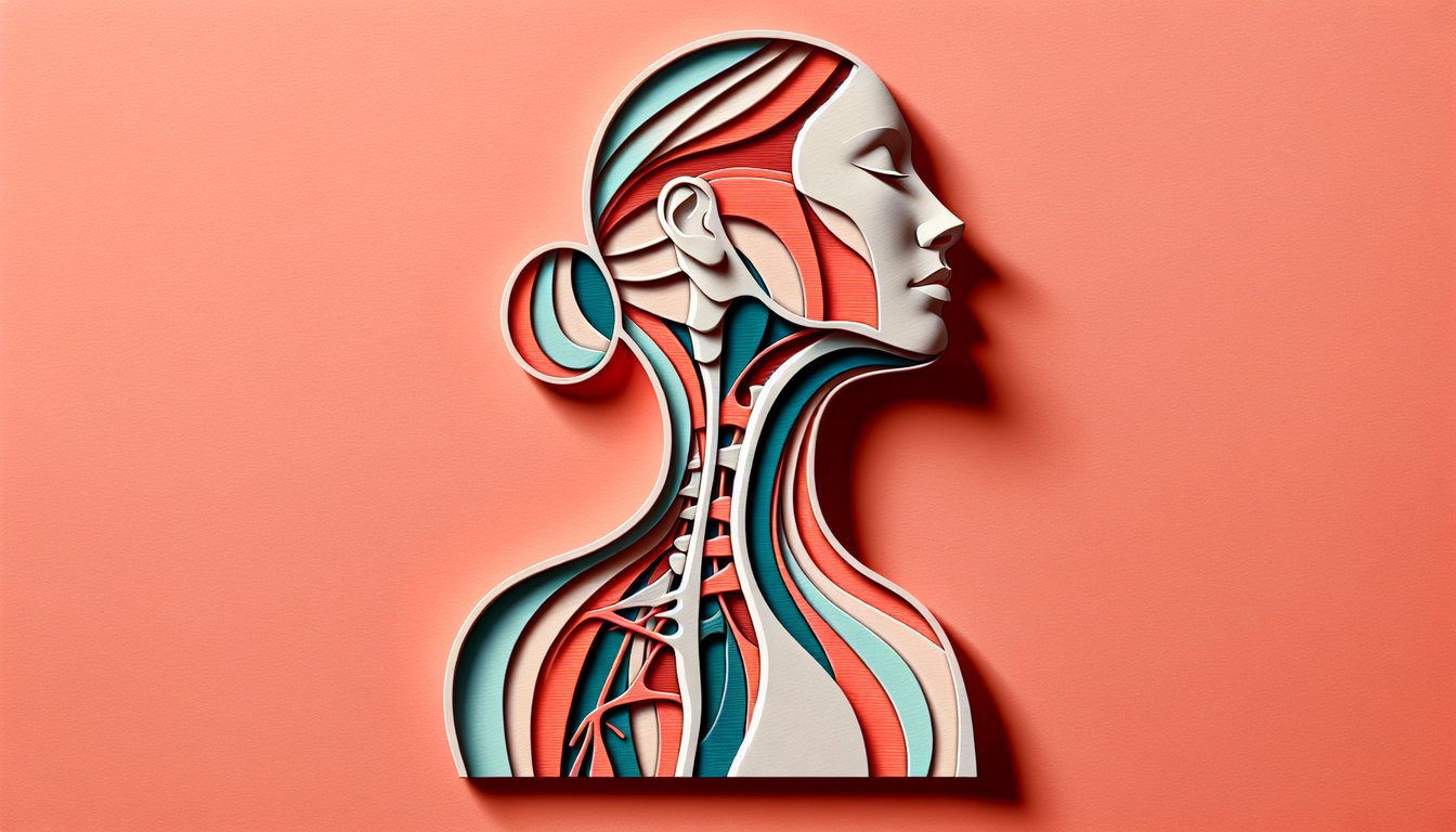
Ready to test your expertise on the anterior and posterior triangles of the neck? Our free scored neck anatomy quiz is designed for med students and anatomy enthusiasts to explore key features of the anterior and posterior neck triangles and master the anterior and posterior triangle of neck. Discover the clinical importance of these regions, their functional significance, and spatial relationships in head and neck practice. With instant feedback and performance tracking, you'll reinforce your grasp of posterior and anterior triangles of the neck in no time. Take the plunge now: identify these vital regions or dive into the neck anatomy quiz !
Which muscle forms the anterior boundary of the posterior triangle?
Trapezius
Splenius capitis
Omohyoid
Sternocleidomastoid
The posterior triangle is bounded anteriorly by the posterior border of the sternocleidomastoid muscle, posteriorly by the anterior border of trapezius, and inferiorly by the clavicle. The sternocleidomastoid divides the neck into anterior and posterior regions and its posterior margin forms the anterior limit of the posterior triangle. It originates from the manubrium and sternal end of clavicle and inserts on the mastoid process. For more details, see Posterior Triangle Anatomy.
Which muscle forms the posterior boundary of the posterior triangle?
Splenius capitis
Trapezius
Sternocleidomastoid
Omohyoid
The posterior triangle's posterior boundary is the anterior border of the trapezius muscle, which extends from the occipital bone to the spine of the scapula. This muscle forms the roof of the posterior triangle and works in shoulder elevation and scapular rotation. The trapezius is a broad, superficial muscle that is easy to palpate along the upper back and neck. For more information, see Posterior Triangle Anatomy.
What is the inferior boundary of the posterior triangle?
Clavicle
Scapula
Hyoid bone
Manubrium of sternum
The inferior boundary of the posterior triangle is formed by the middle third of the clavicle. This bony landmark separates the neck from the upper thorax and serves as an attachment for the trapezius and sternocleidomastoid muscles. Clinically, the clavicle is important in fracturing patterns and in assessing shoulder girdle integrity. See Posterior Triangle Anatomy for more details.
What forms the superior boundary of the anterior triangle of the neck?
Clavicle
Hyoid bone
Inferior border of mandible
Sternal notch
The superior boundary of the anterior triangle is the inferior border of the mandible, which extends from the chin to the angle of the jaw. This border marks the division between the face and neck regions. Subdivisions of the anterior triangle lie below this margin. For further study, refer to Anterior Triangle Anatomy.
What forms the anterior boundary of the anterior triangle of the neck?
Spinous processes
Posterior border of SCM
Midline of neck
Anterior border of trapezius
The anterior boundary of the anterior triangle is the midline of the neck, running from the chin to the sternum. This sagittal plane divides the left and right halves of the neck. Important structures such as the thyroid gland and trachea lie near this midline. See Anterior Triangle Anatomy for more information.
Which nerve lies superficial to the investing layer of deep cervical fascia in the posterior triangle?
Hypoglossal nerve
Vagus nerve
Phrenic nerve
Spinal accessory nerve
The spinal accessory nerve (CN XI) courses superficially across the posterior triangle deep to the investing layer of fascia before penetrating the trapezius muscle. It innervates sternocleidomastoid and trapezius, making it vulnerable to injury during neck dissections. Lesion causes shoulder droop and difficulty elevating the scapula. For details, see Posterior Triangle Anatomy.
Which muscle contributes to the floor of the posterior triangle?
Scalenus medius
Digastric
Omohyoid
Platysma
The floor of the posterior triangle is formed by muscles of the prevertebral layer, including scalenus medius, scalenus posterior, levator scapulae, and splenius capitis. Scalenus medius arises from the transverse processes of C2 - C7 and attaches to the first rib. These muscles separate the triangle from the cervical vertebrae and deep structures. For more, visit Posterior Triangle Anatomy.
Which artery crosses the posterior triangle diagonally?
Third part of subclavian artery
Common carotid artery
External carotid artery
Thyrocervical trunk
The third part of the subclavian artery emerges between the anterior and middle scalene muscles and crosses the apex of the posterior triangle toward the lateral border of the first rib. It gives off branches like the dorsal scapular and suprascapular arteries. This crossing is clinically relevant for vascular procedures and trauma. Learn more at Posterior Triangle Anatomy.
Which vein is formed by the union of the retromandibular and posterior auricular veins in the posterior triangle?
External jugular vein
Internal jugular vein
Anterior jugular vein
Subclavian vein
The external jugular vein is formed by the union of the posterior auricular vein with the posterior branch of the retromandibular vein, coursing superficially over the sternocleidomastoid muscle within the posterior triangle. It drains into the subclavian vein near the clavicle. It is easily visible in slender individuals and is used clinically for venous access. For more, see Posterior Triangle Anatomy.
Which nerve from the cervical plexus supplies sensation to the skin over the clavicle and shoulder in the posterior triangle?
Transverse cervical nerve
Supraclavicular nerves
Great auricular nerve
Lesser occipital nerve
The supraclavicular nerves arise from C3 - C4 roots of the cervical plexus and emerge at the posterior border of the SCM, descending over the posterior triangle to supply skin over the clavicle and shoulder. They have medial, intermediate, and lateral branches. Injury or block of these nerves affects cutaneous sensation in this region. See Posterior Triangle Anatomy.
The subclavian artery in the posterior triangle is divided into how many parts relative to the anterior scalene muscle?
Five
Two
Three
Four
The subclavian artery is classically divided into three parts by its relation to the anterior scalene muscle: medial, posterior, and lateral. This division is important for describing its branches, such as the vertebral, internal thoracic, and thyrocervical trunk. In the posterior triangle, the third (lateral) part lies between the anterior and middle scalene muscles. For more, see Posterior Triangle Anatomy.
Which muscle divides the posterior triangle into the occipital and omoclavicular triangles?
Inferior belly of omohyoid
Levator scapulae
Sternocleidomastoid
Trapezius
The inferior belly of the omohyoid muscle crosses the posterior triangle diagonally and subdivides it into the larger occipital triangle (above) and the smaller omoclavicular (subclavian) triangle (below). This muscle originates from the scapula and attaches to the hyoid bone after a tendinous intersection. Its anatomical position is a landmark during neck dissections. For details, see Posterior Triangle Anatomy.
Which smaller triangle of the anterior triangle contains the carotid sheath?
Submental triangle
Carotid triangle
Muscular triangle
Submandibular triangle
The carotid triangle is bounded by the SCM, posterior belly of digastric, and superior belly of omohyoid. It contains the carotid sheath, which encloses the common and internal carotid arteries, internal jugular vein, and vagus nerve. This region is a key site for vascular access and nerve blocks. More information at Anterior Triangle Anatomy.
Which muscle forms the floor of the submandibular triangle?
Digastric
Mylohyoid
Omohyoid
Sternohyoid
The submandibular triangle is bounded by the inferior border of the mandible and the anterior and posterior bellies of the digastric muscle. Its floor is formed primarily by the mylohyoid muscle, which also constitutes the floor of the oral cavity. The hyoglossus contributes to the floor medially. See Anterior Triangle Anatomy.
Which muscle bellies form the lateral boundaries of the submental triangle?
Anterior bellies of digastric muscles
Sternothyroid
Sternohyoid
Omohyoid
The submental triangle is unpaired and lies between the anterior bellies of the right and left digastric muscles, with the hyoid bone forming the posterior border. Its floor is formed by the mylohyoid muscles and is a site for lymph node drainage. Clinically, it can be examined for submental swellings. More at Anterior Triangle Anatomy.
Which muscle forms the posterior boundary of the submandibular triangle?
Omohyoid
Stylohyoid
Posterior belly of digastric
Anterior belly of digastric
The submandibular (digastric) triangle is bounded by the inferior border of the mandible and the anterior and posterior bellies of the digastric muscle. The posterior belly forms its posteroinferior boundary. The stylohyoid runs alongside it, but does not form the boundary. For more, see Anterior Triangle Anatomy.
At what vertebral level does the common carotid artery bifurcate within the carotid triangle?
C4
C3
C5
C6
The common carotid artery bifurcates into internal and external carotid arteries at the level of the superior border of the thyroid cartilage, which corresponds to the C4 vertebral level. This bifurcation lies in the carotid triangle of the anterior neck. Understanding this landmark is essential for vascular surgery and ultrasonography. Learn more at Anterior Triangle Anatomy.
Which nerve runs over the anterior scalene muscle in the posterior triangle?
Accessory nerve
Phrenic nerve
Vagus nerve
Hypoglossal nerve
The phrenic nerve, formed from C3 - C5, descends obliquely over the anterior scalene muscle before entering the thorax to innervate the diaphragm. It lies deep to the prevertebral fascia but appears in the posterior triangle region atop the anterior scalene. It is a critical structure often visualized during regional anesthesia. See Posterior Triangle Anatomy.
Which nerve lies deep to the posterior belly of the digastric in the submandibular triangle?
Glossopharyngeal nerve
Accessory nerve
Vagus nerve
Hypoglossal nerve
The hypoglossal nerve (CN XII) travels deep to the posterior belly of the digastric muscle and then continues anteriorly to supply the tongue muscles. It is a key landmark in the submandibular triangle and is vulnerable in regional surgeries. Damage causes tongue deviation toward the lesion side. More at Anterior Triangle Anatomy.
Which triangle of the anterior triangle contains the infrahyoid muscles and the thyroid gland?
Muscular triangle
Submandibular triangle
Submental triangle
Carotid triangle
The muscular triangle is bounded by the midline of the neck, superior belly of omohyoid, and anterior border of SCM. It contains infrahyoid muscles like sternohyoid and sternothyroid, as well as the thyroid and parathyroid glands. It is important in thyroid surgery. For more, see Anterior Triangle Anatomy.
Which branch of the thyrocervical trunk crosses the posterior triangle to supply trapezius?
Inferior thyroid artery
Dorsal scapular artery
Suprascapular artery
Transverse cervical artery
The transverse cervical artery, a branch of the thyrocervical trunk, courses laterally across the posterior triangle, deep to the levator scapulae and splenius capitis, to supply the trapezius muscle. It sometimes shares a common trunk with the suprascapular artery. Its location is crucial in flap surgeries. More at Posterior Triangle Anatomy.
Which lymph nodes lie deep to the SCM along the internal jugular vein in the carotid triangle?
Submandibular lymph nodes
Deep cervical lymph nodes
Superficial cervical lymph nodes
Submental lymph nodes
The deep cervical lymph nodes run along the internal jugular vein deep to the SCM muscle, inside the carotid sheath, and extend from the skull base to the thoracic inlet. They are key in head and neck cancer staging and infections. The superior group drains structures of the head and neck. See Anterior Triangle Anatomy.
Which fascia layer encloses the sternocleidomastoid and trapezius muscles and forms the roof of the posterior triangle?
Prevertebral fascia
Investing layer of deep cervical fascia
Carotid sheath
Pretracheal fascia
The investing layer of deep cervical fascia envelops the entire neck, splitting to enclose both the SCM and trapezius muscles, thus forming the roof of the posterior triangle. It is the most superficial layer of deep cervical fascia and provides structural support. It must be incised in surgical approaches. Learn more at Posterior Triangle Anatomy.
Which nerve passes through the carotid sheath within the posterior wall of the carotid triangle?
Vagus nerve
Accessory nerve
Phrenic nerve
Hypoglossal nerve
The vagus nerve (CN X) descends within the carotid sheath between the internal jugular vein and common carotid artery, forming the posterior wall of the carotid triangle. It gives off the superior and recurrent laryngeal nerves in this region. Injury risks include dysphonia and parasympathetic dysfunction. For details, see Anterior Triangle Anatomy.
In the occipital triangle, which nerve crosses the triangle from medial to lateral?
Great auricular nerve
Supraclavicular nerve
Spinal accessory nerve
Phrenic nerve
Within the occipital triangle (superior part of the posterior triangle), the spinal accessory nerve courses from the posterior border of SCM to the deep surface of trapezius, crossing medial to lateral. This location makes it vulnerable in lymph node biopsies. Injury leads to trapezius weakness. More at Posterior Triangle Anatomy.
Which space in the posterior triangle contains the subclavian artery and brachial plexus?
Muscular triangle
Occipital triangle
Carotid triangle
Omoclavicular triangle
The omoclavicular (subclavian) triangle is the inferior part of the posterior triangle, bounded by the clavicle, SCM, and inferior belly of omohyoid. It contains the third part of the subclavian artery and the brachial plexus emerging between the scalene muscles. It is a key area for accessing these structures surgically. See Posterior Triangle Anatomy.
Which triangle of the anterior triangle is unpaired?
Muscular triangle
Carotid triangle
Submandibular triangle
Submental triangle
The submental triangle is the only unpaired subdivision of the anterior triangle, lying between the anterior bellies of the digastric muscles and the hyoid bone. It contains small lymph nodes and the anterior jugular veins. It is clinically important for evaluating submental swellings. More details at Anterior Triangle Anatomy.
Which muscle forms the superior boundary of the muscular triangle?
Thyrohyoid
Superior belly of omohyoid
Inferior belly of omohyoid
Sternohyoid
The muscular triangle is bounded superiorly by the superior belly of the omohyoid muscle, which runs from the hyoid bone to the intermediate tendon near the clavicle. This sector encloses the infrahyoid muscles and the thyroid gland. Knowledge of these boundaries is crucial during thyroidectomy. Learn more at Anterior Triangle Anatomy.
Which artery runs with the hypoglossal nerve in the carotid triangle?
Ascending pharyngeal artery
Lingual artery
Superior thyroid artery
Facial artery
In the carotid triangle, the hypoglossal nerve (CN XII) loops around the occipital artery and then accompanies the lingual artery deep to the hyoglossus muscle toward the tongue. This close relationship is important during surgical exposure of the lingual artery. For more, visit Anterior Triangle Anatomy.
The jugular venous arch connects which veins across the midline in the submental region?
External jugular veins
Internal jugular veins
Facial veins
Anterior jugular veins
The jugular venous arch is a transverse communication between the left and right anterior jugular veins, located just above the manubrium in the submental region. It allows for collateral drainage between the anterior neck veins. Understanding this arch is important in procedures such as central line placement. See Anterior Triangle Anatomy.
Which lymph nodes lie along the external jugular vein at the apex of the posterior triangle?
Deep cervical lymph nodes
Submandibular lymph nodes
Occipital lymph nodes
Superficial cervical lymph nodes
The superficial cervical lymph nodes are located along the course of the external jugular vein, superficial to the SCM and within the posterior triangle's roof. They drain lymph from the scalp, face, and superficial neck. Enlargement can indicate infection or malignancy. For more, see Posterior Triangle Anatomy.
Which branch of the thyrocervical trunk crosses the posterior triangle to supply trapezius?
Suprascapular artery
Inferior thyroid artery
Dorsal scapular artery
Transverse cervical artery
The transverse cervical artery arises from the thyrocervical trunk and travels posterolaterally across the posterior triangle deep to the trapezius, supplying it and neighboring muscles. It often gives off a superficial branch supplying skin and trapezius. It is a key vessel in flap procedures. More at Posterior Triangle Anatomy.
In some individuals, the dorsal scapular artery arises directly from which vessel instead of the thyrocervical trunk?
Second part of subclavian artery
External jugular vein
Third part of subclavian artery
Axillary artery
Although normally a branch of the thyrocervical trunk, the dorsal scapular artery may in some individuals branch directly from the third part of the subclavian artery. This anatomical variation is important during surgical access to the lateral neck and shoulder region. Angiographic identification is used preoperatively. For more, see Posterior Triangle Anatomy.
Which fascia layer forms the floor of the anterior triangle?
Prevertebral layer of deep cervical fascia
Carotid sheath
Pretracheal layer of deep cervical fascia
Investing layer of deep cervical fascia
The pretracheal layer of the deep cervical fascia encloses the thyroid gland, trachea, and esophagus and forms the floor of the muscular triangle, part of the anterior triangle. It blends with the fibrous pericardium in the superior mediastinum. This fascia compartmentalizes infections in the neck. Learn more at Anterior Triangle Anatomy.
Which muscle in the floor of the posterior triangle originates from the transverse processes of C2 - C7?
Splenius capitis
Scalenus medius
Levator scapulae
Scalenus anterior
Scalenus medius originates from the transverse processes of C2 - C7 and forms part of the floor of the posterior triangle, situated between the anterior and posterior scalene muscles. It elevates the first rib during forced inspiration and flexes the neck laterally. Its relationship to the brachial plexus is clinically important in thoracic outlet syndrome. More at Posterior Triangle Anatomy.
The phrenic nerve receives contributions from which cervical spinal nerves?
C1, C2, C3
C3, C4, C5
C4, C5, C6
C2, C3, C4
The phrenic nerve is formed by contributions from the ventral rami of C3 - C5 and descends across the anterior scalene muscle into the thorax to innervate the diaphragm. The mnemonic 'C3, 4, 5 keep the diaphragm alive' is commonly used. Injury leads to respiratory compromise. For details, see Posterior Triangle Anatomy.
Which muscle does the spinal accessory nerve cross to enter the posterior triangle?
Sternocleidomastoid
Splenius capitis
Levator scapulae
Trapezius
After innervating the SCM, the spinal accessory nerve emerges from the posterior border of SCM approximately 2 cm above Erb's point, then crosses the floor of the posterior triangle to reach trapezius. This crossing point is clinical vulnerable during lymph node biopsies. Injury results in trapezius weakness and shoulder droop. More at Posterior Triangle Anatomy.
Which branch of the external carotid artery arises at the level of the superior border of the thyroid cartilage in the carotid triangle?
Ascending pharyngeal artery
Lingual artery
Superior thyroid artery
Facial artery
The superior thyroid artery is the first branch of the external carotid artery, arising at the level of the superior border of the thyroid cartilage within the carotid triangle. It descends to the thyroid gland, giving off infrahyoid and superior laryngeal branches. It is ligated in thyroidectomy surgeries. See Anterior Triangle Anatomy.
Which branch of the vagus nerve runs with the superior thyroid artery in the carotid triangle?
Recurrent laryngeal nerve
Pharyngeal branch
Internal branch of superior laryngeal nerve
External branch of superior laryngeal nerve
The external branch of the superior laryngeal nerve, from the vagus, accompanies the superior thyroid artery into the neck to innervate the cricothyroid muscle. It lies close to the artery within the carotid triangle and is at risk during thyroid surgery, affecting voice pitch. For more, visit Anterior Triangle Anatomy.
Which endocrine gland is found in the muscular triangle?
Parathyroid gland
Pituitary gland
Thyroid gland
Adrenal gland
The thyroid gland is located within the muscular triangle, deep to the infrahyoid muscles and superficial to the trachea. It has two lobes connected by an isthmus that overlies the second to fourth tracheal rings. It is crucial in metabolism and is surgically accessed through this triangle. More information at Anterior Triangle Anatomy.
Which cutaneous branch of the cervical plexus crosses the SCM within the posterior triangle to supply the skin over the parotid and mandibular angle?
Transverse cervical nerve
Supraclavicular nerves
Lesser occipital nerve
Great auricular nerve
The great auricular nerve, arising from C2 - C3 of the cervical plexus, ascends vertically across the SCM within the posterior triangle to provide sensation to the skin over the parotid gland, mastoid process, and angle of the mandible. It is often encountered during neck surgeries. See Posterior Triangle Anatomy.
Which fascia encloses the carotid sheath and contributes to the boundaries of the anterior triangle?
Investing layer of deep cervical fascia
Carotid sheath of deep cervical fascia
Pretracheal fascia
Prevertebral fascia
The carotid sheath is a condensation of the deep cervical fascia that surrounds the common and internal carotid arteries, internal jugular vein, and vagus nerve within the carotid triangle. It forms part of the posterior boundary of the anterior triangle and helps contain infections. For details, refer to Anterior Triangle Anatomy.
Which nerve is most at risk during carotid endarterectomy as it courses through the carotid triangle?
Hypoglossal nerve
Glossopharyngeal nerve
Phrenic nerve
Accessory nerve
The hypoglossal nerve (CN XII) loops around the occipital artery and passes deep to the posterior belly of the digastric within the carotid triangle. It is vulnerable during carotid endarterectomy when exposing the carotid bifurcation. Injury causes ipsilateral tongue deviation and atrophy. See Anterior Triangle Anatomy.
During an interscalene brachial plexus block, anesthetic is injected between which muscles in the posterior triangle?
SCM and trapezius
Middle and posterior scalene muscles
Levator scapulae and splenius capitis
Anterior and middle scalene muscles
An interscalene block targets roots/trunks of the brachial plexus located between the anterior and middle scalene muscles within the posterior triangle. Local anesthetic placed here provides anesthesia to the shoulder and upper limb. Accurate identification minimizes complications. Learn more at Posterior Triangle Anatomy.
Which lymph nodes along the internal jugular vein are designated as Level II in head and neck cancer staging?
Jugulodigastric nodes
Superior deep cervical lymph nodes
Inferior deep cervical lymph nodes
Submental nodes
Level II lymph nodes, or superior deep cervical nodes, lie along the internal jugular vein from the skull base to the inferior border of the hyoid bone. They often include the jugulodigastric node, a key node in oropharyngeal cancer spread. Precise identification is crucial in oncologic neck dissection. For staging details, see Anterior Triangle Anatomy.
The venous angle is formed by the junction of which veins within the neck?
Internal jugular and facial veins
External jugular and subclavian veins
Internal jugular and subclavian veins
Anterior jugular and external jugular veins
The venous angle is created where the internal jugular vein meets the subclavian vein, forming the brachiocephalic vein on each side. It is the site where the thoracic duct (left) and right lymphatic duct empty lymph into the venous system. Knowledge of this anatomy is important in central venous catheter placement. See Posterior Triangle Anatomy.
During neck dissection, which fascial layer must be incised to access the transverse cervical vessels in the posterior triangle?
Pretracheal fascia
Investing layer of deep cervical fascia
Prevertebral fascia
Carotid sheath
The investing layer of deep cervical fascia forms the roof of the posterior triangle. To access the transverse cervical vessels running on the floor, this investing fascia must be incised. It encloses the SCM and trapezius muscles and splits around the parotid gland. Proper fascia management reduces bleeding. More at Posterior Triangle Anatomy.
Injury to the spinal accessory nerve within the occipital triangle results in which clinical deficit?
Inability to shrug the shoulder
Loss of neck flexion
Difficulty swallowing
Loss of tongue movement
The spinal accessory nerve innervates the trapezius muscle, which elevates the shoulder. Injury in the occipital triangle causes trapezius paralysis, leading to inability to shrug and shoulder droop. The SCM remains functional if injury is distal to its innervation. For more, see Posterior Triangle Anatomy.
Which embryological structure gives rise to the scalenes and other prevertebral muscles forming the floor of the posterior triangle?
Branchial arches
Hypaxial myotomes
Neural crest cells
Paraxial mesoderm somites
The prevertebral muscles of the neck, including the scalene group, derive from paraxial mesoderm somites that segment alongside the developing neural tube. These somites differentiate into myotomes that give rise to axial and limb muscles. Neural crest contributes to peripheral nerves but not muscle. Learn more at Posterior Triangle Anatomy.
0
{"name":"Which muscle forms the anterior boundary of the posterior triangle?", "url":"https://www.quiz-maker.com/QPREVIEW","txt":"Which muscle forms the anterior boundary of the posterior triangle?, Which muscle forms the posterior boundary of the posterior triangle?, What is the inferior boundary of the posterior triangle?","img":"https://www.quiz-maker.com/3012/images/ogquiz.png"}
Score11/49
Easy4/15
Medium2/14
Hard3/15
Expert2/5
AI Study Notes
Email these to me
You can bookmark this page to review your notes in future, or fill out the email box below to email them to yourself.
Study Outcomes
Identify Anatomical Boundaries -
Identify the anatomical borders of the anterior and posterior triangles of the neck, including muscle and bony landmarks that define each region.
Describe Triangle Contents -
Describe the major muscles, vessels, and nerves contained within the anterior and posterior neck triangles, emphasizing their spatial relationships.
Distinguish Clinical Differences -
Distinguish between the clinical significance of the anterior versus posterior triangles, focusing on potential sites for surgical access and pathology.
Analyze Nerve Pathways -
Analyze the course of peripheral nerves, such as the accessory nerve and cervical plexus branches, as they traverse the anterior and posterior triangle regions.
Apply Palpation Techniques -
Apply physical examination methods to palpate key landmarks within the anterior and posterior triangles, enhancing accuracy in clinical assessments.
Evaluate Procedural Sites -
Evaluate optimal sites within the neck triangles for common medical procedures, such as nerve blocks or central line placement.
Cheat Sheet
Anterior Triangle Boundaries -
The anterior triangle of the neck is bordered by the anterior border of the sternocleidomastoid, the midline of the neck, and the inferior border of the mandible. It can be subdivided into the submandibular, submental, carotid, and muscular triangles using the digastric and superior belly of omohyoid muscles. Use the mnemonic "My Dog Caught Some Meat" (Mandible, Digastric, Carotid, Sternocleidomastoid, Midline) to remember its boundaries.
Posterior Triangle Boundaries -
The posterior triangle of the neck lies between the posterior border of the sternocleidomastoid, the anterior border of the trapezius, and the middle third of the clavicle. The inferior belly of the omohyoid muscle further divides it into the occipital and supraclavicular (subclavian) triangles. Remember "COT" (Clavicle, Omohyoid, Trapezius) to quickly recall its borders.
Key Anterior Triangle Contents -
Within the carotid triangle of the anterior triangle, you'll find the common carotid artery bifurcating, the internal jugular vein, vagus nerve, hypoglossal nerve, and ansa cervicalis within the carotid sheath. The submandibular triangle contains the submandibular gland and facial artery. A handy mnemonic for cranial nerve XII and the ansa cervicalis is "HA to XII" (Hypoglossal, Ansa, XII).
Key Posterior Triangle Contents -
The posterior triangle houses the spinal accessory nerve (CN XI), cervical plexus branches, roots and trunks of the brachial plexus, subclavian artery, and the external jugular vein. The floor is formed by the scalene muscles, levator scapulae, and splenius capitis. Use "SOAP+B" (Spinal accessory, Omovertebral, Ansa, Phrenic, Brachial plexus) as a memory aid.
Clinical Relevance & Landmarks -
Surface landmarks in the anterior and posterior triangles of the neck guide procedures like central line placement in the internal jugular vein (anterior) and biopsy of cervical lymph nodes (posterior). Beware of the accessory nerve in the posterior triangle; it lies superficially 2 cm above the clavicle at "Erb's point," an important surgical landmark. Clinicians often memorize "J Erb 2" for Jugular access, Erb's point, 2 cm above clavicle.