Unlock hundreds more features
Save your Quiz to the Dashboard
View and Export Results
Use AI to Create Quizzes and Analyse Results

Sign inSign in with Facebook
Sign inSign in with Google

Integumentary System Practice Quiz

Boost Your Histology Skills With Targeted Practice Quiz

Difficulty: Moderate
Grade: Other
Study OutcomesCheat Sheet
Colorful paper art promoting Skin Deep Histology trivia quiz for advanced biology students.

What is the outermost layer of the skin?
Epidermis
Hypodermis
Subcutaneous tissue
Dermis
The epidermis is the outermost protective barrier of the skin composed mainly of keratinocytes. It plays a crucial role in shielding the body from environmental hazards.
Which cell type is primarily responsible for producing the pigment melanin in the skin?
Keratinocytes
Langerhans cells
Fibroblasts
Melanocytes
Melanocytes are specialized cells that produce melanin, the pigment responsible for skin color. Their function is crucial for protecting the skin from UV radiation.
Which of the following is a major function of the skin?
Barrier protection against pathogens and injury
Synthesis of digestive enzymes
Regulation of blood glucose levels
Transport of oxygen throughout the body
The skin provides a critical barrier that protects the body from physical, chemical, and biological harm. Its protective function is key to maintaining overall health.
The primary cell type found in the epidermis is:
Keratinocytes
Adipocytes
Fibroblasts
Melanocytes
Keratinocytes are the most abundant cell type in the epidermis, responsible for producing keratin. This protein provides structural strength and helps form a protective barrier.
Which layer of the epidermis is responsible for the continuous division of cells?
Stratum basale
Stratum lucidum
Stratum corneum
Stratum granulosum
The stratum basale is the deepest layer of the epidermis where basal cells divide to replenish the skin. This process is essential for the constant renewal of the skin surface.
Which epidermal layer is primarily composed of flattened, dead keratinocytes?
Stratum spinosum
Stratum granulosum
Stratum basale
Stratum corneum
The stratum corneum is the outermost layer of the epidermis consisting of dead, flattened keratinocytes. This layer plays a vital role in forming a barrier against environmental hazards.
Which cell type in the epidermis is involved in immune surveillance?
Keratinocytes
Langerhans cells
Fibroblasts
Merkel cells
Langerhans cells are dendritic cells in the epidermis that act as antigen-presenting cells. They play an important role in detecting pathogens and initiating immune responses.
What is the primary histological difference between eccrine and apocrine sweat glands?
Eccrine glands are smaller, more numerous, and produce a watery secretion.
Eccrine glands are mainly found in the armpits and produce a viscous secretion.
Eccrine glands are larger, produce an oily secretion, and are linked with hair follicles.
Eccrine glands are only activated during emotional stress and produce a milky fluid.
Eccrine sweat glands are smaller and more numerous throughout the skin, producing a watery secretion primarily for thermoregulation. In contrast, apocrine glands are larger and produce a thicker secretion in specific areas.
Which type of fibers in the dermis is primarily responsible for the skin's elasticity?
Collagen fibers
Reticular fibers
Elastin fibers
Keratin fibers
Elastin fibers provide the skin with the ability to stretch and recoil. They are responsible for maintaining the elasticity of the skin, unlike collagen fibers which mainly provide strength.
Which of the following receptors is primarily responsible for the perception of light touch in the skin?
Merkel cells
Pacinian corpuscles
Ruffini endings
Meissner's corpuscles
Meissner's corpuscles are specialized nerve endings located in the dermal papillae that detect light touch and fine tactile sensations. They are crucial for tasks requiring delicate touch perception.
Which protein is primarily responsible for providing tensile strength in the dermis?
Collagen
Elastin
Keratin
Actin
Collagen is the predominant protein in the dermis that imparts tensile strength and structural integrity to the skin. Its fibrous nature allows the skin to resist stretching forces.
Which gland in the skin is responsible for secreting an oily substance that helps to lubricate the skin?
Sudoriferous glands
Ceruminous glands
Sebaceous glands
Apocrine glands
Sebaceous glands secrete sebum, an oily substance that lubricates and waterproofs the skin and hair. This secretion helps maintain skin flexibility and prevents dryness.
In which layer of the epidermis are melanocytes predominantly located?
Stratum granulosum
Stratum corneum
Stratum basale
Stratum spinosum
Melanocytes are primarily found in the stratum basale, the deepest layer of the epidermis. Their positioning allows them to transfer melanin to keratinocytes, influencing skin pigmentation.
What histological feature distinguishes thick skin from thin skin?
Higher number of Langerhans cells
Increased density of melanocytes
Presence of the stratum lucidum
Thicker stratum basale
Thick skin, such as that found on the palms and soles, is characterized by the presence of the stratum lucidum. This additional layer provides enhanced protection against abrasion.
Which structure is crucial for anchoring the epidermis to the dermis?
Basement membrane
Epidermal ridges
Dermal papillae
Stratum corneum
The basement membrane serves as a critical interface between the epidermis and dermis, ensuring proper adhesion and structural integrity. It also plays a role in regulating cell behavior and tissue repair.
During wound healing, what process describes the movement and division of keratinocytes to cover the wound area?
Melanogenesis of keratinocytes
Differentiation of keratinocytes into fibroblasts
Migration and proliferation of keratinocytes
Apoptosis of keratinocytes
Re-epithelialization during wound healing involves the migration and proliferation of keratinocytes to restore the continuity of the skin. This process is essential for effective tissue repair and regeneration.
Which mechanism best explains the variation in melanin distribution resulting in different skin tones?
Increased keratin production inhibiting melanin synthesis
Variations in melanosome size, number, and dispersion within keratinocytes
Enhanced sebaceous gland activity affecting pigment deposition
Differences in the density of melanocytes per unit area
Skin tone differences are primarily due to variations in the size, number, and dispersion of melanosomes within keratinocytes. This mechanism explains why individuals with similar numbers of melanocytes can have different pigmentation.
A histological examination revealing a thickened stratum corneum is most indicative of which condition?
Acantholysis
Elastosis
Spongiosis
Hyperkeratosis
Hyperkeratosis refers to the abnormal thickening of the stratum corneum, often due to excessive keratin production. It is a common response to chronic irritation or friction.
Which signaling pathway is critical for regulating the differentiation of keratinocytes in the epidermis?
Sonic hedgehog pathway
MAPK/ERK pathway
Notch signaling
Wnt signaling
Notch signaling plays a central role in the differentiation of keratinocytes within the epidermis. It helps balance cell proliferation and differentiation, which is essential for proper skin formation.
How does the composition of the basement membrane contribute to its role in the skin?
Its specialized extracellular matrix proteins, such as laminin and collagen IV, provide strong adhesion between the epidermis and dermis
Its rich vascular network allows for rapid nutrient delivery
Its thick layer of keratin prevents pathogen entry
Its high concentration of immune cells facilitates defense against infections
The basement membrane is composed of specialized extracellular matrix proteins like laminin and collagen IV, which are essential for anchoring the epidermis to the dermis. This composition ensures tissue integrity and aids in cell signaling during repair processes.
0
{"name":"What is the outermost layer of the skin?", "url":"https://www.quiz-maker.com/QPREVIEW","txt":"What is the outermost layer of the skin?, Which cell type is primarily responsible for producing the pigment melanin in the skin?, Which of the following is a major function of the skin?","img":"https://www.quiz-maker.com/3012/images/ogquiz.png"}

Study Outcomes

  1. Understand the structural organization of the skin layers.
  2. Analyze histological features of the epidermis, dermis, and hypodermis.
  3. Identify key cellular components and tissue types within the skin.
  4. Evaluate the relationship between skin structure and its physiological functions.
  5. Apply histological knowledge to diagnose skin-related exam scenarios.

PAL Histology Integumentary System Cheat Sheet

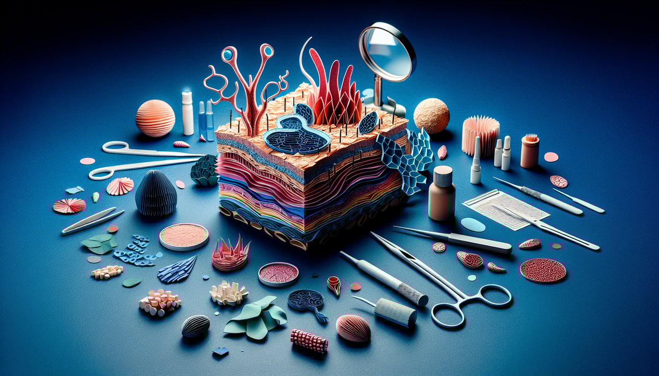
  1. Layers of the Skin - Get ready for a skin‑deep adventure through the three main layers: the epidermis (your outer barrier), dermis (your stretchy middle layer), and hypodermis (your cozy fat padding). Knowing their roles makes you a histology hero in no time. GWU Skin Histology
  2. Epidermal Strata - Dive into the five epic strata of the epidermis: stratum basale, spinosum, granulosum, lucidum (only on palms and soles), and corneum. Each layer has its own superstar cells and functions that keep your skin fresh and fabulous. UB Integument Histology Notes
  3. Key Epidermal Cell Types - Meet the dream team: keratinocytes (make tough keratin), melanocytes (color your world), Langerhans cells (immune sentinels), and Merkel cells (sensory receivers). Together they maintain skin's defense and sensation. DoveMed Skin Structure & Function
  4. Thick vs. Thin Skin - Palm and sole fans rejoice at thick skin (no hair follicles) while the rest of the body rocks thin skin (with hair). Spotting differences helps you ace clinical and lab work. Histology Guide: Skin
  5. Dermis Layers - The dermis has two cool zones: the papillary layer (loose, fingerprint‑forming tissue) and the reticular layer (dense, strength‑giving mesh). Together they provide support and elasticity. UB Integument Histology Notes
  6. Skin Appendages - From hair follicles to sebaceous glands (oil producers) and sweat glands (eccrine for cooling, apocrine for scent), skin appendages keep you insulated, lubricated, and thermo‑regulated. Nurseslabs Integumentary System
  7. Sensory Receptors - Feel it all with Meissner's corpuscles (light touch), Pacinian corpuscles (pressure and vibration), and Ruffini endings (stretch). They turn physical stimuli into neural signals, so you know when you've been tickled. UB Integument Histology Notes
  8. Thermoregulation - Keep your cool (or warm up) via vasodilation, vasoconstriction, and sweat production. Skin acts like a climate‑control system, ensuring your internal thermostat is always on point. Nurseslabs Integumentary System
  9. Keratinization Process - Watch keratinocytes journey from the stratum basale up to the stratum corneum, becoming tougher and more keratinized as they go, before shedding off in a satisfying flake‑off finale. UB Integument Histology Notes
  10. Common Skin Pathologies - Stay vigilant for basal cell carcinoma, which often springs from stratum basale cells after prolonged UV exposure. Early recognition is key to treatment success. UB Integument Histology Notes
Powered by: Quiz Maker