Unlock hundreds more features
Save your Quiz to the Dashboard
View and Export Results
Use AI to Create Quizzes and Analyse Results

Sign inSign in with Facebook
Sign inSign in with Google

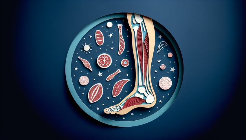
Muscle Labeling Practice Quiz

Master leg muscle labeling with engaging questions

Difficulty: Moderate
Grade: Grade 10
Study OutcomesCheat Sheet
Paper art illustrating a trivia quiz for high school biology students on leg anatomy labeling.

Which bone is the longest bone in the human leg?
Femur
Tibia
Fibula
Patella
The femur is the longest and strongest bone in the human leg and body. Its length and robust structure are essential for supporting the weight of the body.
Which bone is known as the kneecap?
Femur
Patella
Tibia
Fibula
The patella is commonly known as the kneecap and protects the knee joint. It also plays a critical role in knee extension by acting as a fulcrum.
Which bone in the lower leg bears most of the body's weight?
Femur
Tibia
Fibula
Patella
The tibia, or shinbone, bears most of the body's weight in the lower leg. It is a primary weight-bearing bone that supports both the knee and the ankle joints.
Which slender bone is found on the lateral side of the lower leg?
Fibula
Tibia
Patella
Femur
The fibula is the smaller, slender bone located on the lateral side of the lower leg. It primarily serves as a site for muscle attachment and aids in stabilizing the ankle.
Which calf muscle is primarily responsible for plantar flexion of the foot during activities such as standing on tiptoes?
Gastrocnemius
Soleus
Tibialis anterior
Peroneus longus
The gastrocnemius is a prominent calf muscle that facilitates plantar flexion, especially during dynamic actions like tiptoeing or jumping. Its powerful contraction is key to propelling the body upward.
Which muscle is primarily responsible for dorsiflexing the foot?
Gastrocnemius
Tibialis anterior
Soleus
Fibularis longus
The tibialis anterior muscle is the main muscle responsible for dorsiflexion of the foot. Its contraction lifts the foot upward toward the shin, which is crucial during the swing phase of gait.
Which muscle group found in the anterior thigh is essential for knee extension?
Hamstrings
Quadriceps femoris
Adductors
Abductors
The quadriceps femoris is a group of four muscles located in the anterior compartment of the thigh. They work collaboratively to extend the knee during activities such as walking and running.
Which set of muscles includes the biceps femoris, semitendinosus, and semimembranosus?
Hamstrings
Quadriceps
Calf muscles
Gluteal muscles
The hamstrings consist of the biceps femoris, semitendinosus, and semimembranosus muscles. They are located in the posterior compartment of the thigh and are vital for knee flexion and hip extension.
Which muscle, along with the soleus, plays a major role in plantar flexion of the ankle?
Tibialis anterior
Gastrocnemius
Fibularis brevis
Popliteus
The gastrocnemius, together with the soleus, is one of the primary muscles responsible for plantar flexion. This combined action is essential during activities such as running, jumping, and walking.
Which muscle is the longest muscle in the human body, contributing to hip flexion, abduction, and knee flexion?
Sartorius
Rectus femoris
Biceps femoris
Adductor longus
The sartorius is recognized as the longest muscle in the human body, running diagonally across the thigh. It is involved in multiple movements including hip flexion, abduction, and knee flexion.
Which muscle of the quadriceps group originates from the ilium and crosses both the hip and knee joints?
Vastus medialis
Rectus femoris
Vastus lateralis
Vastus intermedius
The rectus femoris is the only muscle in the quadriceps group that originates from the pelvis, specifically the ilium. Its unique course allows it to act on both the hip and knee joints, facilitating hip flexion and knee extension.
Which muscle assists in stabilizing the leg by tensioning the iliotibial band during walking?
Gluteus maximus
Tensor fasciae latae
Adductor magnus
Sartorius
The tensor fasciae latae helps to stabilize the leg by tensing the iliotibial band. This action is crucial for maintaining lateral stability during walking.
Which muscle, found in the lateral compartment of the leg, is primarily responsible for eversion of the foot?
Tibialis posterior
Fibularis longus
Gastrocnemius
Tibialis anterior
The fibularis longus muscle is key for eversion of the foot. Additionally, it helps to support the arches of the foot, ensuring proper foot mechanics during movement.
Which muscle plays a crucial role in 'unlocking' the knee joint at the start of flexion?
Popliteus
Hamstrings
Quadriceps
Gastrocnemius
The popliteus muscle assists in unlocking the knee by initiating the transition from extension to flexion. Its action enables smooth knee bending and is essential for normal gait.
Which muscle, located beneath the gastrocnemius, is essential for sustained plantar flexion during prolonged standing?
Soleus
Tibialis anterior
Fibularis brevis
Popliteus
The soleus muscle, situated deep to the gastrocnemius, is predominantly composed of slow-twitch fibers. This composition allows it to maintain prolonged contractions necessary for standing and postural control.
Which muscle is primarily responsible for medially stabilizing the patella due to its oblique fiber orientation?
Vastus medialis
Vastus lateralis
Rectus femoris
Sartorius
The vastus medialis, particularly its oblique fibers, plays a key role in medially stabilizing the patella. This stabilization helps prevent lateral tracking during knee extension and is important for proper joint alignment.
From which bony landmark does the popliteus muscle originate?
Lateral femoral condyle
Medial femoral condyle
Fibular head
Tibial plateau
The popliteus muscle originates from the lateral femoral condyle. This origin is strategically important as it enables the muscle to effectively unlock the knee during the initiation of flexion.
Which muscle, known for aiding in foot eversion, also contributes to the support of the foot's arch?
Peroneus longus
Peroneus brevis
Tibialis posterior
Tibialis anterior
The peroneus longus plays a dual role by everting the foot and supporting the medial arch through its tendon. Its contribution is vital for maintaining proper foot structure during weight-bearing activities.
Which muscle, located in the deep posterior compartment of the leg, is essential for foot inversion and maintaining the medial longitudinal arch?
Tibialis posterior
Tibialis anterior
Gastrocnemius
Soleus
The tibialis posterior is a key muscle in the deep posterior compartment that facilitates foot inversion. It also supports the medial longitudinal arch, making it crucial for overall foot stability.
A lesion of the femoral nerve most notably results in weakness of which muscle involved in knee extension?
Biceps femoris
Rectus femoris
Adductor longus
Sartorius
The rectus femoris is part of the quadriceps group and is innervated by the femoral nerve, making it crucial for knee extension. Damage to this nerve can lead to significant weakness in the muscle, impairing the ability to extend the knee properly.
0
{"name":"Which bone is the longest bone in the human leg?", "url":"https://www.quiz-maker.com/QPREVIEW","txt":"Which bone is the longest bone in the human leg?, Which bone is known as the kneecap?, Which bone in the lower leg bears most of the body's weight?","img":"https://www.quiz-maker.com/3012/images/ogquiz.png"}

Study Outcomes

  1. Identify the major anatomical structures of the leg.
  2. Label key muscles accurately on a diagram of the leg.
  3. Analyze the spatial relationships between different leg muscles.
  4. Apply anatomical terminology to describe leg structures.
  5. Evaluate understanding of leg anatomy to build exam confidence.

Muscle Labeling Quiz: Leg Muscle Review Cheat Sheet

  1. Master the Leg Compartments - The leg is split into three key zones: anterior (dorsiflexors), posterior (plantar flexors), and lateral (fibular) compartments. Each group of muscles works as a team to power movements like walking, running, and kicking. Understanding these zones is your first step to acing muscle anatomy! Kenhub quizzes & diagrams
  2. kenhub.com
  3. Quadriceps Femoris Group - This powerhouse quartet (rectus femoris, vastus lateralis, medialis, intermedius) is your go-to for straightening the knee and flexing the hip. When you kick a ball or climb stairs, these muscles are doing the heavy lifting! Study their origins, insertions, and actions to see how they work in concert. Detailed Wiki Overview
  4. en.wikipedia.org
  5. Hamstring Heroes - Nestled at the back of your thigh are the biceps femoris, semitendinosus, and semimembranosus. These three amigos bend the knee and extend the hip, making them crucial for running, jumping, and even standing up straight. Don't forget to note their common tendon attach‑ ment at the ischial tuberosity! Try a Hamstring Quiz
  6. parallelcoaching.co.uk
  7. Calf Camp - Meet the gastrocnemius and soleus, your dynamic duo for plantar flexion (think toe‑pointing). From calf raises to sprinting, these muscles propel you forward and up. Sketching their fibers can help you remember which one crosses the knee! Calf Anatomy Quiz
  8. parallelcoaching.co.uk
  9. Sartorius Spotlight - The sartorius is the body's longest muscle, winding diagonally across your thigh like a tailor's ribbon. It assists in hip flexion, abduction, and external rotation - hence its nickname, "the tailors muscle." Trace its path from the ASIS to the medial tibia to lock in your understanding! Sartorius Deep Dive
  10. en.wikipedia.org
  11. Interactive Labeling Practice - Quizzes and drag‑and‑drop diagrams are a surefire way to cement muscle names and locations in your mind. Repetition equals retention, so challenge yourself daily and watch your confidence grow. Turn study time into play time! Kenhub Interactive
  12. kenhub.com
  13. Unlabeled Diagram Drills - Once you've seen the labels, remove them and quiz yourself on blank sketches. This level‑up challenge forces you to recall names purely from muscle shape and position. Flip between labeled and unlabeled for maximum brain burn! Blank Diagram Challenge
  14. kenhub.com
  15. Adductors vs. Abductors - Adductors pull the thigh toward the midline, while abductors send it away - both vital for stabilizing your pelvis during movement. Mix up exercises like side‑leg raises and squeeze ball drills to feel these muscle groups in action. Visualizing their angles helps lock in their roles! Adductor/Abductor Quiz
  16. parallelcoaching.co.uk
  17. Tibialis Anterior Trek - Sitting along the front of your shin, the tibialis anterior lifts your foot up (dorsiflexion) and twists it inward (inversion). Strong tibialis muscles are your secret weapon against shin splints and ankle sprains. Add toe‑drag walks to your routine to feel them engage! Shin Muscle Guide
  18. kenhub.com
  19. Peroneus (Fibularis) Focus - The longus, brevis, and tertius muscles on the outer shin evert and assist in plantar flexion of the foot. They're key players in ankle stability during side‑to‑side moves. Strengthen them with resistance band foot eversion drills for injury‑proof ankles! ankle stability drills
  20. kenhub.com
Powered by: Quiz Maker