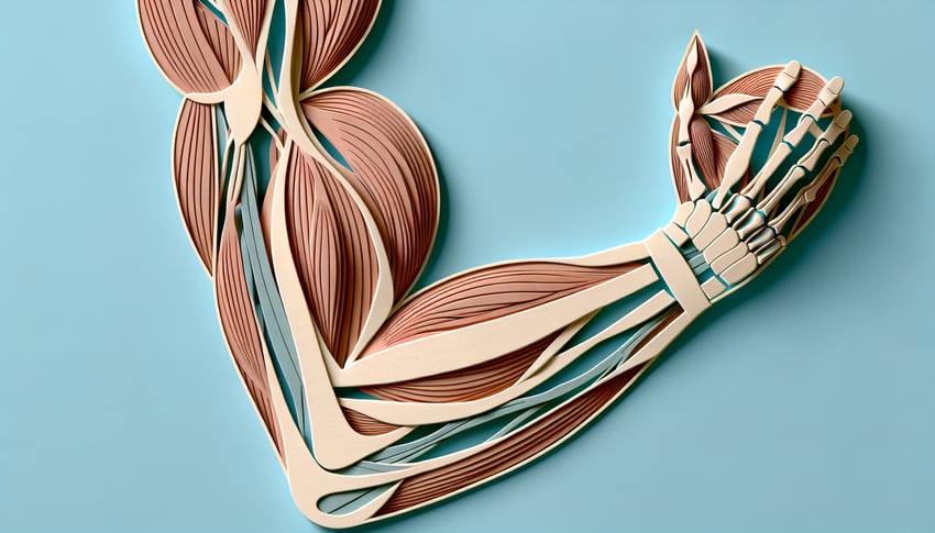
Ready to flex your knowledge with our Ultimate Muscles of the Arm Quiz? This free muscles of the arm quiz challenges fitness enthusiasts, anatomy buffs, and curious learners to master muscles of the arm labeling and arm muscle labeling - from biceps and triceps to detailed forearm muscles quiz sections. You'll sharpen your muscle identification skills, practice precise arm labeling, and gain confidence in your anatomy expertise. If you love exploring detailed structures, try our related muscle anatomy quiz or test your skeletal savvy with an upper extremity bones quiz . Dive in now and prove you know your arm muscles inside and out!
Which muscle lies on the anterior aspect of the upper arm and has two heads originating from the scapula?
Biceps brachii
Coracobrachialis
Triceps brachii
Brachialis
The biceps brachii is located on the front of the upper arm with a long head from the supraglenoid tubercle and a short head from the coracoid process, allowing it to flex the elbow and supinate the forearm. It is the most prominent muscle when the arm is flexed. Other muscles in the anterior compartment do not originate from both of these scapular landmarks. For more information, see Biceps brachii.
Which deep muscle of the upper arm lies directly beneath the biceps brachii and is the primary flexor of the elbow?
Pronator teres
Brachioradialis
Brachialis
Coracobrachialis
The brachialis lies deep to the biceps brachii on the anterior humerus and attaches to the ulnar tuberosity, making it the strongest single flexor of the elbow joint. It operates regardless of forearm position. Other muscles like brachioradialis assist in flexion but are more superficial. More details can be found at Brachialis muscle.
Which muscle originates from the lateral supracondylar ridge of the humerus and is most active during rapid elbow flexion?
Extensor carpi radialis longus
Brachialis
Brachioradialis
Pronator teres
The brachioradialis originates from the lateral supracondylar ridge of the humerus and inserts on the radial styloid, making it uniquely positioned to flex the elbow especially during rapid movements or when the forearm is in a neutral position. It is innervated by the radial nerve. For further reading, see Brachioradialis muscle.
Which muscle is the main extensor of the elbow and has three distinct heads?
Triceps brachii
Biceps brachii
Brachioradialis
Anconeus
The triceps brachii has three heads - long, lateral, and medial - that converge into a single tendon inserting on the olecranon of the ulna, and it is the primary muscle responsible for elbow extension. The anconeus assists but is much smaller. Learn more at Triceps brachii muscle.
Which small triangular muscle assists the triceps in extending the elbow and stabilizes the elbow joint?
Brachialis
Coracobrachialis
Supinator
Anconeus
The anconeus is a small muscle on the posterior aspect of the elbow that assists the triceps brachii in extending the forearm and helps stabilize the elbow joint during pronation and supination. It also may spread synovial fluid within the joint. More on this can be found at Anconeus.
Which muscle originates from the coracoid process of the scapula and flexes the arm at the shoulder joint?
Coracobrachialis
Deltoid
Pectoralis minor
Biceps brachii
The coracobrachialis arises from the coracoid process, travels medial to the humerus, and inserts on the mid-shaft, acting to flex and adduct the arm at the shoulder. It is innervated by the musculocutaneous nerve. For details, refer to Coracobrachialis muscle.
Which head of the biceps brachii originates from the coracoid process of the scapula?
Short head
Lateral head
Long head
Medial head
The short head of the biceps brachii arises from the coracoid process, while the long head originates on the supraglenoid tubercle of the scapula. These two heads join to flex the elbow and supinate the forearm. See Biceps brachii for more.
Which muscle flexes and supinates the forearm, often forming a prominent bulge when the elbow is flexed?
Biceps brachii
Coracobrachialis
Brachioradialis
Brachialis
The biceps brachii flexes the elbow and supinates the forearm, creating the classic 'bicep bulge' when contracted. While the brachialis is a strong flexor, it does not contribute to supination. Read more at Biceps brachii.
Which head of the triceps brachii originates from the infraglenoid tubercle of the scapula?
Lateral head
Accessory head
Long head
Medial head
The long head of the triceps brachii originates from the infraglenoid tubercle of the scapula, allowing it to assist in shoulder extension as well as elbow extension. The lateral and medial heads originate on the posterior humerus. More at Triceps brachii.
Which nerve innervates the coracobrachialis muscle?
Median nerve
Musculocutaneous nerve
Ulnar nerve
Radial nerve
The coracobrachialis is innervated by the musculocutaneous nerve, which also pierces this muscle on its way to supply the biceps brachii and brachialis. The radial nerve supplies posterior arm muscles. See Musculocutaneous nerve.
What is the insertion point of the brachialis muscle?
Olecranon
Radial tuberosity
Ulnar tuberosity
Coronoid process of the ulna
The brachialis originates on the anterior humerus and inserts on the ulnar tuberosity and coronoid process, making it the strongest flexor of the elbow regardless of forearm rotation. For more, visit Brachialis muscle.
The radial nerve travels in which groove on the humerus?
Sulcus for the ulnar nerve
Pronator teres channel
Spiral (radial) groove
Intertubercular groove
The radial nerve and profunda brachii artery wind around the posterior humerus in the spiral (radial) groove. This location makes them vulnerable in midshaft fractures. Details at Spiral groove.
Which branch of the brachial artery supplies the posterior compartment of the arm?
Radial recurrent artery
Inferior ulnar collateral artery
Profunda brachii (deep brachial) artery
Superior ulnar collateral artery
The profunda brachii, or deep brachial artery, arises from the brachial artery and travels with the radial nerve to supply the muscles of the posterior compartment of the arm. More at Profunda brachii artery.
The bicipital aponeurosis protects underlying structures at the elbow; which structures are most directly shielded by it?
Brachial artery and median nerve
Radial artery and radial nerve
Ulnar artery and ulnar nerve
Cephalic vein and lateral cutaneous nerve
The bicipital aponeurosis is a fascial extension of the biceps brachii tendon that fans medially over the cubital fossa, protecting the underlying brachial artery and median nerve. It also blends with the antebrachial fascia. See Bicipital aponeurosis.
After innervating the biceps brachii and brachialis, the musculocutaneous nerve continues as which sensory nerve?
Intercostobrachial nerve
Lateral cutaneous nerve of forearm
Superficial radial nerve
Medial cutaneous nerve of forearm
After supplying the anterior arm muscles, the musculocutaneous nerve emerges laterally as the lateral cutaneous nerve of the forearm, providing sensory innervation to the lateral forearm. For reference, see Musculocutaneous nerve.
Which muscle is NOT innervated by the musculocutaneous nerve?
Brachialis
Biceps brachii
Coracobrachialis
Pronator teres
Pronator teres is innervated by the median nerve, not the musculocutaneous nerve. The musculocutaneous nerve supplies coracobrachialis, biceps brachii, and brachialis. More at Pronator teres.
After a midshaft fracture of the humerus, a lesion in the spiral groove typically spares which muscle due to its nerve branch arising proximal to the injury?
Lateral head of triceps brachii
Anconeus
Long head of triceps brachii
Brachioradialis
The radial nerve gives off a branch to the long head of the triceps brachii before it enters the spiral groove; thus, an injury within the groove often spares that head while affecting the lateral and medial heads. For more details, see Triceps brachii.
Injury to the profunda brachii artery would most directly compromise blood flow to which compartment of the arm?
Medial compartment
Anterior compartment
Lateral compartment
Posterior compartment
The profunda brachii (deep brachial) artery is the main arterial supply to the posterior compartment of the arm, running alongside the radial nerve in the spiral groove. Disruption affects triceps perfusion. More at Profunda brachii artery.
The bicipital aponeurosis fans out medially to blend with which fascia at the elbow?
Antebrachial fascia
Brachial fascia
Axillary fascia
Clavipectoral fascia
The bicipital aponeurosis extends medially from the biceps tendon to merge with the antebrachial fascia, strengthening the anterior forearm fascia and protecting underlying neurovascular structures. See Bicipital aponeurosis.
The profunda brachii artery branches off from which major vessel?
Brachial artery
Subclavian artery
Axillary artery
Radial artery
The profunda brachii, or deep brachial artery, arises from the posterolateral aspect of the brachial artery near its origin and supplies the posterior arm. For more, see Profunda brachii artery.
Which structure forms the roof of the cubital fossa?
Triceps brachii tendon
Pronator teres tendon
Brachialis muscle
Bicipital aponeurosis
The bicipital aponeurosis forms the superficial roof of the cubital fossa, protecting the brachial artery and median nerve underlying it. This fascia is continuous with the antebrachial fascia. See Cubital fossa.
The lateral intermuscular septum serves as an origin for which head of the triceps brachii?
Medial head
Lateral head
Accessory head
Long head
The lateral head of the triceps brachii originates from the posterior humeral shaft above the radial groove and adjacent lateral intermuscular septum. The medial head is deep, while the long head originates on the scapula. More at Triceps brachii.
Entrapment of the musculocutaneous nerve within the coracobrachialis leads to sensory loss in which region?
Dorsal hand
Medial arm
Lateral forearm
Medial forearm
The musculocutaneous nerve provides sensation to the lateral forearm after piercing the coracobrachialis. Compression there leads to sensory loss along the lateral aspect of the forearm. See Musculocutaneous nerve.
Which muscle's long head tendon lies within the intertubercular (bicipital) groove of the humerus?
Latissimus dorsi
Coracobrachialis
Triceps brachii
Biceps brachii
The long head of the biceps brachii travels through the intertubercular groove of the humerus en route from the supraglenoid tubercle to the radial tuberosity. This positioning helps stabilize the shoulder. More information: Bicipital groove.
After an injury to the radial nerve within the spiral groove, which head of the triceps brachii continues to function due to its innervation branch arising before the lesion?
Medial head of triceps brachii
Anconeus
Lateral head of triceps brachii
Long head of triceps brachii
The radial nerve gives off its branch to the long head of the triceps brachii before entering the spiral groove, so an injury within the groove typically spares the long head while paralyzing the lateral and medial heads. This nuance is important in upper limb nerve injury assessment. See Triceps brachii for details.
0
{"name":"Which muscle lies on the anterior aspect of the upper arm and has two heads originating from the scapula?", "url":"https://www.quiz-maker.com/QPREVIEW","txt":"Which muscle lies on the anterior aspect of the upper arm and has two heads originating from the scapula?, Which deep muscle of the upper arm lies directly beneath the biceps brachii and is the primary flexor of the elbow?, Which muscle originates from the lateral supracondylar ridge of the humerus and is most active during rapid elbow flexion?","img":"https://www.quiz-maker.com/3012/images/ogquiz.png"}
Score6/25
Easy4/8
Medium1/8
Hard1/8
Expert0/1
AI Study Notes
Email these to me
You can bookmark this page to review your notes in future, or fill out the email box below to email them to yourself.
Study Outcomes
Identify Major Arm Muscles -
Pinpoint and label key muscles such as the biceps brachii, triceps brachii, and brachialis on an arm diagram.
Differentiate Forearm Muscle Groups -
Distinguish between the flexor and extensor muscle groups of the forearm and place them correctly in your labels.
Apply Anatomical Terms in Labeling -
Use precise anatomical terminology for muscle origins, insertions, and actions to enhance the accuracy of your arm muscle labeling.
Recall Functions of Arm and Forearm Muscles -
Memorize and state the primary actions of each muscle, including flexion, extension, pronation, and supination.
Analyze Muscle Coordination in Arm Movements -
Understand how different arm and forearm muscles work together during common movements like lifting, pushing, and pulling.
Evaluate Your Labeling Accuracy -
Test your knowledge with the muscles of the arm quiz to receive immediate feedback and a score that highlights your mastery and areas for improvement.
Cheat Sheet
Mastering the Biceps Brachii -
The biceps brachii has two heads (long and short) that originate on the scapula and insert on the radial tuberosity, making it the primary elbow flexor. Remember "SLiDER" (Short head - Coracoid, Long head - Supraglenoid) to ace your muscles of the arm labeling. Its role in supination is tested often in any muscles of the arm quiz, so visualize the twisting motion of the forearm.
Understanding the Triceps Brachii -
The triceps brachii extends the elbow and consists of long, lateral, and medial heads that converge on the olecranon process. A simple mnemonic "3 Heads => 1 Tee" helps you recall the three origins and single insertion point. In arm muscle labeling drills, look for the posterior compartment bulk and olecranon landmarks.
Spotlighting Brachialis and Brachioradialis -
The brachialis lies deep to the biceps and is a pure flexor of the elbow, while the brachioradialis spans from the lateral supracondylar ridge to the styloid process of the radius. Use "Beer Raising" to remember brachioradialis action when lifting a glass - perfect for forearm muscles quiz practice. Labeling these correctly is key to distinguishing deep versus superficial flexors.
Deep Flexors & Pronators of the Forearm -
In the anterior compartment, muscles like pronator teres, flexor carpi radialis, and palmaris longus work in concert for wrist flexion and pronation. The phrase "Pass, Fail, Pass" (Palmaris, Flexor carpi radialis, Pronator teres) helps when tackling arm muscle labeling tasks. These deep flexors often appear in forearm muscles quiz questions on tendinous arches.
Key Extensors in the Posterior Compartment -
Extensor carpi radialis longus/brevis, extensor digitorum, and extensor carpi ulnaris share the common extensor tendon at the lateral epicondyle. Recall "Lynx, Dogs, Unfortunately" (Longus, Digitorum, Ulnaris) to streamline your muscles of the arm quiz answers. Spotting the muscle bellies along the dorsal forearm is crucial for accurate arm labeling.