Ready to ace your EEG board prep? Our free EEG practice quiz is your ticket to mastering the essentials of electroencephalography technical sciences and solidifying your eeg exam prep strategy. You'll tackle carefully crafted electroencephalography practice questions, refine electrode placement skills with a review of the 10-20 EEG system fundamentals , and even explore a neuropsych exam sample to sharpen diagnostic insights. Perfect for neurotechnologists seeking a thorough board review, this interactive test pinpoints strengths, reveals knowledge gaps, and fuels your confidence. Test your skills now - jump in and start your success story!
What is the primary physiological signal measured by an EEG?
Glucose metabolism rates
Electrical activity from cortical neurons
Magnetic fields produced by ion channels
Hemoglobin oxygenation changes
EEG records the summed electrical potentials generated by cortical neurons, primarily pyramidal cells, through electrodes placed on the scalp. It does not directly measure magnetic fields, which is the domain of MEG. Changes in blood oxygenation and glucose metabolism are measured by fMRI and PET respectively. For more detail, see NCBI - EEG overview.
Which electrode placement system is most commonly used in clinical EEG?
10 - 20 system
12-lead system
International 94 system
5 - 10 system
The International 10 - 20 system is the standardized method for electrode placement in EEG, ensuring reproducibility across studies and clinical recordings. The 5 - 10 system is a high-density variant but less common clinically. The 12-lead system refers to ECG, not EEG. For standards, see American Clinical Neurophysiology Society.
What is the normal frequency range of the alpha rhythm in adults?
4 - 7 Hz
8 - 13 Hz
14 - 30 Hz
0.5 - 4 Hz
Alpha rhythm in the awake, relaxed adult typically falls between 8 and 13 Hz, most prominent over occipital regions with eyes closed. Theta (4 - 7 Hz) and beta (14 - 30 Hz) are different frequency bands. Delta is 0.5 - 4 Hz and is seen in deep sleep. See ScienceDirect - Alpha Waves.
During an EEG recording, a high-pass filter set at 1 Hz will primarily:
Enhance DC offsets
Attenuate slow potentials below 1 Hz
Attenuate fast potentials above 1 Hz
Remove 60 Hz line noise
A 1 Hz high-pass filter attenuates frequency components below 1 Hz, reducing drift and slow artifacts. It does not affect frequencies above its cutoff. Line noise is at 50/60 Hz and requires a notch filter. DC offsets would be reduced, not enhanced. More at NCBI - EEG filtering.
What artifact is commonly produced by muscle activity during EEG recording?
Electro-oculographic (EOG) artifact
Electromyographic (EMG) artifact
Pulse artifact
Galvanic skin response
EMG artifacts stem from muscle tension, producing high-frequency noise especially over frontal and temporal electrodes. EOG is from eye movements, lower frequency. Galvanic skin response and pulse artifacts are separate phenomena. See Epilepsy Society - EMG artifact.
Which maneuver is used to activate generalized epileptiform discharges during routine EEG?
Photoplethysmography
Passive head rotation
Deep breath hold
Hyperventilation
Hyperventilation reduces CO?, provoking generalized spike-wave discharges, especially in absence epilepsy. Breath hold is similar but less effective. Photoplethysmography measures blood volume changes, not used for activation. Head rotation is not standard. Read more at NCBI - Activation procedures.
In EEG, the term 'montage' refers to:
Configuration of electrode pairs for display
Type of amplifier used
Patient positioning
Filter settings applied
A montage defines which electrode pair differences are calculated and how traces are displayed. Amplifier type and filter settings are separate parameters. Patient positioning is not called a montage. For details see NCBI - EEG Montage.
Which of the following is a normal EEG feature in drowsiness?
Posterior dominant rhythm
Theta activity
Sleep spindles
K-complexes
Theta activity (4 - 7 Hz) becomes prominent in stage 1 drowsiness. Posterior dominant alpha rhythm decreases. Sleep spindles and K-complexes are stage 2 sleep features. See Sleep Review - EEG in Sleep.
A 60 Hz notch filter in EEG is primarily used to:
Eliminate power line interference
Enhance muscle artifacts
Remove DC offsets
Suppress eye-blink artifacts
The 60 Hz notch filter is designed to remove mains power line noise in North America. It does not affect DC offsets or EMG/EOG artifacts. For guidelines, see AES Standards on Notch Filters.
Which of these describes the typical voltage amplitude of an EEG signal?
1 - 5 mV
1 - 10 V
200 - 500 mV
10 - 100 ?V
Scalp EEG potentials usually range from 10 to 100 microvolts. Millivolt and volt ranges are far above physiological EEG amplitudes. Detailed amplitude ranges are found at NCBI - EEG amplitude.
What is a common contraindication for using EEG electrodes with silver-silver chloride (Ag/AgCl)?
Infants under 1 year
Patients allergic to silver
Patients with pacemakers
Patients with chloride sensitivity
Ag/AgCl electrodes can release chloride ions, contraindicated in chloride-sensitive individuals. Silver allergy is rare, pacemakers do not interfere, and infants can use these electrodes safely. More at Medscape - EEG electrodes.
In EEG terminology, what does 'bipolar montage' mean?
Each channel uses a single common reference
Each channel represents the difference between two adjacent electrodes
A single bipolar amplifier is used
All electrodes referenced to the ear lobe
Bipolar montage computes voltages as differences between pairs of adjacent electrodes. A referential montage uses one common reference. Ear lobe referencing is a type of referential montage. See NCBI - EEG Montages.
Which of the following rhythms is most prominent over the occipital region with eyes closed?
Mu rhythm
Delta rhythm
Beta rhythm
Alpha rhythm
Occipital alpha rhythm appears when the eyes are closed and the subject is relaxed. Mu rhythm is over sensorimotor cortex, beta is faster (>14 Hz), and delta is slow (<4 Hz). See ScienceDirect - Alpha Waves.
What typical sampling rate is recommended for clinical EEG to accurately capture frequencies up to 70 Hz?
256 Hz
100 Hz
50 Hz
70 Hz
By the Nyquist theorem, to capture up to 70 Hz, sampling must be at least 140 Hz. Clinically, 256 Hz is standard for margin and better resolution. 50 Hz and 70 Hz are too low. See NCBI - EEG sampling rates.
Which of the following is NOT a standard EEG frequency band?
Theta (4 - 7 Hz)
Sigma (12 - 16 Hz)
Delta (0.5 - 4 Hz)
Alpha (8 - 13 Hz)
Standard EEG bands are delta, theta, alpha, beta, and gamma. Sigma refers specifically to sleep spindles in stage 2 sleep but is not a canonical frequency band. See NCBI - EEG bands.
Which of the following best describes a referential montage?
Alternating references between channels
Each channel compares two active electrodes
Reference moved to each electrode sequentially
Each electrode is measured against a single reference
A referential montage uses one common reference electrode for all active electrodes. Bipolar montage compares adjacent electrodes. There is no alternating or sequential scheme in standard practice. See NCBI - EEG Montages.
What is the main advantage of digital over analog EEG systems?
Lower electrode impedance
Improved storage and signal processing capabilities
Elimination of all artifacts
Reduced need for patient preparation
Digital EEG allows for large data storage, advanced filtering, and computerized analysis. Electrode impedance, artifact presence, and patient prep remain similar to analog. Read more at NCBI - Digital EEG.
Which of the following best characterizes triphasic waves seen in metabolic encephalopathy?
Generalized spike-and-wave every 3 seconds
Occipital rhythmic theta bursts
Unilateral, fast beta complexes
Bilateral, synchronous with broad initial negative phase, then positive, then negative deflection
Triphasic waves in metabolic encephalopathy are generalized, symmetric, and have a characteristic three-phase morphology (negative-positive-negative) with a broad initial negative phase. They are distinct from epileptic spike-and-wave discharges. For more, see NCBI - Triphasic waves.
Which EEG pattern is most suggestive of Creutzfeldt - Jakob disease (CJD)?
Periodic sharp wave complexes at 1 Hz
Generalized alpha coma
Frontal intermittent rhythmic delta activity
Lateralized polymorphic delta activity
CJD often shows periodic sharp wave complexes occurring about once per second. Alpha coma is post-anoxic, FIRDA occurs in diffuse encephalopathy but not specific, and lateralized delta suggests focal lesions. See NCBI - EEG in CJD.
What is the primary purpose of conducting an impedance check before starting an EEG recording?
Calibrate amplifier gain
Ensure low-resistance contact for signal quality
Measure patient skin temperature
Detect presence of pacemaker leads
Low electrode - scalp impedance (<5 k?) is essential to minimize noise and ensure good signal quality. It does not calibrate gain, measure temperature, or detect pacemakers. More at ACNS - Impedance Guidelines.
Which artifact appears as a slow wave coinciding with the cardiac cycle on EEG?
Blink artifact
Ballistocardiographic artifact
Jaw clench artifact
Electrode pop artifact
Ballistocardiographic artifact arises from head movement with each heartbeat, producing rhythmic slow waves. Blinks, jaw clench, and electrode pops have different morphologies and timings. See Journal of Clinical Neurophysiology.
In neonates, which EEG pattern indicates normal discontinuous background activity?
Hypsarrhythmia
Trace alternant
Burst suppression
Generalized slowing
Trace alternant is a normal neonatal sleep pattern with bursts of activity alternating with quiescence. Hypsarrhythmia is epileptic, burst suppression is pathological, and generalized slowing is non-specific. For neonatal norms, see NCBI - Neonatal EEG.
Which filter setting would you adjust to better visualize slow waves in an EEG?
Use a stronger notch filter
Raise the high-pass filter cutoff
Lower the high-pass filter cutoff
Lower the low-pass filter cutoff
Lowering the high-pass cutoff allows slower frequencies to pass, enhancing slow wave visibility. Raising it removes more slow waves. Low-pass filters affect fast frequencies. Notch filters target narrow-band noise. See NCBI - EEG filters.
Which electrode artifact is characterized by large slow waves during eye blinks?
Electrodermal artifact
Electro-oculogram (EOG) artifact
Electrode movement artifact
Electrocardiogram artifact
EOG artifacts from eyelid movement generate large slow deflections, especially in frontal leads. ECG appears as rhythmic spikes. Electrodermal is unrelated, and electrode movement has abrupt shifts. More at NCBI - EEG artifacts.
What clinical information can video-EEG monitoring provide beyond routine EEG?
Correlation of clinical behavior with EEG events
Real-time blood gas analysis
Metabolic rate of glucose
Direct measurement of intracranial pressure
Video-EEG synchronizes patient behavior and EEG changes, crucial for seizure classification. It does not measure intracranial pressure, blood gases, or metabolism. For uses, see NCBI - Video EEG.
Which of these antiseizure medications can attenuate the EEG alpha rhythm?
Lamotrigine
Gabapentin
Benzodiazepines
Levetiracetam
Benzodiazepines enhance GABAergic inhibition, slowing the EEG and diminishing alpha power. Levetiracetam and lamotrigine have less pronounced EEG effects, and gabapentin minimally alters rhythms. See NCBI - AED effects on EEG.
Which neuronal population contributes most to the scalp-recorded EEG signal?
Thalamic relay neurons
Basal ganglia interneurons
Cortical pyramidal cells
Cerebellar Purkinje cells
Scalp EEG arises mainly from synchronous postsynaptic potentials in aligned cortical pyramidal cells. Thalamic and deep structures contribute less to scalp potentials. Purkinje cells are deep in the cerebellum and not a primary source. More at NCBI - EEG sources.
What does the term 'interictal' refer to in EEG studies?
EEG during a seizure
EEG activity between seizures
EEG under anesthesia
EEG after medication administration
Interictal refers to the time between clinical seizure events and the EEG patterns therein, important for detecting spikes or slow waves. Ictal is during seizures. Anesthesia and medication effects are separate contexts. Reference: Epilepsy Foundation.
Which of the following is characteristic of hypsarrhythmia on EEG?
Chaotic high-voltage slow waves and spikes
Continuous alpha rhythm prominence
Regular 3 Hz spike-and-wave complexes
Periodic lateralized epileptiform discharges
Hypsarrhythmia is an interictal pattern in infantile spasms showing chaotic, high-amplitude slow waves and multifocal spikes. 3 Hz spike-and-wave occurs in absence seizures. PLEDs suggest focal pathology. See NCBI - Hypsarrhythmia.
In routine EEG, under which condition is photic stimulation least likely to provoke a response?
Eyes open and fixed on light
Eyes closed and relaxed
At flash frequencies between 10 - 20 Hz
During hyperventilation
With eyes closed and relaxed, the visual cortex is less ready to respond to photic stimuli. Eyes open increases response, hyperventilation may further activate, and 10 - 20 Hz often matches alpha frequencies to provoke photic driving. For details see NCBI - Photic stimulation.
Which electrode layout offers the highest spatial resolution for EEG?
12-lead ECG system
EEG with only 8 electrodes
High-density 64+ electrode array
Standard 10 - 20 system
High-density EEG (64+ channels) provides finer spatial sampling of cortical activity. The standard 10 - 20 system has fewer electrodes. ECG systems are cardiac, and 8-electrode setups have poor spatial resolution. See NCBI - High-density EEG.
Which of these changes is seen in EEG after administration of barbiturates?
Suppression of all cortical rhythms
Exclusive delta frequency enhancement
Emergence of 3 Hz spike-and-wave complexes
Increased beta activity and generalized slowing at high doses
Barbiturates often increase fast beta activity at sedative doses and cause diffuse slowing at higher doses. They do not completely suppress rhythms nor induce spike-and-wave. Delta may increase as part of generalized slowing but not exclusively. Reference: NCBI - AED effects on EEG.
Which intracranial EEG feature best localizes a mesial temporal lobe seizure onset?
Low-voltage fast activity in hippocampal depth electrodes
Generalized 3 Hz spike-and-wave
High-amplitude spikes over lateral neocortex
Periodic lateralized epileptiform discharges in frontal lobe
Early seizure onset in mesial temporal regions often appears as low-voltage high-frequency activity recorded with depth electrodes in the hippocampus. Lateral spikes and generalized patterns are less specific. See NCBI - iEEG in temporal lobe epilepsy.
What EEG pattern is characteristic of anti-NMDA receptor encephalitis?
Hypsarrhythmia
Triphasic waves
Extreme delta brush
Periodic lateralized discharges
Extreme delta brush, consisting of rhythmic delta waves with superimposed beta bursts, is strongly associated with anti-NMDA receptor encephalitis. Triphasic waves occur in metabolic encephalopathy, hypsarrhythmia in infantile spasms. For more see NCBI - Extreme delta brush.
During presurgical mapping, direct cortical stimulation at 60 Hz produces after-discharges. What does this signify?
After-discharges indicate that the stimulation current is above the excitability threshold, risking seizure induction, and requiring intensity reduction. They are not artifacts or mapping success markers. See NCBI - Cortical stimulation.
Which metric in high-frequency oscillation (HFO) analysis correlates best with epileptogenic zone identification?
Power of delta waves
Rate of ripples (80 - 200 Hz) occurrence
Amplitude of gamma oscillations (30 - 80 Hz)
Duration of beta bursts
Ripples in the 80 - 200 Hz band, when frequent, are highly localized to epileptogenic tissue. Gamma, delta, and beta metrics are less specific. See NCBI - HFOs in epilepsy.
What does current source density (CSD) analysis in EEG aim to achieve?
Improve spatial localization by estimating transmembrane currents
Filter out high-frequency noise
Quantify functional connectivity only
Increase electrode impedance
CSD transforms scalp potentials into estimates of local transmembrane current flow, enhancing spatial resolution. It is not a noise filter, connectivity metric, or impedance adjuster. For methods see ScienceDirect - CSD.
Which advanced source imaging method uses Bayesian inference to localize EEG generators?
Variational Bayesian equivalent current dipole (VB-ECD)
VB-ECD employs Bayesian frameworks to estimate dipole sources with uncertainty quantification. LORETA and sLORETA use minimum-norm approaches without Bayesian priors. Beamforming typically doesn't use Bayesian inference. For details see NCBI - Bayesian EEG imaging.
In magnetoencephalography (MEG), which orientation of neuronal current dipole produces the strongest signal?
Tangential to the scalp surface
Along the sulcal walls
Perpendicular to cortical layers
Radial to the scalp surface
MEG is most sensitive to currents tangential to the skull, such as those in the walls of sulci. Radial sources produce little external magnetic field. Cortical orientation relative to layers is less directly descriptive. See ScienceDirect - MEG.
Which statistical test is most appropriate for comparing spectral power between two conditions in the same subjects' EEG data?
Paired t-test
Independent samples t-test
One-way ANOVA without repeated measures
Chi-square test
A paired t-test compares two related samples, such as the same subjects under two conditions. Independent t-tests are for unrelated groups, chi-square for categorical data, and ANOVA without repeated measures ignores within-subject correlation. See NCBI - EEG statistics.
What phenomenon does the Nyquist theorem describe in EEG signal acquisition?
Relationship between voltage and frequency
Maximum electrode impedance allowable
Optimal filter order for bandpass
Minimum sampling rate must be twice the highest frequency of interest
The Nyquist theorem states that to avoid aliasing, sampling rate must exceed twice the maximum signal frequency. It does not set impedance, filter order, or voltage-frequency relations. Reference: NCBI - EEG sampling.
Which method is used to correct eye-blink artifacts in EEG using independent component analysis (ICA)?
Subtract the average waveform from all channels
Use a high-pass filter at 30 Hz
Identify and remove the component with blink topography
Apply a 60 Hz notch filter to the raw data
ICA separates mixed signals; the component reflecting blinks is identified by its frontal distribution and waveform, then removed or corrected. Notch/high-pass filters cannot selectively remove blinks. Average subtraction is not effective. See NCBI - ICA for EEG.
Which EEG finding is most characteristic of juvenile myoclonic epilepsy?
Focal temporal spikes
1 Hz periodic complexes
4 - 6 Hz polyspike-and-wave
Continuous delta activity
Juvenile myoclonic epilepsy commonly demonstrates bursts of polyspike-and-wave discharges at 4 - 6 Hz, often upon awakening. 1 Hz periodic complexes are CJD, delta indicates encephalopathy, and temporal spikes localize focal epilepsy. More at NCBI - JME.
What is the purpose of re-referencing EEG data to a common average reference (CAR)?
Enhance high-frequency noise
Eliminate muscle artifacts completely
Adjust sampling rate automatically
Reduce bias by subtracting mean across all electrodes
CAR uses the average of all electrode signals as reference, which can reduce spatial bias and common mode noise. It doesn't specifically enhance noise, eliminate artifacts entirely, or change sampling rate. Reference: NCBI - EEG referencing.
Which pathology is suggested by continuous generalized periodic sharp and slow wave complexes at 1 - 2 Hz on EEG?
Subacute sclerosing panencephalitis
Frontal lobe tumor
Absence epilepsy
Hippocampal sclerosis
SSPE produces generalized periodic complexes at about 1 - 2 Hz. Absence has 3 Hz spike-wave, hippocampal sclerosis is structural, and frontal lobe tumors yield focal slowing or spikes. See NCBI - SSPE.
Which advanced EEG analysis technique focuses on time - frequency decomposition using wavelets?
Principal component analysis (PCA)
Cross-correlation analysis
Fast Fourier transform (FFT)
Continuous wavelet transform (CWT)
CWT provides time - frequency resolution by convolving wavelets with EEG signals. FFT gives frequency only. PCA reduces dimensions, and cross-correlation assesses similarity over time. For methods see NCBI - Wavelet EEG.
In the context of EEG source localization, what does the term 'ill-posed inverse problem' refer to?
Scalp EEG cannot record cortical activity
Infinite source configurations can produce the same scalp potentials
Forward model is insufficiently defined
No solution exists for mapping sources
The inverse problem is ill-posed because multiple dipole arrangements can yield identical scalp recordings, requiring constraints or priors for unique solutions. It's not that no solution exists, but that it's non-unique. Forward models are well-defined. See NCBI - Inverse problem.
Which regularization technique is commonly used in minimum-norm EEG source imaging to handle the ill-posed inverse problem?
Tikhonov regularization
Fourier smoothing
Unregularized least squares
Lasso regression without penalty
Tikhonov (ridge) regularization adds a penalty term to stabilize solutions of the minimum-norm problem. Lasso uses L1 norms, unregularized least squares is unstable, and Fourier smoothing is not a standard inverse solver. See ScienceDirect - Tikhonov.
Which measure is best for assessing directed functional connectivity in EEG time series?
Mutual information
Coherence
Granger causality
Phase-locking value
Granger causality tests whether past values of one time series improve prediction of another, indicating directionality. Coherence and phase-locking are undirected synchrony measures; mutual information is also nondirectional. See NCBI - Granger in EEG.
In EEG microstate analysis, what is typically interpreted as a microstate?
Slow oscillation during sleep
Artifact from eye movement
Quasi-stable topographic map lasting ~100 ms
Transient high-frequency burst
EEG microstates are brief periods (~60 - 120 ms) with stable scalp potential patterns, thought to reflect basic building blocks of cognition. They are distinct from high-frequency bursts, artifacts, or sleep waves. See NCBI - Microstates.
Which property of EEG electrodes is critical for minimizing distortion in high-frequency recordings?
Low capacitance at electrode - skin interface
Use of unshielded leads
Large electrode surface area
High impedance above 100 k?
Low interface capacitance prevents roll-off of high-frequency signals. High impedance and unshielded leads increase noise, and large electrodes can smear spatial detail. For technical specs see IEEE - EEG electrode properties.
0
{"name":"What is the primary physiological signal measured by an EEG?", "url":"https://www.quiz-maker.com/QPREVIEW","txt":"What is the primary physiological signal measured by an EEG?, Which electrode placement system is most commonly used in clinical EEG?, What is the normal frequency range of the alpha rhythm in adults?","img":"https://www.quiz-maker.com/3012/images/ogquiz.png"}
Score17/51
Easy4/17
Medium5/15
Hard6/14
Expert2/5
AI Study Notes
Email these to me
You can bookmark this page to review your notes in future, or fill out the email box below to email them to yourself.
Study Outcomes
Analyze EEG waveforms -
Interpret alpha, beta, delta, and theta rhythms to distinguish normal from pathological patterns in your eeg board prep practice.
Apply standardized electrode placement -
Demonstrate correct 10 - 20 system positioning to ensure accurate recordings during the EEG practice quiz.
Identify common artifacts -
Recognize and address muscle, movement, and external interference artifacts to improve signal quality for effective eeg exam prep.
Interpret clinical case questions -
Analyze sample scenarios from your neurotechnologist board review to sharpen critical thinking and problem-solving skills.
Recall technical standards -
Review regulatory guidelines and quality assurance criteria essential for electroencephalography practice questions and professional compliance.
Enhance test-taking strategies -
Practice time management and question analysis techniques to boost confidence and performance on the EEG board prep quiz.
Cheat Sheet
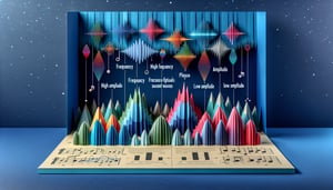
Brainwave Frequency Bands -
Review the five primary EEG rhythms - delta (0.5 - 4 Hz), theta (4 - 8 Hz), alpha (8 - 13 Hz), beta (13 - 30 Hz), and gamma (>30 Hz) - and their associated behavioral states. Use the mnemonic "Darn The Aardvark Bought Grapes" to recall the order from slowest to fastest. Recognizing these bands is essential for accurate EEG board prep and EEG exam prep.
10 - 20 Electrode Placement System -
Memorize the international 10 - 20 electrode placement system, where distances between nasion, inion, and preauricular points are measured at 10% or 20% intervals to ensure standardized lead positions such as Fp1, F7, C3, or O2. Mastering this protocol ensures reliable recordings and is a cornerstone of electroencephalography practice questions. Visualizing head templates from ACNS or IFCN resources can speed up your recall during the exam.
Montage Configurations -
Compare montage configurations - referential (single reference), bipolar (adjacent electrodes), and Laplacian (localized derivatives) - to understand how each impacts signal amplitude and spatial resolution. Refer to ACNS guidelines to choose the optimal montage for focal versus generalized activity, a frequent focus in neurotechnologist board review. Practicing switching montages on sample datasets can solidify your decision-making skills.
Filter Settings & Artifact Management -
Fine-tune filter settings - commonly a 1 Hz high-pass to remove drift, a 70 Hz low-pass to exclude muscle noise, and a 50/60 Hz notch to reject line interference - following manufacturer specs or IFCN standards. Practice differentiating physiological artifacts (e.g., EOG, EMG) from true cerebral signals using video-EEG correlates, a skill often tested in EEG practice quizzes. Remember, improper filter use can mask or mimic pathology.
Seizure Pattern Recognition -
Identify hallmark seizure patterns such as the 3 Hz spike-and-wave complex in absence epilepsy or rhythmic temporal lobe spike bursts in focal seizures, and note their typical localization and spread. Use annotated waveform examples from journals like Epilepsia to reinforce pattern recognition and boost confidence in your eeg board prep success. Regularly timing waveform segments can help you gauge frequency and duration under exam conditions.