Unlock hundreds more features
Save your Quiz to the Dashboard
View and Export Results
Use AI to Create Quizzes and Analyse Results

Sign inSign in with Facebook
Sign inSign in with Google

Dental Radiography Quiz: Master Exposure Times

Ready to ace conventional radiography exposure times? Take the quiz!

Difficulty: Moderate
2-5mins
Learning OutcomesCheat Sheet
Paper cut dental radiography quiz with x ray sensor tooth timers layered over golden yellow background

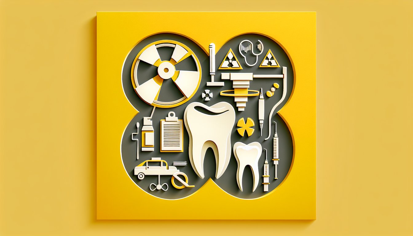
Think you've mastered the nuances of a dental radiography quiz? Designed for student hygienists, dental assistants, aspiring radiographers, and seasoned pros, this free challenge will test your understanding of exposure times dental radiography and spotlight essential conventional radiography exposure times. As you tackle each question, you'll reinforce dental radiography trivia, deepen your grasp of image quality versus patient dose, and learn tips to assist with dental radiography workflows in real clinical settings. For extra practice, dive into our dental x ray practice test or refine angle control with the foreshortening dental x ray guide. You'll get instant feedback and a personalized score to track your progress - perfect for exam prep or continuing education. Ready to elevate your confidence? Start the quiz now!

What unit is used to measure X-ray exposure time in dental radiography?
Rads
Seconds
Amperes
Volts
Exposure time in dental X-ray units is measured in seconds, which determines how long the X-ray tube emits radiation. Proper timing helps ensure diagnostic image quality without excessive patient dose. Adjusting exposure time directly affects film density or digital receptor response. RadiologyInfo.org
Which component of the X-ray machine restricts the size and shape of the X-ray beam?
Collimator
Anode
Generator
Converter
The collimator limits the X-ray beam to the area of interest, reducing patient exposure and scatter radiation. By shaping the beam, it improves image contrast and sharpness. Rectangular collimation is often recommended in dentistry for further dose reduction. ADA Guidelines
Increasing milliamperage (mA) in the X-ray unit will primarily affect which aspect of the image?
Image density
Focal spot size
Image contrast
Beam energy
Milliamperage controls the quantity of X-ray photons produced per unit time, which directly influences image density (darkness). Higher mA increases the number of photons and thus a darker image if exposure time remains constant. It does not change the energy (quality) of the beam. Radiology Cafe
In intraoral radiography, which film speed requires the least exposure time?
F-speed
D-speed
E-speed
C-speed
F-speed film is approximately 60% faster than D-speed, meaning it requires significantly less exposure time to produce a diagnostic image. Reduced exposure times decrease patient dose and motion artifacts. E-speed is intermediate but less common than F-speed. ADA Film Speed Recommendations
Which radiographic technique aligns the film sensor parallel to the long axis of the tooth?
Bisecting-angle technique
Paralleling technique
Panoramic technique
Occlusal technique
The paralleling technique places the image receptor parallel to the tooth's long axis and directs the X-ray beam perpendicular to both. This method reduces distortion and improves consistency. It is preferred over bisecting for periapical images. Radiology Assistant
How does increasing kilovoltage (kVp) affect the X-ray beam?
Decreases focal spot size
Increases image density only
Increases patient dose only
Increases beam penetration
Higher kVp increases the energy of X-ray photons, enhancing beam penetration through tissues. This reduces subject contrast but decreases required exposure time for adequate density. Patient dose may be constant or reduced if exposure time is adjusted. RadiologyInfo.org
Which device in extraoral radiography uses screens to reduce exposure time?
Lead apron
Grid
Intensifying screen
Collimator
Intensifying screens in the film cassette emit light when struck by X-rays, exposing the film with less radiation. This significantly reduces patient exposure and motion artifacts. Different screen speeds alter the degree of exposure reduction. Radiology Assistant
What happens to image density if exposure time is doubled while all other factors remain constant?
Density remains the same
Density decreases
Contrast increases
Density increases
Doubling exposure time doubles the number of X-ray photons reaching the receptor, resulting in a darker (higher density) image. This direct proportional relationship helps calibrate exposures. Contrast remains primarily controlled by kVp. RadiologyInfo.org
Which factor is NOT directly controlled by the radiographer during exposure?
Atomic number of the tooth
kVp
Exposure time
mA
The atomic number of tissues is inherent and cannot be altered by the radiographer. kVp, mA, and exposure time are adjustable settings on the X-ray unit to control beam quality and quantity. These choices optimize image quality and patient safety. ADA Guidelines
What effect does using a longer PID (position-indicating device) have on the image?
Decreases image quality
Has no effect
Increases magnification
Reduces magnification
A longer PID increases the distance between the focal spot and the receptor, reducing beam divergence and magnification. This produces sharper images with less distortion. However, patient dose remains similar if exposure factors are adjusted. Radiology Assistant
Which film speed designation is slower and requires more exposure time than F-speed?
S-speed
T-speed
D-speed
E-speed
D-speed film is older and slower than F-speed, requiring approximately twice the exposure time for equivalent density. E-speed is intermediate but still slower than F-speed. Modern digital sensors are even faster, reducing exposure further. ADA Film Speed Recommendations
Which technique error is characterized by a clear unexposed area on the film receptor?
Cone cutting
Elongation
Double exposure
Foreshortening
Cone cutting results from misalignment of the PID and receptor, leaving a clear, unexposed region. Proper alignment and centering of the X-ray beam prevent this error. It is instantly recognizable and requires retake. Radiology Assistant
If you increase the kVp by 15 while halving the exposure time, what remains approximately constant?
Image contrast
Image density
Focal spot size
Patient dose
The 15 kVp rule states that increasing kVp by 15 allows you to half the exposure time to maintain similar image density. While density stays constant, contrast decreases slightly due to higher beam energy. Patient dose may be reduced if time is halved. RadiologyInfo.org
When switching from D-speed film to F-speed film, what approximate exposure time adjustment is needed?
Decrease by 20%
No change
Increase by 60%
Decrease by 60%
F-speed film is roughly 60% faster than D-speed, allowing a 60% reduction in exposure time for comparable density. This results in less patient dose and improved workflow. Exact adjustment depends on the X-ray unit and receptor system. ADA Film Speed Guidelines
A pediatric patient requires 50% less exposure time than an adult. If the adult premolar film is 0.2 seconds, what time should you use for the child?
0.15 seconds
0.1 seconds
0.2 seconds
0.05 seconds
Children often require about half the exposure time of adults due to smaller enamel and dentin thickness. Halving 0.2 seconds yields 0.1 seconds. Proper adjustment reduces dose and prevents overexposure. ADA Pediatric Radiology
Which factor is adjusted to compensate for scatter radiation when using a grid in extraoral radiography?
Increase exposure time
Decrease mA
Decrease kVp
Use faster film
Grids absorb scatter and improve contrast but also absorb some primary photons, requiring increased exposure time or mA to compensate for lower receptor exposure. kVp adjustments are less common when using a grid. RadiologyInfo.org
What is the recommended collimator shape for intraoral dental radiography?
Circular
Elliptical
Square
Rectangular
Rectangular collimation reduces patient exposure by limiting the X-ray beam to the receptor size, cutting scatter and dose up to 60%. Circular collimators are less effective for dose reduction. Rectangular is recommended by most dental associations. ADA Guidelines
Halving the distance between the X-ray tube and the receptor without changing exposure settings will do what to receptor exposure?
Have no effect
Increase it fourfold
Reduce it by half
Double it
According to the inverse square law, reducing distance by half increases intensity by four times, making the image much darker if settings remain unchanged. Proper distance and exposure adjustments are crucial for safety and image quality. RadiologyInfo.org
What error can result from using excessive vertical angulation in the bisecting-angle technique?
Underexposure
Cone cutting
Foreshortening
Elongation
Too much vertical angulation causes the image receptor to capture a shorter projection of the tooth, appearing foreshortened. Elongation occurs with insufficient angulation. Accurate angulation is essential for diagnostic images. Radiology Assistant
Which effect does using a digital sensor instead of film typically have on required exposure time?
Triples it
Decreases it by 50 - 70%
Increases it by 50%
No change
Digital sensors are more sensitive to X-rays than film, allowing exposure time reductions of 50 - 70% while maintaining image quality. This lowers patient dose and improves workflow. Specific reduction depends on sensor type. RadiologyInfo.org
What adjustment should be made if a periapical film appears too dark at normal settings?
Use a grid
Increase mA
Increase kVp
Reduce exposure time
A dark image indicates overexposure; reducing exposure time will decrease receptor exposure and correct density. Changing mA or kVp would alter other image characteristics. Grids are not used intraorally. RadiologyInfo.org
Which focal spot size reduces the penumbra effect and increases image sharpness?
Size has no effect
Smaller focal spot
Larger focal spot
Medium focal spot
Smaller focal spots reduce the geometric unsharpness (penumbra) at object edges, improving image resolution. Larger spots produce more blur. Most dental units use small focal spots (0.4 - 0.7 mm). RadiologyInfo.org
When does reciprocity failure become a concern in dental film exposures?
High kVp
Short exposure times
Small focal spot
Long exposure times
Reciprocity failure occurs when very long exposures reduce film sensitivity, causing underdevelopment despite adequate exposure. Modern films and digital sensors minimize this effect, but it remains a factor for exposures over several seconds. RadiologyInfo.org
What is the recommended maximum half-value layer (HVL) of aluminum filtration for dental X-ray units operating above 70 kVp?
1.5 mm Al
2.5 mm Al
0.5 mm Al
3.5 mm Al
Regulations require at least 2.5 mm aluminum HVL for units above 70 kVp to filter low-energy photons and reduce patient dose. This ensures beam quality and patient safety. Lower HVL is permissible for lower kVp units. ADA Filtration Standards
According to the inverse square law, if source-to-receptor distance is increased by 1.5 times, what happens to receptor exposure?
Decreases to 44% of original
Decreases to 75% of original
Increases by 150%
No change
Inverse square law states exposure varies with the square of distance. (1/1.5²)=1/2.25?0.44, so exposure falls to 44%. Accurate distance control is critical for consistent image density and patient dose. RadiologyInfo.org
What is the primary purpose of beam filtration in dental X-ray units?
Increase mA
Improve film speed
Increase focal spot size
Remove low-energy photons
Filtration removes low-energy (soft) X-rays that add patient dose without improving image quality. By hardening the beam, it increases the average photon energy and improves penetration. Regulatory bodies mandate minimum filtration levels. ADA Filtration Guidelines
In panoramic radiography, what happens if the patient's head is positioned too far forward?
Mandibular ramus is superimposed
Distortion is minimal
Anterior teeth appear narrowed and blurred
Anterior teeth appear widened
When the patient's head is too far forward, the teeth lie outside the focal trough, causing them to appear narrowed and blurred. Proper head positioning within the focal plane is crucial for diagnostic panoramic images. Radiology Assistant
Which factor most directly affects image contrast in digital radiography?
mA
kVp
Focal spot size
Exposure time
Kilovoltage alters the energy spectrum of X-rays; higher kVp produces a more penetrating beam with lower subject contrast. mA and time affect density, not contrast. Sensor dynamic range further influences apparent contrast. RadiologyInfo.org
What is the purpose of a compensating filter in extraoral cephalometric imaging?
Focus beam
Reduce kVp
Even out exposure across anatomical curvature
Increase scatter
Compensating filters (wedge-shaped) balance the exposure between dense and less dense areas, such as the skull and neck, producing uniform image density. This improves diagnostic detail across varying tissue thicknesses. RadiologyInfo.org
When processing film manually, which factor must be adjusted to compensate for lower darkroom temperature?
Increase development time
Increase kVp
Increase exposure time
Decrease rinse time
Lower developer temperatures slow chemical reactions, producing underdeveloped films. Increasing development time compensates for the cooler environment. Exposure settings remain unchanged. RadiologyInfo.org
What is the typical exposure time range for an adult panoramic radiograph?
0.2 - 0.5 seconds
30 - 40 seconds
1 - 2 seconds
10 - 20 seconds
Adult panoramic exposures generally last between 10 and 20 seconds as the tube rotates around the patient's head. These longer times require patient stability but yield a full-arch image. Intraoral exposures are much shorter. Radiology Assistant
In a dual-film screen cassette system, what adjustment is required when switching to single-film use?
Reduce kVp
Increase exposure by 50%
No change
Decrease exposure by 50%
Removing one screen-film combination reduces overall sensitivity, so exposure must be increased by about 50% to maintain density. Dual screens are faster than single. kVp remains the same. RadiologyInfo.org
Which characteristic of high-frequency X-ray generators improves exposure efficiency?
Higher mA capacity
Larger focal spot
Lower kVp variability
Near-constant voltage output
High-frequency generators produce almost constant voltage, minimizing voltage ripple and maximizing X-ray production during the exposure. This increases efficiency and allows shorter exposure times. Conventional units have more ripple. RadiologyInfo.org
How does automatic exposure control (AEC) function in dental panoramic systems?
Shifts patient position
Adjusts focal spot size
Changes tube voltage during exposure
Terminates exposure when sensor reaches target density
AEC uses a sensor behind the image receptor to measure transmitted radiation and automatically ends the exposure once the preset density is achieved. This ensures consistent image quality despite patient thickness variations. It does not alter kVp or mA during exposure. RadiologyInfo.org
In CBCT imaging, which parameter most directly reduces patient dose while maintaining image quality?
Increasing kVp
Reducing field of view (FOV)
Increasing voxel size
Increasing mA
Reducing the FOV limits the scan region, directly lowering radiation dose to surrounding tissues without compromising resolution in the area of interest. Adjusting voxel size or kVp affects image noise or penetration but has less impact on overall patient dose. RadiologyInfo.org
What is the principle behind organ-dose modulation in advanced dental X-ray units?
Increasing exposure time
Shifting focal spot away from organs
Using higher kVp for organ protection
Altering mA based on tube angle to spare radiosensitive organs
Organ-dose modulation reduces mA when the tube rotates over sensitive regions (e.g., thyroid), decreasing dose without affecting image quality. The unit raises mA when over less sensitive areas to maintain overall density. This dynamic approach optimizes patient safety. RadiologyInfo.org
0
{"name":"What unit is used to measure X-ray exposure time in dental radiography?", "url":"https://www.quiz-maker.com/QPREVIEW","txt":"What unit is used to measure X-ray exposure time in dental radiography?, Which component of the X-ray machine restricts the size and shape of the X-ray beam?, Increasing milliamperage (mA) in the X-ray unit will primarily affect which aspect of the image?","img":"https://www.quiz-maker.com/3012/images/ogquiz.png"}

Study Outcomes

  1. Analyze Exposure Time Differences -

    Distinguish between exposure times required for dental radiography and conventional radiography, identifying the factors that influence shorter or longer settings.

  2. Apply Optimal Patient Positioning -

    Implement proper positioning protocols to ensure consistent and accurate image capture during dental radiography procedures.

  3. Recall Standard Exposure Parameters -

    Memorize the typical exposure times for both intraoral and extraoral imaging techniques to enhance daily practice efficiency.

  4. Interpret Exposure Impact on Image Quality -

    Evaluate how adjustments in exposure times affect radiographic density, contrast, and diagnostic usefulness.

  5. Assess Radiation Dose Management -

    Determine how to balance exposure times to minimize patient radiation while maintaining diagnostic image quality.

Cheat Sheet

  1. Digital vs. Conventional Exposure Times -

    Digital sensors in dental radiography often require exposure times 50 - 90% shorter than that required for conventional radiography (American Dental Association, 2020). Remember the mnemonic "Digital Dashes, Film Lingers" to recall that a 0.2-second digital bitewing equates to about 1.6 seconds with film. Mastering this difference will boost your score on any dental radiography quiz and reinforce safety.

  2. Inverse Square Law & Distance -

    The intensity of X-rays falls off with the square of the distance (I ∝ 1/d²), so doubling the tube-to-sensor distance cuts exposure to one-quarter (NCRP Report No. 145). Use the simple phrase "Double Distance, Quarter Dose" to lock in this formula. This concept is a staple in exposure times dental radiography questions and critical for dose management.

  3. Optimal kVp and mA Settings -

    Balancing kilovoltage peak (kVp) and milliamperage (mA) adjusts contrast and exposure: higher kVp lowers patient dose but reduces contrast, while higher mA increases image density (University of Washington School of Dentistry). A handy trick is "High kV for Low Dose, High mA for Bright Image." Understanding these trade-offs helps assist with dental radiography technique selection.

  4. Paralleling Technique & Sensor Alignment -

    The paralleling technique places the sensor parallel to the tooth axis and the beam perpendicular, reducing distortion (ADA Guidelines). Think "Parallel Equals Precise" to remember error-minimized positioning. Perfecting this is key trivia for positioning questions in your dental radiography quiz.

  5. ALARA Principle & Safety Protocols -

    "As Low As Reasonably Achievable" (ALARA) guides all exposure decisions, promoting thyroid collars, lead aprons, and proper collimation (International Atomic Energy Agency). Use the acronym ALARA to recall equipment checks, patient shielding, and beam collimation before every shot. This principle underpins every conventional radiography exposure times guideline and keeps patients safe.

Powered by: Quiz Maker