Unlock hundreds more features
Save your Quiz to the Dashboard
View and Export Results
Use AI to Create Quizzes and Analyse Results

Sign inSign in with Facebook
Sign inSign in with Google

Pon a Prueba tus Conocimientos de los Huesos del Cuerpo Humano

Piensa rápido: ¿puedes nombrar los huesos del esqueleto humano?

Difficulty: Moderate
2-5mins
Learning OutcomesCheat Sheet
Esqueleto humano estilo papel recortado con nombres de huesos sobre fondo celeste para quiz de anatomia

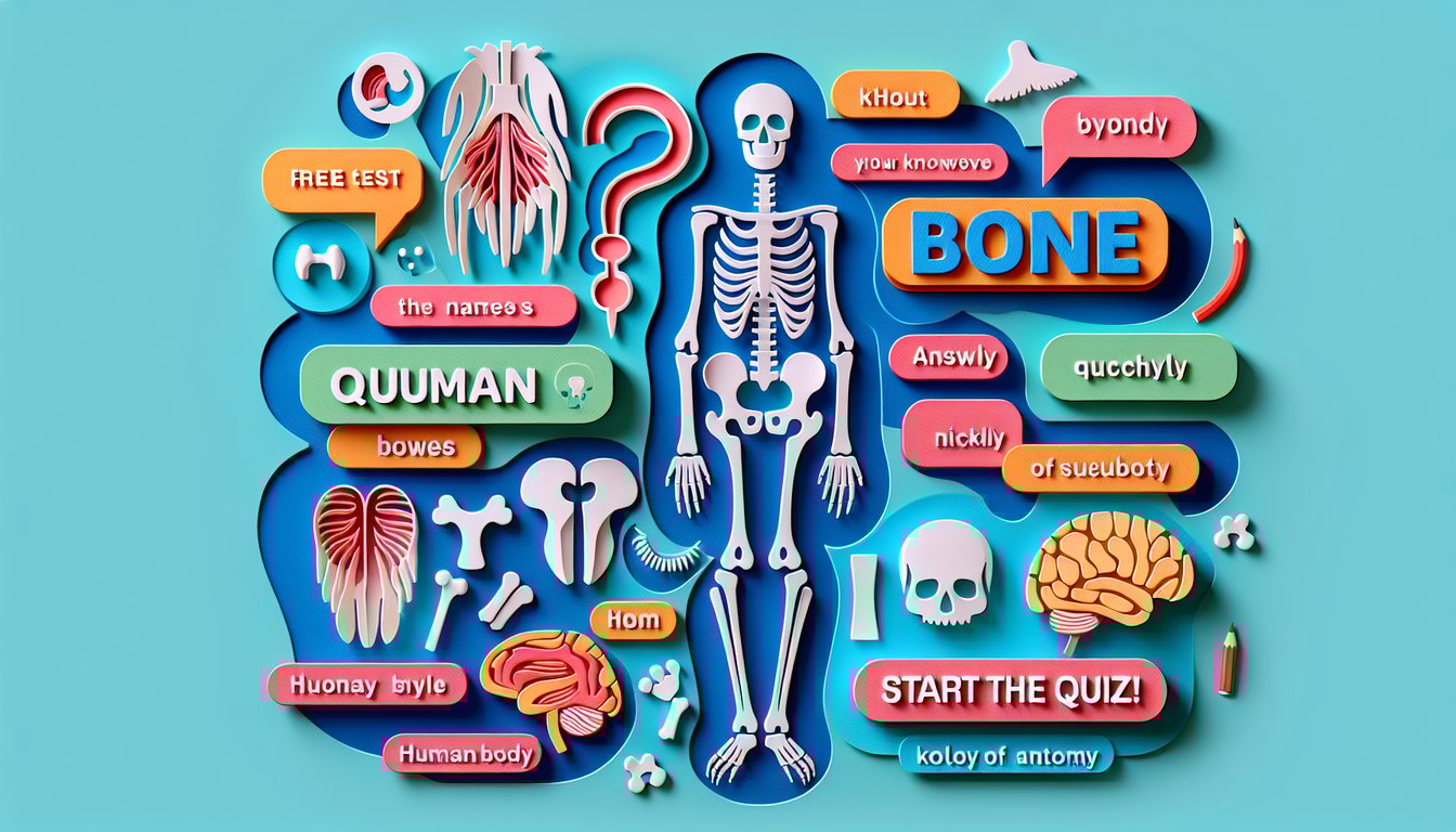
¡Bienvenido al Quiz Gratis: Nombres de Huesos del Cuerpo Humano! Pon a prueba tus conocimientos sobre los nombres de huesos del cuerpo humano y descubre cuánto dominas la anatomía de los huesos humanos. A lo largo de preguntas rápidas y dinámicas, repasaremos los huesos del esqueleto humano y las partes del esqueleto humano esenciales para tu aprendizaje. Con cada pregunta, reforzarás la memoria y la terminología de los nombres de huesos humanos. Esta prueba es ideal para estudiantes y aficionados a la anatomía, así que atrévete a jugar y mide tu nivel. Si quieres más práctica, explora nuestro test de huesos del cuerpo o repasa con un cuestionario de partes del cuerpo humano . ¡Empieza ahora y demuestra tu experiencia!

Which bone is the longest in the human body?
Femur
Humerus
Tibia
Fibula
The femur, or thigh bone, is the longest and strongest bone in the human body, extending from the hip to the knee. It supports most of the body’s weight when standing and walking. Its robust structure is key for locomotion and load-bearing. source
Which bone protects the brain?
Skull
Mandible
Sternum
Pelvis
The skull, composed of several fused bones, provides a protective cavity for the brain. It also supports the structures of the face and houses the organs for smell, hearing, and vision. The cranial vault is specifically designed to absorb impacts and protect neural tissue. source
Which bone is commonly called the collarbone?
Clavicle
Scapula
Humerus
Sternum
The clavicle, or collarbone, is a long slender bone that connects the sternum to the scapula. It acts as a strut to keep the upper limb away from the thorax and allows for a wide range of shoulder motion. Its subcutaneous position makes it a common site for fractures. source
Which bone forms the upper arm?
Humerus
Radius
Ulna
Scapula
The humerus is the long bone in the upper arm, extending from the shoulder to the elbow. It articulates proximally with the scapula and distally with the radius and ulna. This bone provides attachment sites for muscles that move the shoulder and elbow joints. source
Which bone is commonly known as the shinbone?
Tibia
Fibula
Femur
Patella
The tibia, or shinbone, is the larger and stronger of the two bones in the lower leg. It bears most of the body's weight and articulates with the femur at the knee and the talus at the ankle. Its anterior border lies just beneath the skin, making it a common site for injury. source
Which bone is the kneecap?
Patella
Fibula
Tibia
Femur
The patella, or kneecap, is a small triangular sesamoid bone embedded in the quadriceps tendon. It protects the knee joint and increases the leverage of the thigh muscles during leg extension. Its position facilitates smooth movement of the quadriceps over the knee. source
Which bone is part of the pelvic girdle and is also known as the hip bone?
Ilium
Ischium
Pubis
Sacrum
The ilium is the broad, flaring portion of the hip bone and forms the upper third of each pelvic bone. It provides attachment for abdominal and thigh muscles. The iliac crest is a notable landmark for clinicians during physical examinations. source
Which bone is known as the breastbone?
Sternum
Clavicle
Manubrium
Xiphoid process
The sternum is a flat bone at the center of the chest, commonly called the breastbone. It connects to the ribs via costal cartilages, forming the anterior portion of the rib cage and protecting vital organs such as the heart and lungs. The manubrium, body, and xiphoid process are its three parts. source
Which bone forms the forearm on the thumb side?
Radius
Ulna
Humerus
Carpals
The radius is one of the two long bones of the forearm, located on the lateral (thumb) side. It articulates with the humerus at the elbow and with the carpal bones at the wrist. The radius rotates around the ulna to allow pronation and supination of the forearm. source
Which bone forms the lower jaw?
Mandible
Maxilla
Zygomatic bone
Vomer
The mandible is the largest and strongest facial bone, forming the lower jaw. It holds the lower teeth and provides attachment for muscles involved in chewing and facial expression. Its unique structure allows for movement at the temporomandibular joint. source
Which bone forms the forehead region of the skull?
Frontal bone
Parietal bone
Occipital bone
Temporal bone
The frontal bone forms the forehead and the upper part of the eye sockets. It articulates with the parietal, sphenoid, ethmoid, and nasal bones. It protects the frontal lobes of the brain and supports facial structures. source
Which bone is commonly called the shoulder blade?
Scapula
Clavicle
Humerus
Sternum
The scapula, or shoulder blade, is a flat triangular bone that connects the humerus with the clavicle. It provides attachment for several muscles responsible for shoulder and arm movements. Its glenoid cavity forms the socket for the shoulder joint. source
Which bone in the foot is known as the heel bone?
Calcaneus
Talus
Navicular
Cuboid
The calcaneus is the largest tarsal bone and forms the heel of the foot. It articulates with the talus and cuboid bones and serves as the attachment for the Achilles tendon. It bears much of the body’s weight when standing and walking. source
Which bone forms the posterior part of the hard palate?
Palatine bone
Vomer
Maxilla
Zygomatic bone
The palatine bones form the posterior portion of the hard palate and contribute to the walls of the nasal cavity and the floor of the orbits. They articulate with the maxillae, sphenoid, ethmoid, and inferior nasal conchae. Their shape allows separation of the oral and nasal cavities. source
Which bone is commonly known as the cheekbone?
Zygomatic bone
Maxilla
Sphenoid bone
Lacrimal bone
The zygomatic bone forms the prominence of the cheek and part of the lateral wall and floor of the orbit. It articulates with the maxilla, temporal, sphenoid, and frontal bones. This bone supports facial muscles and contributes to the cheek’s contour. source
Which small bone at the base of the sternum is the lowest part of that bone?
Xiphoid process
Manubrium
Body of sternum
Costal cartilage
The xiphoid process is the smallest and most inferior portion of the sternum. It begins as cartilage and ossifies in adulthood. It serves as an attachment point for abdominal muscles. source
Which bone is located at the sides and base of the skull?
Temporal bone
Parietal bone
Occipital bone
Ethmoid bone
The temporal bones are situated at the sides and base of the skull. They house the structures of the ear and form part of the cranial floor. Each temporal bone articulates with the parietal, occipital, sphenoid, and zygomatic bones. source
Which carpal bone is most commonly fractured when falling on an outstretched hand?
Scaphoid
Lunate
Pisiform
Hamate
The scaphoid is the most frequently fractured carpal bone due to its location in the anatomical snuffbox. Falls onto an outstretched hand transmit force directly to this bone. Fractures can compromise blood supply, leading to avascular necrosis if not treated promptly. source
Which bone in the neck is the only bone that does not articulate with any other bone?
Hyoid bone
Atlas
Mandible
Clavicle
The hyoid bone is a U-shaped bone in the anterior neck that does not articulate with any other bone. It is suspended by muscles and ligaments and supports the tongue and its muscles. It also serves as an attachment point for neck and pharyngeal muscles. source
Which bone is the smallest in the human body?
Stapes
Incus
Malleus
Hyoid
The stapes, located in the middle ear, is the smallest bone in the human body. It transmits sound vibrations from the incus to the oval window of the inner ear. Its stirrup-like shape is essential for hearing conduction. source
Which bone forms the lateral malleolus at the ankle?
Fibula
Tibia
Calcaneus
Talus
The fibula is the slender bone located lateral to the tibia in the lower leg. Its distal end forms the lateral malleolus, which stabilizes the ankle joint. It also provides attachment points for muscles of the lower leg. source
Which bone is formed by the fusion of five sacral vertebrae?
Sacrum
Coccyx
Lumbar vertebra
Ilium
The sacrum is formed by the fusion of five sacral vertebrae and connects the spine to the pelvis. It supports the weight of the upper body when sitting and standing. It also transmits forces between the axial skeleton and the lower limbs. source
Which bone is shaped like a butterfly and forms part of the base of the skull?
Sphenoid bone
Ethmoid bone
Occipital bone
Frontal bone
The sphenoid bone is a complex bone with a central body and paired wings, resembling a butterfly. It forms part of the base of the skull and contributes to the orbits. It houses the sella turcica, which supports the pituitary gland. source
Which bone is the second cervical vertebra with a structure known as the dens?
Axis
Atlas
C3
Sacrum
The axis (C2) is the second cervical vertebra and features the odontoid process or dens, which projects upward to articulate with the atlas (C1). This joint allows the head to rotate from side to side. The dens provides a pivot that stabilizes the atlanto-axial joint. source
Which bone forms the lower part of the nasal septum?
Vomer
Perpendicular plate of ethmoid
Septal cartilage
Palatine bone
The vomer is a thin, flat bone that forms the inferior portion of the nasal septum. It articulates with the sphenoid, ethmoid, maxillae, and palatine bones. It separates the left and right airways in the nasal cavity. source
Which bone supports body weight when sitting and forms the lower part of the pelvis?
Ischium
Ilium
Pubis
Sacrum
The ischium is the curved bone forming the base of each half of the pelvis and bears body weight when sitting. It articulates with the ilium and pubis to form the hip bone. The ischial tuberosity is the prominent area where hamstring muscles attach. source
Which bone contains the foramen magnum allowing passage of the spinal cord?
Occipital bone
Temporal bone
Parietal bone
Sphenoid bone
The occipital bone forms the posterior part of the cranial vault and contains the foramen magnum, through which the spinal cord connects to the brainstem. It articulates with the atlas (C1) vertebra. Its external occipital protuberance serves as a muscle attachment. source
Which bone is the most superior vertebra that supports the skull?
Atlas
Axis
C3
Occipital bone
The atlas (C1) is the first cervical vertebra and supports the skull by articulating with the occipital condyles. It lacks a vertebral body and allows the nodding motion of the head. Its ring-like structure provides stability and range of motion. source
Which bone articulates with the manubrium of the sternum on the anterior thorax?
Clavicle
Scapula
First rib
Humerus
The clavicle articulates medially with the manubrium of the sternum, forming the sternoclavicular joint. This joint is the only skeletal connection between the axial skeleton and the upper limb skeleton and allows a wide range of shoulder movements. Fractures here can disrupt arm function. source
Which bone is the smallest facial bone, located near the tear ducts?
Lacrimal bone
Malar bone
Maxilla
Nasal bone
The lacrimal bones are the smallest and most fragile bones of the face, located in the medial wall of each orbit near the tear ducts. They form part of the nasolacrimal canal, which drains tears into the nasal cavity. Their delicate structure is prone to fractures. source
Which bone houses the cribriform plate that allows olfactory nerve fibers to pass?
Ethmoid bone
Sphenoid bone
Frontal bone
Occipital bone
The ethmoid bone is a light, spongy bone located between the nasal cavity and orbits. Its horizontal cribriform plate contains multiple foramina for the olfactory nerve fibers to transmit smell information from the nasal mucosa to the brain. It also contributes to the medial orbital walls. source
Which bone develops by intramembranous ossification and forms a large portion of the cranial roof?
Parietal bone
Temporal bone
Frontal bone
Occipital bone
The parietal bones form the superior and lateral aspects of the cranial vault. They ossify directly from mesenchymal tissue through intramembranous ossification, without a cartilage precursor. This process is typical for many flat bones of the skull. source
Which bone contains the alveolar processes for the upper teeth?
Maxilla
Mandible
Palatine bone
Vomer
The maxilla is the paired upper jaw bone that holds the upper teeth within its alveolar processes. It contributes to the formation of the orbit, nasal cavity, and hard palate. It also houses the maxillary sinuses. source
0
{"name":"Which bone is the longest in the human body?", "url":"https://www.quiz-maker.com/QPREVIEW","txt":"Which bone is the longest in the human body?, Which bone protects the brain?, Which bone is commonly called the collarbone?","img":"https://www.quiz-maker.com/3012/images/ogquiz.png"}

Study Outcomes

  1. Identificar los huesos principales -

    Podrás reconocer y nombrar los huesos fundamentales del esqueleto humano, desde el cráneo hasta los huesos de las extremidades.

  2. Reconocer los nombres de huesos humanos -

    Aprenderás los nombres de huesos del cuerpo humano más relevantes y mejorarás tu vocabulario anatómico.

  3. Diferenciar las partes del esqueleto humano -

    Serás capaz de distinguir entre las principales secciones del esqueleto, como el axial y el apendicular.

  4. Localizar los huesos en el cuerpo -

    Identificarás la ubicación de cada hueso en distintas regiones del cuerpo y comprenderás su interrelación.

  5. Recordar la anatomía de los huesos humanos -

    Fortalecerás la memoria sobre la estructura ósea mediante preguntas rápidas y ejercicios interactivos.

  6. Aplicar la terminología anatómica -

    Utilizarás correctamente los términos especializados para describir los huesos y sus funciones en contextos educativos y profesionales.

Cheat Sheet

  1. División axial y apendicular -

    El esqueleto humano se organiza en dos grandes partes: el esqueleto axial (cráneo, columna vertebral y caja torácica) y el apendicular (extremidades y cintura pélvica). Esta clasificación es esencial para memorizar los nombres de huesos del cuerpo humano y entender sus funciones de soporte y protección. Según Gray's Anatomy, distinguir estas partes del esqueleto humano facilita el estudio de la anatomía de los huesos humanos.

  2. Huesos del cráneo -

    El cráneo está formado por seis huesos principales: frontal, parietal, temporal, occipital, esfenoides y etmoides. Para recordar estos nombres de huesos humanos, usa el mnemónico "FE-PS-TO" (Frontal, Etmoides, Parietal, Esfenoides, Temporal, Occipital). Este truco, recomendado por la Universidad de Harvard, agiliza la memorización de los huesos del esqueleto humano.

  3. Columna vertebral y sus segmentos -

    La columna vertebral cuenta con 7 vértebras cervicales, 12 dorsales, 5 lumbares, el sacro y el cóccix. Un método mnemotécnico popular es "7-12-5 SC" para los segmentos cervical, dorsal, lumbar, sacro y cóccix. Esta organización estructural, descrita en publicaciones de la Universidad de Oxford, es clave en el estudio de los nombres de huesos del cuerpo humano.

  4. Caja torácica y costillas -

    La caja torácica incluye 12 pares de costillas y el esternón; de ellas, 7 pares son verdaderas, 3 falsas y 2 flotantes. Recuerda la secuencia "7-3-2" para diferenciar cada tipo y su conexión al esternón. La American Thoracic Society recomienda este esquema para familiarizarse con los huesos del esqueleto humano.

  5. Huesos de las extremidades -

    Las extremidades superiores incluyen húmero, radio, cúbito, carpianos, metacarpianos y falanges; las inferiores, fémur, tibia, peroné, tarsianos, metatarsianos y falanges. Un acrónimo útil es "HRCC-MF / FTPTF" para agruparlos rápidamente. Este enfoque práctico, avalado por la Universidad de Cambridge, acelera el repaso de la anatomía de los huesos humanos.

Powered by: Quiz Maker