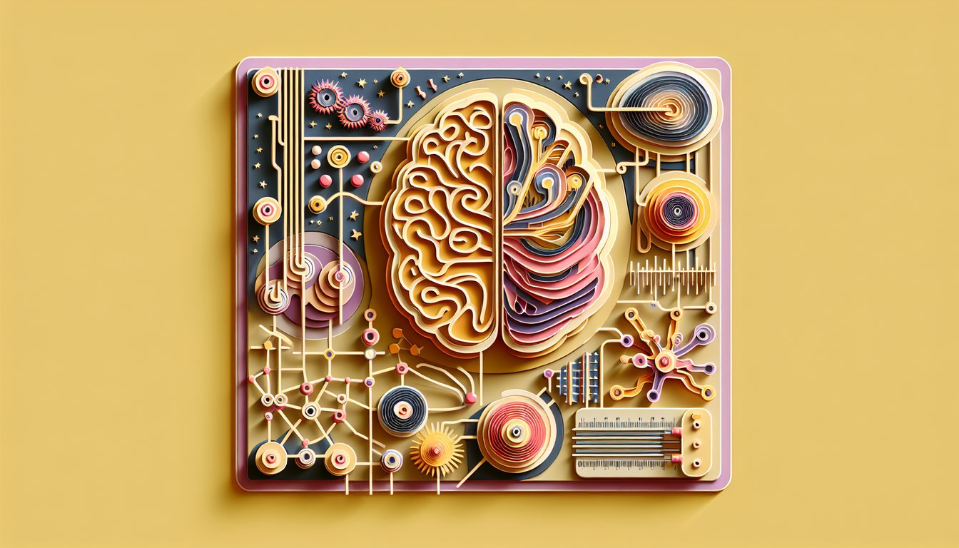
Are you ready to dive into a comprehensive assessment of the neurological system? This interactive challenge is designed for healthcare students, nursing professionals, and curious learners eager to test their knowledge. Through our neurologic system quiz you'll explore cerebellum function assessment, sharpen skills in a cranial nerve quiz, and tackle essential sensory function test scenarios - all in one go. Don't miss your chance to deepen your practice and confidence: take our neurological system quiz now, or switch gears with a quick nervous system quiz for even more insights. Ready to prove your expertise? Jump in today!
Which cranial nerve is responsible for the sense of smell?
Olfactory nerve (CN I)
Optic nerve (CN II)
Trigeminal nerve (CN V)
Vagus nerve (CN X)
The olfactory nerve (CN I) is solely responsible for transmitting smell information from the nasal mucosa to the brain. Other cranial nerves carry visual, facial sensation, or autonomic functions. Damage to CN I can cause anosmia. Learn more about cranial nerve I.
Which lobe of the brain is primarily responsible for coordination and balance?
Frontal lobe
Temporal lobe
Occipital lobe
Cerebellum
The cerebellum is the main structure involved in coordinating voluntary movements and maintaining balance and posture. The other lobes of the cerebrum handle motor planning, auditory processing, and vision. Cerebellar lesions often present with ataxia. More on cerebellar function.
The Romberg test assesses which sensory function?
Proprioception
Vision
Hearing
Taste
The Romberg test evaluates the integrity of the dorsal columns by testing proprioception and position sense. A positive Romberg sign suggests loss of proprioceptive input. Visual input can compensate, so the eyes are closed during the test. Details on Romberg test.
Which instrument is used to test vibration sense?
Monofilament
Tuning fork
Otoscope
Reflex hammer
A vibrating tuning fork is placed over bony prominences to assess vibration sense carried by the dorsal columns. Monofilaments test light touch, reflex hammers test deep tendon reflexes, and otoscopes visualize the ear canal. Vibration testing helps identify dorsal column dysfunction. Vibration sense testing.
What is the normal response of the patellar reflex?
Knee flexion
Knee extension
Hip flexion
Ankle dorsiflexion
Tapping the patellar tendon stretches the quadriceps, resulting in a reflexive knee extension. This deep tendon reflex tests the L2 - L4 spinal segments. Absent or diminished response may indicate peripheral neuropathy or spinal cord injury. Patellar reflex overview.
Which cranial nerve controls lateral eye movement?
Oculomotor nerve (CN III)
Facial nerve (CN VII)
Abducens nerve (CN VI)
Trochlear nerve (CN IV)
The abducens nerve (CN VI) innervates the lateral rectus muscle, which abducts the eye. The oculomotor and trochlear nerves control other extraocular muscles, and the facial nerve controls facial expression. Lesion of CN VI causes medial deviation of the affected eye. Abducens nerve function.
What does a 2+ reflex grade indicate?
Absent reflex
Normal reflex
Hyperreflexia
Clonus
Reflex grading uses a 0 to 4+ scale; 2+ is considered normal. A grade of 0 is absent, 3+ is brisk or slightly hyperactive, and 4+ is clonus. Recognizing normal reflex grades is essential for neurologic assessment. Reflex grading scale.
An inability to perform rapid alternating movements during a neurologic exam is called what?
Aphasia
Dysmetria
Ataxia
Dysdiadochokinesia
Dysdiadochokinesia refers to the inability to perform rapid alternating movements and is a sign of cerebellar dysfunction. Dysmetria is inaccurate targeting, ataxia is generalized incoordination, and aphasia is a language disorder. Cerebellar exam components.
Which test assesses stereognosis during sensory examination?
Vibratory sensation with a tuning fork
Reading a number drawn on the palm
Pinprick sensation
Identifying a coin by touch
Stereognosis is the ability to identify objects by touch alone, such as recognizing a coin. Reading a number drawn on the palm tests graphesthesia. Vibration and pinprick assess different modalities. Sensory modalities overview.
Which reflex specifically tests the S1 spinal level?
Biceps reflex
Triceps reflex
Patellar reflex
Achilles tendon reflex
The Achilles tendon reflex evaluates S1 (and S2) nerve roots by eliciting plantar flexion. The patellar reflex tests L2 - L4, and the biceps and triceps reflexes assess C5 - C6 and C7 - C8 respectively. Deep tendon reflex anatomy.
Which cranial nerve is assessed by asking the patient to shrug their shoulders against resistance?
Glossopharyngeal nerve (CN IX)
Hypoglossal nerve (CN XII)
Vagus nerve (CN X)
Spinal accessory nerve (CN XI)
The spinal accessory nerve (CN XI) innervates the sternocleidomastoid and trapezius muscles, which facilitate shoulder shrug. Testing this nerve involves asking the patient to elevate and rotate the head or shrug shoulders. Accessory nerve assessment.
A positive Babinski sign is indicative of what type of lesion?
Lower motor neuron lesion
Upper motor neuron lesion
Peripheral neuropathy
Cerebellar lesion
An upgoing plantar response (Babinski sign) indicates corticospinal tract (upper motor neuron) dysfunction. Lower motor neuron lesions cause absent or diminished reflexes. Cerebellar lesions cause ataxia but not an extensor plantar reflex. Babinski reflex explained.
The afferent limb of the corneal reflex is mediated by which cranial nerve?
Optic nerve
Trigeminal nerve (ophthalmic branch)
Facial nerve
Oculomotor nerve
The ophthalmic branch of the trigeminal nerve (V1) senses corneal stimulation and sends the afferent signal to the brainstem. The facial nerve provides the efferent limb, causing eyelid closure. Lesions impairing V1 lead to decreased corneal sensation. Corneal reflex pathway.
Brown-Sequard syndrome presents with which pattern of deficits?
Contralateral loss of all modalities below the lesion
Ipsilateral motor and proprioception loss, contralateral pain and temperature loss
Ipsilateral pain and temperature loss only
Bilateral motor loss with preserved sensation
A hemisection of the spinal cord causes ipsilateral corticospinal and dorsal column loss (motor, vibration, proprioception) and contralateral spinothalamic loss (pain and temperature) starting a few segments below the lesion. This is classic for Brown-Sequard syndrome. Brown-Sequard overview.
The dorsal column-medial lemniscal pathway carries which sensory modalities?
Pain and temperature
Light touch only
Vibration and proprioception
Motor signals
The dorsal columns convey fine touch, vibration, and proprioceptive information to the brain via the medial lemniscus. Pain and temperature travel via the spinothalamic tract. Recognition of these pathways is crucial for localizing lesions. Sensory pathways in the spinal cord.
A lesion of the spinothalamic tract will impair which sensations first?
Proprioception
Vibration
Pain and temperature
Discriminative touch
The spinothalamic tract transmits pain and temperature signals. A lesion here first disrupts these modalities. Vibration and proprioception are carried by the dorsal columns, and discriminative touch travels in both. Spinothalamic tract details.
Pronator drift is a clinical sign indicating dysfunction of which tract?
Corticospinal tract
Spinothalamic tract
Vestibulospinal tract
Dorsal columns
Pronator drift reflects subtle upper motor neuron (corticospinal tract) weakness in the arm. Patients hold arms outstretched with palms up; a downward drift with pronation is positive. It helps detect mild hemiparesis. Pronator drift significance.
Unlike sensory ataxia, a patient with cerebellar ataxia will do what during the Romberg test?
Sway with eyes open and closed
Maintain perfect balance
Collapse immediately
Only sway with eyes closed
Cerebellar ataxia causes imbalance regardless of visual input, so patients sway with eyes open and closed. Sensory ataxia (dorsal column dysfunction) manifests only when eyes are closed (positive Romberg). Distinguishing these helps localize pathology. Ataxia differentiation.
The flocculonodular lobe of the cerebellum primarily regulates what function?
Voluntary motor planning
Language processing
Sensory discrimination
Vestibular and eye movements
The flocculonodular lobe, or vestibulocerebellum, integrates vestibular input and coordinates eye movements and balance. The anterior and posterior lobes handle limb coordination and motor planning. Lesions here cause truncal ataxia and nystagmus. Cerebellar lobe functions.
Where do the majority of corticospinal fibers decussate?
Midbrain
Pons
Thoracic spinal cord
Caudal medulla (pyramidal decussation)
About 85 - 90% of corticospinal fibers cross at the pyramidal decussation in the caudal medulla, forming the lateral corticospinal tract. The remainder continue ipsilaterally as the anterior corticospinal tract. This decussation explains contralateral motor control. Corticospinal tract anatomy.
The efferent limb of the corneal reflex is mediated by which cranial nerve?
Oculomotor nerve (CN III)
Glossopharyngeal nerve (CN IX)
Trigeminal nerve (CN V)
Facial nerve (CN VII)
The facial nerve (CN VII) activates orbicularis oculi to close the eyelid in the corneal reflex. The trigeminal nerve provides the afferent sensory limb. Damage to CN VII results in an absent blink on corneal stimulation. Corneal reflex mechanics.
Internuclear ophthalmoplegia is caused by a lesion in which structure?
Edinger - Westphal nucleus
Medial longitudinal fasciculus
Trochlear nucleus
Optic chiasm
Internuclear ophthalmoplegia results from disruption of the medial longitudinal fasciculus, impairing coordinated horizontal eye movement (adduction deficit in the affected eye). The other structures govern visual processing or pupillary constriction. This lesion is often seen in multiple sclerosis. Internuclear ophthalmoplegia details.
A central facial palsy due to an upper motor neuron lesion typically spares which facial region?
Eyebrows
Lower face
Lips
Forehead
Upper motor neuron lesions spare the forehead because of bilateral cortical innervation to the upper facial nucleus. Only the contralateral lower facial muscles are weakened. Peripheral (lower motor neuron) lesions affect the entire half of the face. Facial nerve UMN vs LMN.
A Marcus Gunn pupil indicates a lesion in which part of the pupillary light reflex pathway?
Facial nerve
Efferent limb of the oculomotor nerve
Afferent limb of the optic nerve
Sympathetic chain
A Marcus Gunn pupil (afferent pupillary defect) occurs when the optic nerve (afferent limb) is damaged; shining light in the affected eye causes paradoxical dilation. The efferent limb (oculomotor nerve) remains intact, so both pupils constrict when light is shown in the unaffected eye. Marcus Gunn pupil mechanism.
0
{"name":"Which cranial nerve is responsible for the sense of smell?", "url":"https://www.quiz-maker.com/QPREVIEW","txt":"Which cranial nerve is responsible for the sense of smell?, Which lobe of the brain is primarily responsible for coordination and balance?, The Romberg test assesses which sensory function?","img":"https://www.quiz-maker.com/3012/images/ogquiz.png"}
Score10/24
Easy3/7
Medium2/7
Hard4/7
Expert1/3
AI Study Notes
Email these to me
You can bookmark this page to review your notes in future, or fill out the email box below to email them to yourself.
Study Outcomes
Understand cerebellum functions -
Describe the cerebellum's role in coordinating movement, maintaining posture, and ensuring balance through targeted quiz questions.
Identify cranial nerves -
Match each cranial nerve's number to its primary motor, sensory, or mixed functions for accurate clinical application.
Analyze sensory pathways -
Distinguish key sensory modalities and trace their anatomical pathways to recognize sites of lesions.
Apply assessment techniques -
Perform cranial nerve exams, coordination tests, and reflex evaluations to accurately assess neurologic system function.
Distinguish normal and abnormal findings -
Interpret quiz results to differentiate typical neurological responses from pathological signs during patient exams.
Evaluate and direct learning -
Leverage quiz feedback to pinpoint knowledge gaps and develop targeted study strategies for advanced neurological assessment skills.
Cheat Sheet
Cerebellar Coordination Testing -
In the assessment of the neurological system, cerebellum function assessment often starts with finger - nose - finger and heel - shin maneuvers to check for dysmetria and intention tremor. A quick mnemonic, "FNF for Fine, HS for Heel - Shin," helps you remember these core tests (American Academy of Neurology guidelines). Observing irregular, overshooting movements pinpoints cerebellar lesions.
Cranial Nerve Examination -
The cranial nerve quiz component relies on systematic testing of CN I - XII for motor, sensory and reflex functions, from smell (I) to tongue movement (XII). Use the classic mnemonic "Oh, Oh, Oh, To Touch And Feel Very Green Vegetables, AH" to recall nerve names and order (Harrison's Principles of Internal Medicine). Document any asymmetry or deficits to localize lesions.
Sensory Pathway Mapping -
A thorough sensory function test differentiates spinothalamic (pain and temperature) from dorsal column (vibration and proprioception) pathways. Recall "Pain Travels Lateral, Vibes Dorsal" to remember tract location in the spinal cord (Gray's Anatomy for Students). Mapping the sensory level helps identify spinal cord or peripheral nerve involvement.
Deep Tendon Reflex Grading -
Reflex assessment uses a reflex hammer to grade responses from 0 (absent) to 4+ (clonus), testing biceps, triceps, patellar and Achilles reflexes. Consistency across sides aids in spotting upper motor neuron (hyperreflexia) versus lower motor neuron (hyporeflexia) disorders (Harrison's Principles of Internal Medicine). Note any asymmetry, as it can signal radiculopathy or neuropathy.
Gait and Balance Evaluation -
Gait analysis and the Romberg test complete your neurologic system quiz by assessing cerebellar, vestibular and proprioceptive integration. Instruct patients to walk heel-to-toe and stand with feet together, eyes closed - loss of balance suggests proprioceptive or cerebellar dysfunction (World Health Organization exam standards). Observing an ataxic or spastic gait refines localization for targeted review.