Take the Muscular Tissue Quiz and Test Your Knowledge
Think you know muscle tissue? Try this muscular system quiz now!
Curious about how your muscles really work? Step into our free Muscular Tissue Quiz and put your knowledge to the test! Ideal for students, budding healthcare pros, and fitness fans, this interactive muscular tissue quiz challenges you with practical muscle tissue questions and a concise muscular tissue test that covers everything from fiber types to contraction processes. You'll sharpen your grasp of anatomy muscular tissue quiz essentials and gain confidence in the muscular system quiz fundamentals. Ready for the challenge? Click here to start the muscle tissue quiz and dive deeper with our focused skeletal muscle quiz - let's power up your learning journey!
Study Outcomes
- Identify muscular tissue types -
Recognize the three primary muscle tissue categories - skeletal, cardiac, and smooth - and their locations in the body.
- Differentiate muscle fiber characteristics -
Distinguish between type I and type II fibers based on structure, function, and energy metabolism.
- Explain contraction mechanics -
Describe the sliding filament theory and the roles of actin, myosin, and ATP in muscle contraction.
- Apply anatomical terminology -
Use correct anatomical and physiological terms when answering muscle tissue questions in the quiz.
- Analyze muscle system functions -
Interpret how different muscle types contribute to movement, posture, and involuntary actions.
- Evaluate quiz performance -
Assess your understanding of muscular tissue concepts and identify areas for further study.
Cheat Sheet
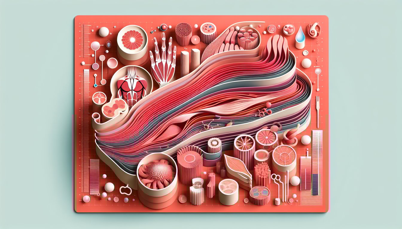
- Muscle Tissue Types -
Review the three main muscle tissue types - skeletal, cardiac, and smooth - and their distinct structures and functions. A handy mnemonic, "SCS," can help you remember: Skeletal moves the body, Cardiac pumps the heart, Smooth lines organs. Understanding these classifications is essential for acing your muscular tissue quiz.
- Sliding Filament Theory -
Focus on how actin and myosin filaments interact during contraction, powered by the cross-bridge cycle. Remember the four steps: attachment, power stroke, detachment, and re-cocking, all driven by ATP hydrolysis (ATP → ADP + Pi). Visualize the sarcomere shortening when filaments slide past one another to cement the concept.
- Muscle Fiber Types -
Differentiate fast-twitch (Type IIa/IIb) and slow-twitch (Type I) fibers in terms of speed, fatigue resistance, and metabolic pathways. For instance, Type I fibers are rich in mitochondria for endurance, whereas Type IIb rely on glycolysis for rapid bursts. Charting their properties side-by-side can simplify recall during a muscle tissue test.
- Neuromuscular Junction Mechanics -
Examine the sequence: motor neuron releases acetylcholine, ACh binds to receptors, muscle fiber depolarizes, and an action potential triggers contraction. A simple mnemonic is "See ACh and CEe" (Calcium Entry prior to Excitation-Contraction coupling). Grasping this pathway is key for muscle tissue questions on synaptic transmission.
- Energy Systems in Muscles -
Memorize the three ATP-generating systems: phosphagen (creatine phosphate), anaerobic glycolysis, and aerobic respiration. Use the formula PCr + ADP → ATP + Cr for the phosphagen system, and note that glycolysis yields lactate under anaerobic conditions. This trio of pathways forms the metabolic backbone of the muscular system quiz.