Unlock hundreds more features
Save your Quiz to the Dashboard
View and Export Results
Use AI to Create Quizzes and Analyse Results

Sign inSign in with Facebook
Sign inSign in with Google

Muscle Action Quiz: Are You Ready to Challenge Yourself?

Ready for a muscle function quiz? Dive into this muscle anatomy challenge!
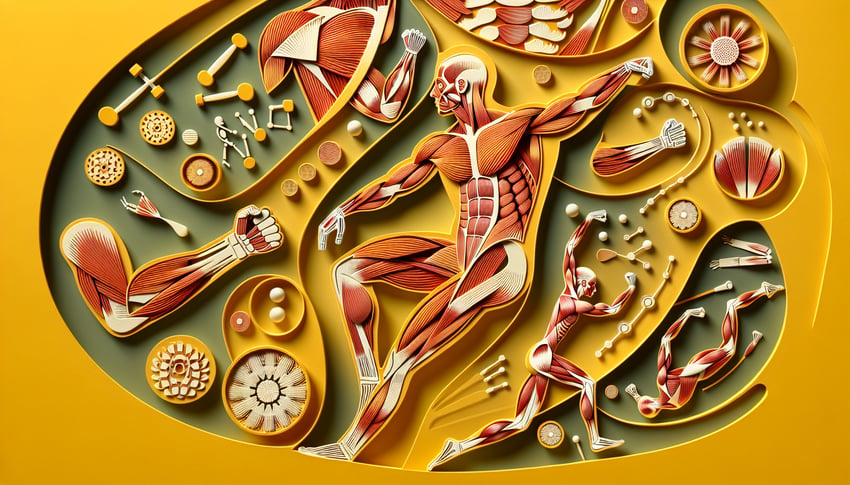
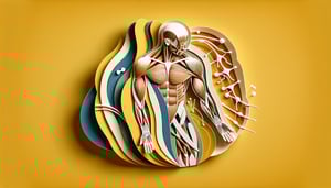
Difficulty: Moderate
2-5mins
Learning OutcomesCheat Sheet
Paper art illustration for muscle action quiz on a golden yellow background

Think you know your muscles? Take our free muscle action quiz to test your anatomy expertise and learn how origin, insertion, and movement power every motion. You'll tackle sections in the muscle function quiz, muscle groups quiz, and a complete muscular system quiz, then sharpen recall in a quick muscle anatomy quiz. Explore detailed origin action and insertion of muscles insights or focus on your legs with our lower limb muscles quiz . Whether you're studying for an exam or fine-tuning workout know-how, this challenge will boost your confidence and deepen your muscle mastery. With instant feedback and progress tracking, jump in now and master anatomy!

Which type of muscle tissue is responsible for voluntary movement?
Smooth muscle
Skeletal muscle
Connective tissue
Cardiac muscle
Skeletal muscle is under voluntary control and responsible for conscious movement. Cardiac muscle contracts involuntarily in the heart, smooth muscle controls organs without conscious effort, and connective tissue is not contractile. source
Which muscle is the primary extensor of the elbow joint?
Brachialis
Biceps brachii
Brachioradialis
Triceps brachii
The triceps brachii extends the elbow by contracting its long, lateral, and medial heads. The biceps brachii and brachialis flex the elbow, while brachioradialis assists flexion. source
The insertion of the biceps brachii is on which bone?
Coronoid process of the ulna
Radial tuberosity of the radius
Olecranon of the ulna
Deltoid tuberosity of the humerus
The biceps brachii inserts on the radial tuberosity, allowing it to flex the elbow and supinate the forearm. The olecranon is for the triceps, and the deltoid tuberosity and coronoid process are insertion sites for other muscles. source
The origin of the gastrocnemius muscle is on which part of the femur?
Medial and lateral condyles
Linea aspera
Greater trochanter
Intertrochanteric crest
The gastrocnemius has two heads originating from the medial and lateral femoral condyles. The other landmarks serve as attachment for different thigh muscles. source
Which muscle is responsible for plantarflexion of the foot?
Peroneus tertius
Tibialis anterior
Gastrocnemius
Extensor digitorum longus
Gastrocnemius plantarflexes the foot at the ankle joint. Tibialis anterior dorsiflexes, peroneus tertius everts, and extensor digitorum longus extends the toes and dorsiflexes the foot. source
Which muscle abducts the arm at the shoulder?
Pectoralis major
Deltoid
Latissimus dorsi
Teres major
The deltoid muscle abducts the arm after the initial 15° by the supraspinatus. Pectoralis major, latissimus dorsi, and teres major adduct or medially rotate the arm. source
The deltoid muscle is innervated by which nerve?
Median nerve
Musculocutaneous nerve
Axillary nerve
Radial nerve
The axillary nerve arising from C5 - C6 roots innervates the deltoid. The musculocutaneous, radial, and median nerves supply other muscle groups in the arm and forearm. source
Which movement does the pectoralis major primarily perform?
Extension of the elbow
Plantarflexion of the foot
Adduction and medial rotation of the humerus
Flexion of the knee
Pectoralis major adducts and medially rotates the humerus at the shoulder. It does not act on the elbow, foot, or knee joints. source
Which muscle originates from the iliac crest and inserts onto the lower ribs?
Rectus abdominis
Transversus abdominis
Internal oblique
External oblique
The internal oblique originates from the iliac crest and inserts on lower ribs and linea alba. External oblique originates from ribs, rectus abdominis from pubis, and transversus abdominis from iliac crest but inserts on linea alba. source
The popliteus muscle performs what action at the knee?
Extends the knee
Abducts the leg
Unlocks the knee by medially rotating the tibia
Laterally rotates the femur
The popliteus medially rotates the tibia to unlock the knee at the start of flexion. It does not extend, laterally rotate the femur, or abduct the leg. source
Which muscle is the prime mover for hip flexion?
Gluteus maximus
Hamstrings
Iliopsoas
Adductor magnus
Iliopsoas, formed by psoas major and iliacus, is the strongest hip flexor. Gluteus maximus extends the hip, hamstrings extend the hip and flex the knee, and adductor magnus adducts the thigh. source
The latissimus dorsi inserts on which structure of the humerus?
Greater tubercle
Radial groove
Intertubercular sulcus
Deltoid tuberosity
Latissimus dorsi inserts into the floor of the intertubercular (bicipital) sulcus of the humerus, allowing extension, adduction, and medial rotation. The other sites are for different muscles. source
Which muscle assists in forced inspiration by elevating the ribs?
Internal intercostals
Serratus posterior superior
Transversus thoracis
Serratus posterior inferior
Serratus posterior superior elevates ribs 2 - 5 during forced inspiration. Serratus posterior inferior depresses ribs, internal intercostals aid forced expiration, and transversus thoracis compresses the thoracic cavity. source
What is the antagonist of the tibialis anterior in dorsiflexion?
Tibialis posterior
Gastrocnemius
Flexor hallucis longus
Peroneus longus
Gastrocnemius plantarflexes the foot, opposing dorsiflexion by tibialis anterior. Peroneus longus everts and plantarflexes, tibialis posterior inverts and plantarflexes, and flexor hallucis longus flexes the big toe. source
Which muscle originates from the supraorbital ridge and elevates the eyebrows?
Procerus
Corrugator supercilii
Frontalis
Orbicularis oculi
Frontalis originates from the galea aponeurotica and inserts on the skin of the eyebrows to raise them. Orbicularis oculi closes eyelids, corrugator supercilii draws eyebrows medially, and procerus wrinkles the nose. source
The coracobrachialis muscle originates from _____ and inserts on _____.
Acromion; radial tuberosity
Clavicle; olecranon
Coracoid process; medial humerus
Scapular spine; deltoid tuberosity
Coracobrachialis originates on the coracoid process of the scapula and inserts on the medial humeral shaft, aiding flexion and adduction of the arm. The other pairings describe different muscles. source
Which muscle's contraction causes internal rotation of the humerus?
Teres minor
Subscapularis
Infraspinatus
Supraspinatus
Subscapularis medially rotates the humerus. Infraspinatus and teres minor externally rotate, and supraspinatus abducts the arm. source
The linea alba is an insertion site for which muscle?
Transversus abdominis
Iliacus
Quadratus lumborum
Psoas major
Transversus abdominis inserts onto the linea alba and pubic crest, helping compress the abdomen. The other muscles act on the hip or back. source
Which muscle is innervated by the long thoracic nerve and helps stabilize the scapula?
Rhomboid major
Levator scapulae
Serratus anterior
Trapezius
Serratus anterior is innervated by the long thoracic nerve and holds the scapula against the thoracic wall. Rhomboids and levator scapulae are innervated by dorsal scapular nerve, trapezius by accessory nerve. source
The quadratus lumborum originates from the iliac crest and inserts on _____.
Sacrum
12th rib and lumbar transverse processes
Greater trochanter
Xiphoid process
Quadratus lumborum inserts on the 12th rib and transverse processes of L1 - L4, aiding lateral flexion of the vertebral column. The other structures are not its insertion points. source
Which posterior forearm muscle is responsible for extension of the index finger?
Extensor indicis
Extensor digitorum
Extensor digiti minimi
Extensor carpi ulnaris
Extensor indicis specifically extends the index finger. Extensor digitorum extends all four fingers, extensor carpi ulnaris extends the wrist, and extensor digiti minimi extends the little finger. source
What action does the gluteus minimus perform at the hip?
Flexion and adduction
Abduction and medial rotation
Extension and lateral rotation
Adduction and medial rotation
Gluteus minimus abducts the thigh and medially rotates the hip. Extension and lateral rotation are functions of gluteus maximus; flexion and adduction involve other muscle groups. source
Which statement best describes the significance of pennation angle in the gastrocnemius muscle?
Pennation angle does not affect muscle force
Lower pennation favors greater physiological cross-sectional area
Higher pennation results in faster contraction velocity
Increased pennation allows more fibers in parallel, enhancing force production
A larger pennation angle enables more fibers to pack in parallel, increasing force at the expense of contraction speed. This is key in gastrocnemius to generate powerful plantarflexion. source
Which arrangement of rotator cuff muscles is most critical for preventing superior migration of the humeral head?
Synergistic action of infraspinatus and teres minor
Balanced force couple between supraspinatus and deltoid
Teres minor and subscapularis force couple
Subscapularis dominance over other cuff muscles
The supraspinatus counters the upward pull of the deltoid, stabilizing the humeral head in the glenoid. This force couple is vital to prevent superior migration. source
At what elbow joint angle does the biceps brachii generate its maximal active force according to the length-tension relationship?
Approximately 90° of flexion
Halfway between full extension and 45°
Full extension
Approximately 120° of flexion
The biceps brachii produces maximal active force near 90° elbow flexion, where optimal overlap of actin-myosin occurs. Full extension and very deep flexion reduce overlap and force. source
0
{"name":"Which type of muscle tissue is responsible for voluntary movement?", "url":"https://www.quiz-maker.com/QPREVIEW","txt":"Which type of muscle tissue is responsible for voluntary movement?, Which muscle is the primary extensor of the elbow joint?, The insertion of the biceps brachii is on which bone?","img":"https://www.quiz-maker.com/3012/images/ogquiz.png"}

Study Outcomes

  1. Identify key muscle actions -

    Clearly pinpoint the primary movements of major muscle groups through the muscle action quiz and muscle anatomy quiz format.

  2. Describe muscle origins and insertions -

    Detail the specific attachment points of muscles to bones to understand how muscle function drives movement.

  3. Analyze biomechanical movement patterns -

    Break down the roles muscles play during different exercises in this muscle function quiz to enhance your understanding of mechanics.

  4. Differentiate between synergists and antagonists -

    Recognize how muscles work together and oppose each other to produce smooth and controlled motion in a muscular system quiz context.

  5. Apply knowledge to practical scenarios -

    Use quiz insights to predict muscle involvement in everyday movements and exercise routines effectively.

  6. Evaluate muscle group coordination -

    Assess how different muscle groups interact during complex actions to improve biomechanics and injury prevention strategies.

Cheat Sheet

  1. Origins and Insertions -

    The origin is the stable attachment site, while the insertion moves toward it; for example, the biceps brachii originates on the scapula and inserts on the radial tuberosity (Gray's Anatomy). Remember the mnemonic "O-I: Origin Immobile" to avoid mixing them up. This foundational concept underpins every muscle action quiz question.

  2. Muscle Action Types -

    Muscles can contract concentrically (shortening), eccentrically (lengthening under load), or isometrically (static tension) (Journal of Biomechanics). A quick formula to estimate concentric torque is τ = F × r, where F is force and r is the moment arm. Use "C-E-I" (Cats Eat Ice cream) to recall Concentric, Eccentric, Isometric.

  3. Lever Systems in the Body -

    The human musculoskeletal system uses first-, second-, and third-class levers to amplify force or speed (Kinesiology textbooks). First-class levers (like the head nod) follow F-A-R; second-class levers (calf raises) are A-R-F; third-class levers (elbow flexion) are A-F-R. A handy trick: "FAR, ARF, AFR" corresponds to Force, Axis, Resistance order.

  4. Sliding Filament Mechanism -

    The cross-bridge cycle involves myosin heads pulling actin filaments, powered by ATP hydrolysis (ATP + H₂O → ADP + Pi + energy) as detailed in Molecular Biology of the Cell. Visualize it like a ratchet: ATP binds, releases, and rebinds to slide filaments. This core process explains how muscle contraction generates movement.

  5. Prime Movers, Antagonists, Synergists -

    During elbow flexion, the biceps brachii acts as the prime mover, the triceps brachii is the antagonist, and the brachialis acts as a synergist stabilizing the action (American College of Sports Medicine). Think "P-A-S" - the Prime mover Acts, while its Antagonist Slows, and the Synergist Supports. Identifying these roles is essential for any muscle groups quiz.

Powered by: Quiz Maker