Unlock hundreds more features
Save your Quiz to the Dashboard
View and Export Results
Use AI to Create Quizzes and Analyse Results

Sign inSign in with Facebook
Sign inSign in with Google

Lower Extremity Bone Quiz: Identify Every Bone of the Lower Limb

Ready to Test Your Anatomy? Dive into the Bones of the Lower Limb Quiz!

Difficulty: Moderate
2-5mins
Learning OutcomesCheat Sheet
Paper art showing femur tibia fibula patella foot silhouettes on dark blue background for lower limb bones quiz

Calling all anatomy enthusiasts and future clinicians! Ready to challenge yourself with a lower extremity bone quiz designed to test your knowledge of every bone in the lower limb? This free bones of the lower limb quiz not only sharpens your grasp on the biomechanics but also prepares you for any anatomy lower limb quiz or skeletal system quiz you might face. As you identify the tibia, fibula, tarsals, and more, you'll boost your confidence and retention. For a deeper dive, explore our muscles of the lower extremity quiz or master ankle details with our ankle and lower leg anatomy guide. Jump in now and elevate your anatomy game!

Which bone is the longest in the human body?
Tibia
Humerus
Femur
Fibula
The femur is the longest and strongest bone in the human body, extending from the hip to the knee. It plays a key role in supporting body weight and enabling bipedal locomotion. Its length also helps determine overall stature. https://en.wikipedia.org/wiki/Femur
What bone is commonly called the kneecap?
Talus
Navicular
Patella
Calcaneus
The patella is a triangular sesamoid bone embedded in the quadriceps tendon and forms the anterior surface of the knee joint, providing leverage for knee extension. It protects the knee joint and increases the mechanical advantage of the quadriceps muscle. https://en.wikipedia.org/wiki/Patella
Which bone forms the heel of the foot?
Talus
Calcaneus
Cuboid
Navicular
The calcaneus is the largest tarsal bone and forms the heel, bearing much of the body's weight during walking and standing. It serves as an attachment for the Achilles tendon. https://en.wikipedia.org/wiki/Calcaneus
Which tarsal bone articulates directly with both the tibia and fibula to form the ankle joint?
Talus
Calcaneus
Cuboid
Navicular
The talus sits just above the calcaneus and articulates with the distal ends of the tibia and fibula to form the talocrural (ankle) joint. It transmits body weight from the tibia to the foot. https://en.wikipedia.org/wiki/Talus_(bone)
How many metatarsal bones are present in one human foot?
4
6
5
7
Each foot contains five metatarsal bones numbered I to V from the medial to lateral side, forming the arch and connecting the tarsals to the toes. They play a critical role in weight distribution and locomotion. https://en.wikipedia.org/wiki/Metatarsus
Which bone is located lateral to the tibia in the lower leg?
Femur
Fibula
Ulna
Patella
The fibula runs parallel and lateral to the tibia in the lower leg. Though it is not weight-bearing, it provides muscle attachment sites and stabilizes the ankle joint. https://en.wikipedia.org/wiki/Fibula
Which bone primarily bears the weight of the body from the knee to the ankle?
Talus
Femur
Tibia
Fibula
The tibia, or shinbone, is the main weight-bearing bone of the lower leg, transmitting forces from the femur down to the foot. It articulates proximally with the femur and distally with the talus. https://en.wikipedia.org/wiki/Tibia
How many phalanges does the great toe (hallux) have?
1
2
4
3
The great toe, or hallux, has two phalanges (proximal and distal), whereas each of the other toes has three phalanges. This structure aids in balance during gait. https://en.wikipedia.org/wiki/Phalanges_of_the_foot
Which landmark on the proximal femur serves as the attachment point for the gluteus medius muscle?
Linea aspera
Greater trochanter
Medial condyle
Lesser trochanter
The greater trochanter projects laterally from the junction of the neck and shaft of the femur and serves as the attachment site for the gluteus medius (and minimus) muscles, important for hip abduction. https://en.wikipedia.org/wiki/Femur#Greater_and_lesser_trochanters
The tibial plateau refers to the articular surface found on which bone?
Femur
Fibula
Patella
Tibia
The tibial plateau is the flat proximal surface of the tibia that articulates with the femoral condyles to form part of the knee joint. It is divided into medial and lateral condyles. https://en.wikipedia.org/wiki/Tibia#Proximal_tibia
What ridge runs obliquely across the posterior surface of the tibia providing attachment for muscle fibers?
Interosseous border
Linea aspera
Tibial tuberosity
Soleal line
The soleal line on the posterior tibia marks the origin of the soleus muscle. It runs obliquely from the fibular border toward the medial side of the tibia. https://en.wikipedia.org/wiki/Tibia#Soleal_line
Which bone is triangular, lies anterior to the talus, and helps maintain the medial longitudinal arch?
Medial cuneiform
Lateral cuneiform
Cuboid
Navicular
The navicular bone is boat-shaped and sits anterior to the talus and medial to the cuneiforms. It supports the head of the talus and contributes to the medial longitudinal arch. https://en.wikipedia.org/wiki/Navicular_bone
The medial malleolus is a prominent process on which lower extremity bone?
Fibula
Talus
Calcaneus
Tibia
The medial malleolus is the distal medial projection of the tibia and forms the medial aspect of the ankle mortise, articulating with the talus. https://en.wikipedia.org/wiki/Tibia#Distal_tibia
Which metatarsal bone commonly has a tuberosity that is prone to avulsion fractures?
Third metatarsal
Second metatarsal
First metatarsal
Fifth metatarsal
The base of the fifth metatarsal has a prominent tuberosity where the peroneus brevis tendon attaches. Avulsion fractures here, often called 'dancer's fractures', are common. https://en.wikipedia.org/wiki/Metatarsal_fracture#Fifth_metatarsal_fracture
The linea aspera is a longitudinal ridge found on which aspect of the femur?
Anterior shaft
Posterior shaft
Medial epicondyle
Lateral condyle
The linea aspera runs along the posterior mid-shaft of the femur, serving as an attachment for adductor muscles and the intermuscular septa of the thigh. https://en.wikipedia.org/wiki/Femur#Linea_aspera
Which tarsal bone lies directly lateral to the navicular and anterior to the calcaneus?
Lateral cuneiform
Talus
Cuboid
Medial cuneiform
The cuboid is the most lateral of the distal tarsal bones, articulating posteriorly with the calcaneus and medially with the lateral cuneiform and navicular. https://en.wikipedia.org/wiki/Cuboid_bone
At birth, how many primary ossification centers are present in the femur?
Four
One
Three
Two
At birth, only the primary ossification center in the diaphysis of the femur is present. Secondary centers in the proximal head and distal condyles appear later in infancy. https://en.wikipedia.org/wiki/Ossification
The sustentaculum tali is a bony shelf on which foot bone?
Calcaneus
Talus
Navicular
Cuboid
The sustentaculum tali is a medial projection of the calcaneus that supports the talus and serves as a pulley for the flexor hallucis longus tendon. https://en.wikipedia.org/wiki/Sustentaculum_tali
A Jones fracture involves the base of which metatarsal bone?
First metatarsal
Third metatarsal
Fifth metatarsal
Second metatarsal
A Jones fracture is a transverse fracture at the metaphyseal - diaphyseal junction of the fifth metatarsal base, often challenging to heal due to limited blood supply. https://en.wikipedia.org/wiki/Metatarsal_fracture#Jones_fracture
The patellar surface is located on which part of the femur?
Posterior proximal
Lateral proximal
Medial shaft
Anterior distal
The patellar surface (trochlear groove) is a smooth area on the anterior distal femur between the condyles, articulating with the patella during knee flexion and extension. https://en.wikipedia.org/wiki/Femur#Distal_structure
What is the name of the socket in the pelvis that articulates with the head of the femur?
Glenoid fossa
Trochlea
Acetabulum
Olecranon fossa
The acetabulum is a deep, concave structure formed by the ilium, ischium, and pubis. It articulates with the femoral head to form the hip joint. https://en.wikipedia.org/wiki/Acetabulum
Which ligament of the ankle stabilizes the medial aspect by connecting the tibia to multiple tarsal bones?
Posterior talofibular ligament
Deltoid ligament
Calcaneofibular ligament
Anterior talofibular ligament
The deltoid (medial) ligament is a strong, triangular band that attaches the medial malleolus of the tibia to the talus, calcaneus, and navicular, providing medial stability to the ankle. https://en.wikipedia.org/wiki/Deltoid_ligament
The medial and lateral condyles of the tibia articulate with which femoral structures?
Patellar facets
Greater trochanter
Femoral trochanters
Femoral condyles
The medial and lateral tibial condyles articulate with the corresponding femoral condyles, forming the tibiofemoral part of the knee joint. Proper alignment is crucial for knee mechanics. https://en.wikipedia.org/wiki/Knee_joint
Which feature on the posterior aspect of the calcaneus serves as the attachment for the Achilles tendon?
Sustentaculum tali
Peroneal trochlea
Calcaneal tuberosity
Calcaneonavicular canal
The calcaneal tuberosity is the posterior prominence of the calcaneus where the Achilles (calcaneal) tendon inserts, transmitting the powerful force of the gastrocnemius and soleus muscles. https://en.wikipedia.org/wiki/Calcaneal_tuberosity
Which anatomical feature on the femoral head provides attachment for the ligamentum teres and transmits a branch of the obturator artery?
Fovea capitis
Trochanteric fossa
Intertrochanteric line
Linea aspera
The fovea capitis is a small pit on the head of the femur where the ligamentum teres attaches, carrying a small branch of the obturator artery to the femoral head. This vascular contribution is important for femoral head viability. https://en.wikipedia.org/wiki/Femur#Head
0
{"name":"Which bone is the longest in the human body?", "url":"https://www.quiz-maker.com/QPREVIEW","txt":"Which bone is the longest in the human body?, What bone is commonly called the kneecap?, Which bone forms the heel of the foot?","img":"https://www.quiz-maker.com/3012/images/ogquiz.png"}

Study Outcomes

  1. Identify Lower Limb Bones -

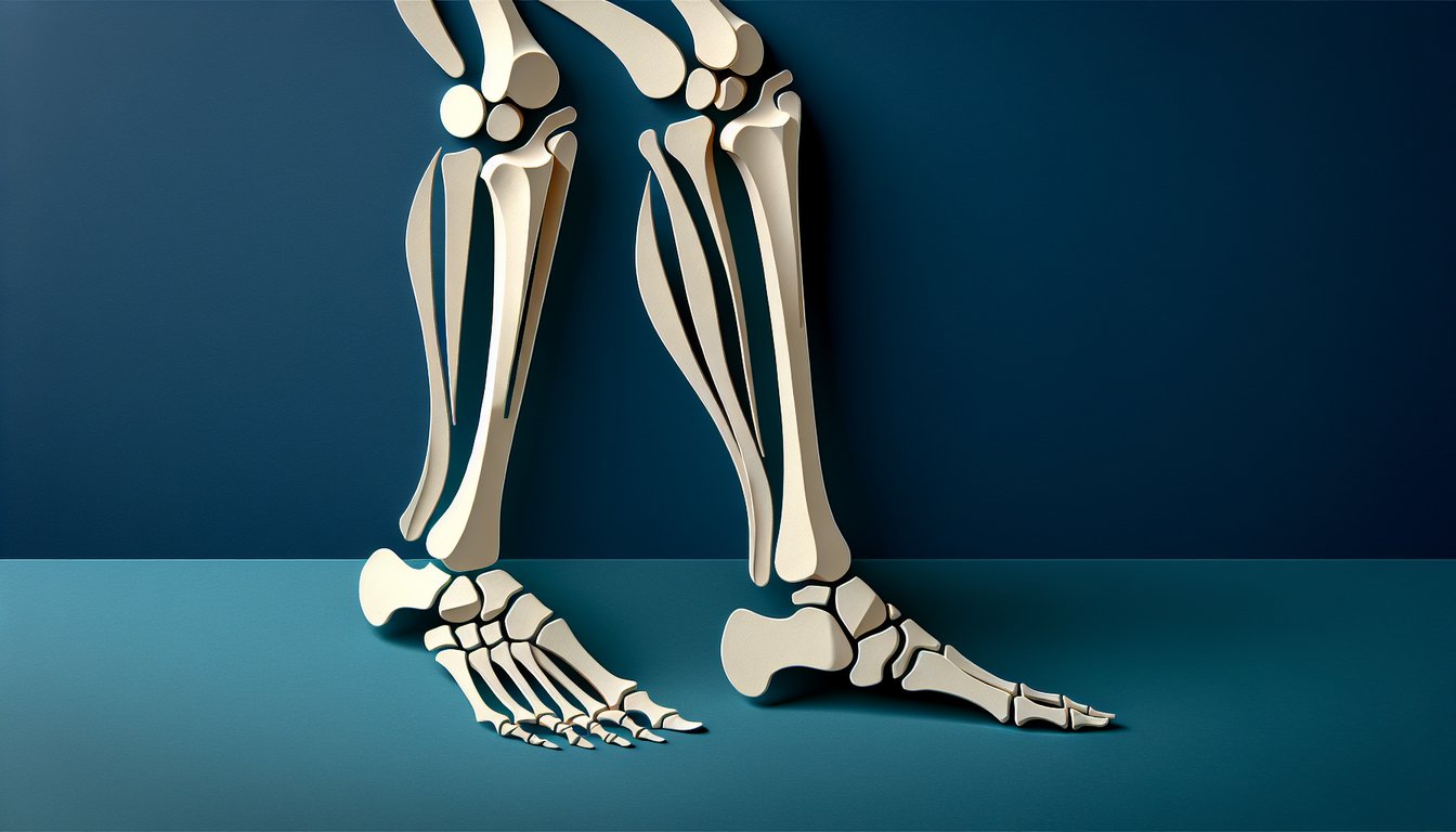
    Pinpoint the names and anatomical positions of the pelvis, femur, tibia, fibula, and foot bones in the lower extremity bone quiz.

  2. Differentiate Bone Regions -

    Distinguish between the bones of the thigh, lower leg, and foot by recognizing their unique shapes and landmark features.

  3. Recall Key Anatomical Landmarks -

    Memorize crucial landmarks such as the iliac crest, greater trochanter, medial malleolus, and navicular to enhance your skeletal system knowledge.

  4. Apply Correct Terminology -

    Use precise anatomical language when referring to structures in the lower extremity anatomy test, reinforcing professional communication skills.

  5. Analyze Skeletal Articulations -

    Examine how bones connect at joints and interpret their relationships to understand movement mechanics and stability.

  6. Assess Knowledge Gaps -

    Leverage quiz results to identify areas for additional review and strengthen your mastery of the human lower limb anatomy lower limb quiz.

Cheat Sheet

  1. Pelvic Girdle Composition -

    The pelvic girdle consists of the ilium, ischium, and pubis, which fuse by age 25 (Gray's Anatomy, 42nd ed.). Use the mnemonic "IIP" (Ilium, Ischium, Pubis) to recall their arrangement and articulations. Reviewing acetabular landmarks will help you ace the lower extremity bone quiz.

  2. Femoral Landmarks and Angles -

    The femur is the longest bone, featuring the head, neck (angle of inclination ~125°), and medial/lateral condyles (StatPearls, 2023). Remember "HNC" (Head - Neck - Condyles) to identify these parts quickly on radiographs. Understanding the coxa vara/valga variations is essential for any anatomy lower limb quiz.

  3. Tibia vs. Fibula Roles -

    The tibia bears ~90% of body weight, while the fibula provides lateral stability (Journal of Anatomy, 2021). A handy mnemonic is "Tib for Time, Fib for Fun" to distinguish weight-bearing functions. Don't forget to review the tibial plateau and fibular head for common injury sites.

  4. Tarsal Bone Mnemonic -

    There are seven tarsals: talus, calcaneus, navicular, medial/intermediate/lateral cuneiforms, and cuboid. Use "Tiger Cubs Need MILC" to remember their sequence from proximal-medial to distal-lateral. Mastering these names will boost your score on any bones of the lower limb quiz.

  5. Metatarsals and Phalanges Structure -

    The forefoot contains 5 metatarsals and 14 phalanges, forming MTP, PIP, and DIP joints. A quick fact: "5 + 14 = Foot's 19" helps you recall the total count. Reviewing joint ranges of motion (e.g., 65° MTP dorsiflexion) reinforces knowledge for the lower extremity anatomy test.

Powered by: Quiz Maker