Take the Scapula Parts Quiz: Master Shoulder Girdle Anatomy
Ready to challenge your scapulothoracic & glenohumeral knowledge? Start the quiz now!

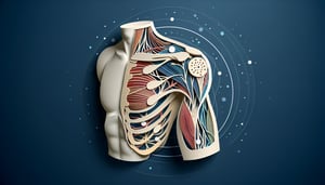
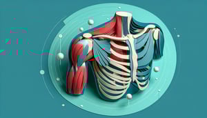
This scapula parts quiz helps you identify landmarks, borders, and articulations of the shoulder blade, so you can check gaps and build recall before an exam or lab. When you're done, get extra practice with the shoulder joint quiz to reinforce how the scapula works with the humerus and clavicle.
Study Outcomes
- Identify Scapula Landmarks -
Learn to recognize and label major scapular features such as the spine, acromion, coracoid process, and glenoid cavity when preparing for the scapula parts quiz.
- Differentiate Shoulder Girdle Joints -
Distinguish between the sternoclavicular, acromioclavicular, and scapulothoracic joints, enhancing your shoulder girdle anatomy quiz accuracy.
- Explain Scapulothoracic Mechanics -
Understand the movement patterns of the scapula against the thoracic wall, including elevation, depression, protraction, and retraction.
- Describe Glenohumeral Movements -
Detail the primary motions of the glenohumeral joint such as abduction, adduction, rotation, and flexion for improved quiz performance.
- Analyze Sternoclavicular and Acromioclavicular Joints -
Explore the structural differences and functional roles of these joints in shoulder stability and motion.
- Apply Knowledge to Quiz Challenges -
Use your understanding of scapular anatomy and shoulder girdle mechanics to confidently tackle each question in the scapula parts quiz.
Cheat Sheet
- Scapular Borders and Angles -
When preparing for the scapula parts quiz, focus on the three borders (superior, medial, lateral) and three angles (superior, lateral/glenoid, inferior). A handy mnemonic "SLI" helps recall Superior, Lateral, and Inferior angles in sequence. Visual models from Gray's Anatomy reinforce spatial orientation and landmark identification.
- Sternoclavicular Joint Mechanics -
The saddle-shaped sternoclavicular joint allows elevation, depression, protraction, and retraction of the clavicle, key for clavicular motion in the shoulder girdle anatomy quiz. With about 45° of elevation and 15° of depression, rotational movements support full overhead reach. Reviewing diarthrodial classification from the American Academy of Orthopaedic Surgeons sharpens accuracy for your sternoclavicular joint quiz sections.
- Acromioclavicular Joint Stability -
The acromioclavicular joint is a plane synovial articulation reinforced by the conoid and trapezoid ligaments of the coracoclavicular complex, often highlighted in the acromioclavicular joint test. Recognizing ligamentous grading I - III in AC separation aids clinical decision-making. Cross-referencing University of California biomechanics modules or Netter's Atlas enhances memorization.
- Scapulothoracic Movement Patterns -
Although not a true synovial joint, the scapulothoracic articulation permits upward/downward rotation, protraction/retraction, and elevation/depression, central to any scapulothoracic anatomy quiz. A memorable cue "UP-DE-PRO" stands for Upward Rotation, Depression, and Protraction. Reviewing kinematics in the Journal of Anatomy clarifies how these motions integrate during arm abduction.
- Glenohumeral Joint and Scapulohumeral Rhythm -
The ball-and-socket glenohumeral joint allows flexion, extension, abduction, and rotation with stability from the rotator cuff muscles - vital for the glenohumeral joint anatomy quiz. Remember the 2:1 scapulohumeral rhythm: every 3° of arm elevation comprises 2° at the GH joint and 1° of scapular rotation. Incorporating this formula from orthopedic texts ensures mastery of dynamic shoulder mechanics.