Gastrointestinal Histology Quiz: Test Your GI Pathology Mastery
Start the GI pathology quiz: tackle histology MCQs and prove your expertise!
Calling all medical students, pathologists-in-training, and curious science buffs! Ready to explore the hidden world of digestive tissues? Our free gastrointestinal histology quiz offers a fun, interactive way to solidify your GI pathology knowledge and prepare you for real-world diagnosis. Through comprehensive GI histology MCQ and gastrointestinal pathology questions, you'll master mucosal organization, specialized cell types, and disease-related alterations. Dive into our gi histology quiz and expand your learning with the gastrointestinal anatomy quiz . Take the challenge now and prove your expertise in this gastrointestinal pathology quiz! Get instant feedback, track your progress, and bolster your diagnostic confidence in GI disorders.
Study Outcomes
- Identify gut wall layers -
Distinguish the mucosa, submucosa, muscularis, and serosa in gastrointestinal histology quiz questions to recognize each layer's unique features.
- Differentiate epithelial cell types -
Recognize squamous, columnar, goblet, and enteroendocrine cells in GI histology MCQ items to pinpoint their roles in secretion and protection.
- Apply pathological criteria to cases -
Use histological markers like villous atrophy, dysplasia, and fibrosis to tackle gastrointestinal pathology quiz scenarios effectively.
- Analyze structure-function relationships -
Correlate tissue microanatomy with digestive and absorptive functions to answer GI pathology MCQ items confidently.
- Recall specialized GI terminology -
Reinforce key terms such as lamina propria, Peyer's patches, and Brunner's glands to enhance performance on gastrointestinal pathology questions.
Cheat Sheet
- GI Wall Layers Mnemonic -
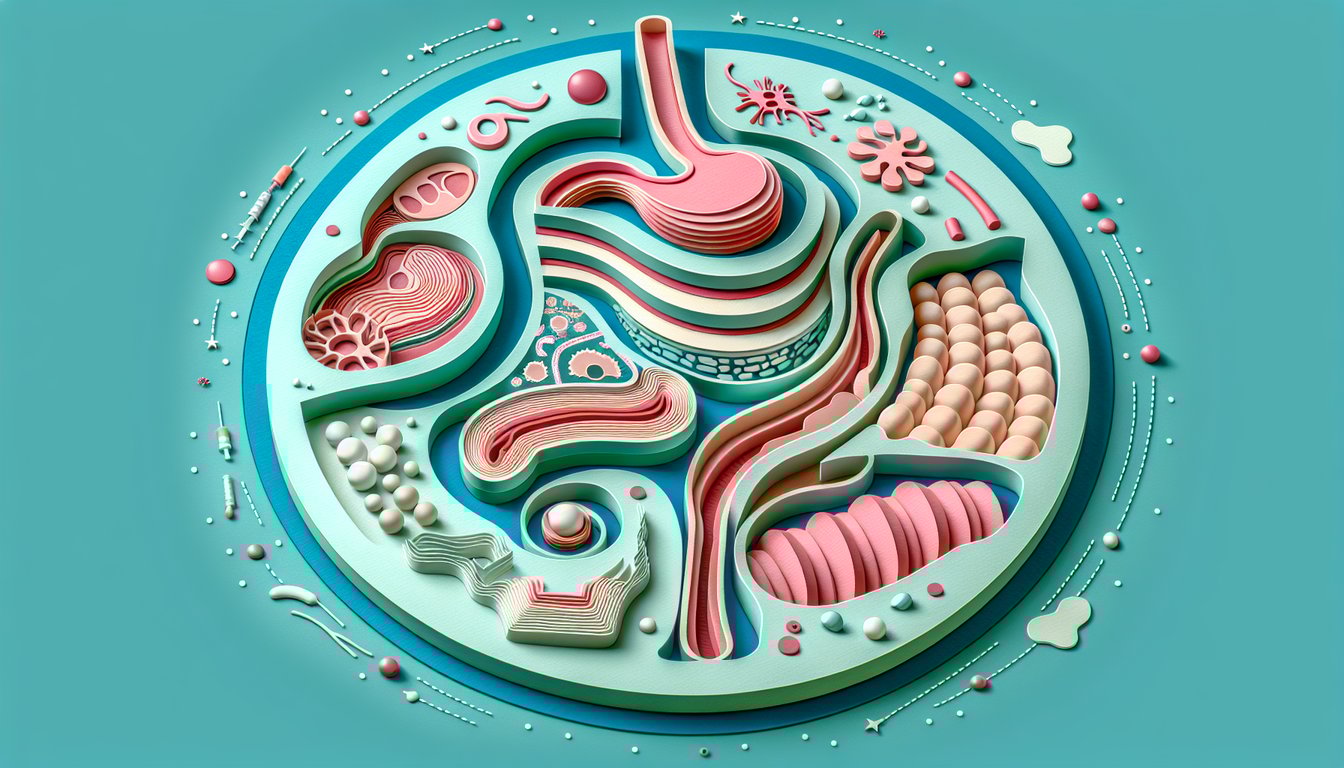
Remember the four main layers - Mucosa, Submucosa, Muscularis externa, Serosa - using the mnemonic "MSMS." The mucosa contains the epithelium, lamina propria, and muscularis mucosae, while the submucosa houses glands and vessels. Mastering these layers will boost your confidence in any gastrointestinal histology quiz.
- Regional Epithelial Variations -
Esophagus features stratified squamous epithelium, while the stomach and intestines shift to simple columnar; use "Squamous Eats, Columnar Dines" to recall this. Note that the duodenum adds Brunner's glands and the ileum contains Peyer's patches for immune defense. Spotting these differences is a common GI pathology MCQ strategy.
- Gastric Gland Cell Types -
In gastric pits, chief cells secrete pepsinogen and parietal cells release HCl and intrinsic factor; think "Pep's Perfect HCl." Mucous neck cells protect the lining with bicarbonate-rich mucus. Recognizing cell morphology on slides is crucial for acing GI pathology MCQ walls.
- Small Intestine Surface Boosters -
Villi, microvilli, and plicae circulares increase surface area by ~600-fold - picture a shaggy carpet of absorption. Duodenum has Brunner's glands, jejunum focuses on absorption, and ileum houses lymphoid Peyer's patches. These zonal features often appear in the gastrointestinal histology quiz to test your detail recall.
- Large Intestine Hallmarks -
The colon lacks villi but is packed with goblet cells for mucus secretion and taenia coli muscle bands for peristalsis. Crypt architecture becomes deeper, and lymphoid aggregates can be seen in the lamina propria. Spotting absence versus presence of villi is a favorite trick in GI histology MCQ sections.