Unlock hundreds more features
Save your Quiz to the Dashboard
View and Export Results
Use AI to Create Quizzes and Analyse Results

Sign inSign in with Facebook
Sign inSign in with Google

Think You Know Your Joints? Take the Anatomy Quiz Now!

Ready to ace this joints quiz anatomy? Start the joint quiz and explore types of joints now!

Difficulty: Moderate
2-5mins
Learning OutcomesCheat Sheet
Paper cutout bones and joints illustration on dark blue background free joints quiz challenge your anatomy skills

Ready to dive into the world of articulation? Our Free Ultimate Joints Quiz: Test Your Anatomy Skills is the perfect way to see how well you know the bending, sliding, and twisting machines inside your body. This quick joints quiz challenges you on synovial, fibrous, and cartilaginous connections, so you can master every types of joints quiz and track your progress. Whether you're brushing up on joints quiz anatomy or getting serious about an anatomy joints quiz, you'll gain instant feedback on your form and function smarts. Love a good bone challenge? Try our fun quiz on bones for an extra test! Embrace your inner anatomist - take the joint quiz now and flex those brain muscles!

Which of the following best describes the type of joint the knee is?
Saddle joint
Ball and socket joint
Hinge joint
Pivot joint
The knee functions primarily as a hinge joint, allowing flexion and extension with slight rotational capacity. It is stabilized by ligaments such as the ACL and PCL. Unlike ball-and-socket joints, it does not permit multi-axial rotation. source
What type of cartilage covers the articulating surfaces of bones in a synovial joint?
Fibrocartilage
Dense regular connective tissue
Articular cartilage
Elastic cartilage
Articular cartilage is a form of hyaline cartilage that covers bone ends in synovial joints to reduce friction and absorb shock. It is avascular and relies on synovial fluid for nutrient exchange. Damage to this cartilage can lead to osteoarthritis. source
Which structure produces synovial fluid within a joint capsule?
Synovial membrane
Meniscus
Bursa
Articular cartilage
The synovial membrane lines the inner surface of the joint capsule and secretes synovial fluid. Synoviocytes within this membrane are responsible for producing lubricating components and nutrients for the joint. The fluid reduces friction and nourishes articular cartilage. source
Which type of synovial joint allows rotation around a single axis, such as the atlantoaxial joint?
Pivot joint
Condyloid joint
Saddle joint
Hinge joint
Pivot joints permit rotation around a single longitudinal axis. The atlantoaxial joint between C1 and C2 is a classic example, allowing head rotation. Other joint types permit different movement patterns. source
The shoulder joint is classified as which type of synovial joint?
Plane joint
Pivot joint
Ball and socket joint
Hinge joint
The glenohumeral (shoulder) joint is a ball-and-socket joint, allowing multi-axial movement including flexion, abduction, rotation, and circumduction. Its wide range of motion comes at the expense of stability. The rotator cuff and labrum help maintain joint integrity. source
Which classification includes fibrous, cartilaginous, and synovial joints?
Functional classification
Histological classification
Mobility classification
Structural classification
Structural classification categorizes joints by material binding bones and presence of joint cavity. It divides joints into fibrous, cartilaginous, and synovial types. Functional classification, by contrast, is based on mobility. source
What is the primary function of bursae in relation to joints?
Facilitate bone growth
Produce synovial fluid
Connect muscles to bones
Reduce friction
Bursae are fluid-filled sacs that reduce friction between tendons, ligaments, and bones. They allow smooth gliding of overlying tissues. Inflammation of a bursa leads to bursitis, causing pain and limited movement. source
Which movement decreases the angle between two bones?
Abduction
Flexion
Adduction
Extension
Flexion is the movement that decreases the angle between two bones at a joint. For example, bending the elbow brings the forearm closer to the upper arm. Extension is the opposite motion, increasing the angle. source
Which movement takes a body part away from the midline?
Adduction
Circumduction
Abduction
Flexion
Abduction moves a limb away from the body's midline, such as lifting the arm laterally. Adduction pulls it back toward the midline. Circumduction combines flexion, abduction, extension, and adduction. source
The sternoclavicular joint is uniquely classified as which type of synovial joint?
Condyloid joint
Saddle joint
Plane joint
Ball and socket joint
The sternoclavicular joint is a saddle-shaped joint that allows multi-directional movement. It acts as the only bony connection between the upper limb and axial skeleton. Despite its shape, it functions similarly to a ball-and-socket joint. source
What is the anatomical name for the ankle joint?
Subtalar joint
Talocalcaneal joint
Tibiotalar joint
Tibiofibular joint
The ankle joint, also known as the tibiotalar joint, is formed by the tibia and talus bones. It permits dorsiflexion and plantarflexion. The subtalar joint below allows inversion and eversion of the foot. source
What term describes the fibrocartilaginous pads within the knee joint?
Labra
Ligaments
Menisci
Discs
The menisci are crescent-shaped fibrocartilaginous pads that cushion and stabilize the knee joint. There are medial and lateral menisci, each attaching to the tibial plateau. They help distribute load and improve joint congruity. source
Which structure contributes most to dynamic stability in the glenohumeral joint?
Glenoid labrum
Axillary nerve
Acromion
Rotator cuff tendons
The rotator cuff tendons dynamically stabilize the humeral head within the glenoid fossa during shoulder movements. They generate compressive forces and fine-tune joint alignment. The glenoid labrum deepens the socket but contributes less to active stabilization. source
Which synovial joint permits dorsiflexion and plantarflexion of the foot?
Subtalar joint
Tarsometatarsal joint
Talocrural joint
Transverse tarsal joint
The talocrural joint is the primary hinge joint between the tibia, fibula, and talus that allows dorsiflexion and plantarflexion. The subtalar joint below permits inversion and eversion. Combined, these joints enable complex foot movements. source
Which synovial joint is an example of a condyloid joint?
Humeroulnar joint
Radiocarpal joint
Carpometacarpal joint of the thumb
Proximal radioulnar joint
The radiocarpal (wrist) joint is a condyloid joint that permits flexion, extension, abduction, and adduction. Its oval articular surfaces allow biaxial movement. Saddle joints, like the thumb CMC, allow more freedom of movement. source
Which ligament prevents anterior translation of the tibia on the femur?
Medial collateral ligament
Anterior cruciate ligament
Posterior cruciate ligament
Lateral collateral ligament
The anterior cruciate ligament (ACL) resists anterior displacement of the tibia relative to the femur and contributes to rotational stability. Injury to the ACL is common in pivoting sports. The PCL prevents posterior translation. source
What term describes an immovable joint?
Synarthrosis
Amphiarthrosis
Diarthrosis
Hinge
Synarthrosis refers to a joint with no appreciable movement, such as cranial sutures. Diarthroses are freely movable (synovial joints), while amphiarthroses are slightly movable (e.g., intervertebral discs). source
The hip joint is formed by the articulation of the femoral head and the acetabulum of the pelvis. This is an example of which type of synovial joint?
Pivot joint
Plane joint
Saddle joint
Ball and socket joint
The hip is a ball-and-socket joint, allowing movement in multiple planes including flexion, extension, abduction, adduction, and rotation. Its deep socket provides greater stability than the shoulder. The acetabular labrum further deepens the joint. source
During forearm pronation, what movement occurs at the proximal radioulnar joint?
The carpal bones shift laterally
The humerus rotates medially
The ulna pivots around the radius
The radial head rotates within the radial notch of the ulna
In forearm pronation, the radius crosses over the ulna as the radial head spins within the radial notch of the ulna. The ulna remains relatively stationary. This pivot action is characteristic of a pivot joint. source
The pubic symphysis is classified as which type of joint?
Fibrous suture
Fibrocartilaginous disc
Synovial hinge
Cartilaginous symphysis
The pubic symphysis is a secondary cartilaginous joint (symphysis) where bones are united by fibrocartilage. It allows slight movement for childbirth. It lacks a synovial cavity. source
The humeroulnar component of the elbow joint is classified as which type of synovial joint?
Pivot joint
Plane joint
Hinge joint
Condyloid joint
The humeroulnar articulation is a hinge joint permitting flexion and extension. It is formed by the trochlea of the humerus and the trochlear notch of the ulna. Lateral and medial collateral ligaments reinforce its stability. source
What fibrocartilaginous structure deepens the glenoid cavity of the shoulder joint?
Rotator cuff tendon
Coracoid process
Glenoid labrum
Acromion
The glenoid labrum is a fibrocartilaginous rim attached to the margin of the glenoid cavity. It deepens the socket and increases joint stability. Tears in the labrum are common in shoulder dislocations. source
Which of the following is an example of a plane (gliding) synovial joint?
Elbow joint
Hip joint
Shoulder joint
Intercarpal joints
Intercarpal joints are plane synovial joints where flat or slightly curved surfaces glide past each other. They allow limited gliding movements between carpal bones. This is unlike hinge or ball-and-socket joints. source
Which type of collagen is most abundant in ligaments?
Type I collagen
Type II collagen
Type III collagen
Type IV collagen
Type I collagen fibers are the primary structural component of ligaments, providing high tensile strength. Type II is predominately found in cartilage. Type III is in reticular fibers, and type IV in basement membranes. source
According to the concave-convex rule, when a convex surface moves on a concave surface, how do roll and glide occur?
Only roll occurs
Only glide occurs
Roll and glide occur in the same direction
Roll and glide occur in opposite directions
The concave-convex rule states that when a convex surface moves on a fixed concave surface, rolling and gliding occur in opposite directions. This maintains joint congruity and prevents subluxation. The opposite applies when a concave surface moves on a fixed convex surface. source
Which layer of the joint capsule provides mechanical support and stability?
Endosteum
Fibrous capsule
Periosteum
Synovial membrane
The fibrous capsule is the outer layer of a joint capsule, composed of dense irregular connective tissue. It provides mechanical support and resists dislocation. The inner synovial membrane produces fluid but does not offer structural strength. source
Sharpey's fibers have what function in joint anatomy?
Anchor the periosteum to bone
Lubricate the synovial cavity
Absorb synovial fluid
Connect muscle to bone
Sharpey's fibers are collagen fibers that anchor the periosteum to underlying bone. They help transmit muscular and ligamentous forces to the skeleton. They are not involved in synovial fluid dynamics or muscle attachments directly. source
Which unique structure divides the temporomandibular joint cavity into two compartments?
Articular disc
Bursa
Meniscus
Labrum
The temporomandibular joint contains an articular disc of fibrocartilage that separates the joint space into superior and inferior compartments. Each compartment allows different movements: gliding in the upper and hinge in the lower. Tears or displacement of this disc cause dysfunction. source
In the talocrural joint, which direction does the roll and glide occur during dorsiflexion according to the concave-convex rule?
Roll posteriorly and glide anteriorly
Both roll and glide occur anteriorly
Roll anteriorly and glide posteriorly
Both roll and glide occur posteriorly
In dorsiflexion at the talocrural joint, the convex talus rolls anteriorly on the concave tibia and fibula while gliding posteriorly. This opposite roll-glide relationship follows the concave-convex rule. Dysfunction leads to limited ankle motion. source
Which mechanoreceptor in joint capsules is primarily sensitive to joint position and sustained pressure?
Pacinian corpuscles
Golgi tendon organs
Ruffini endings
Free nerve endings
Ruffini endings are slowly adapting mechanoreceptors in joint capsules that respond to sustained pressure and joint position changes. Pacinian corpuscles detect rapid changes and vibration. Golgi tendon organs are in tendons and sense tension. source
0
{"name":"Which of the following best describes the type of joint the knee is?", "url":"https://www.quiz-maker.com/QPREVIEW","txt":"Which of the following best describes the type of joint the knee is?, What type of cartilage covers the articulating surfaces of bones in a synovial joint?, Which structure produces synovial fluid within a joint capsule?","img":"https://www.quiz-maker.com/3012/images/ogquiz.png"}

Study Outcomes

  1. Identify Joint Types -

    Recognize and name the six major types of synovial and other joints covered in the joints quiz, such as hinge, pivot, and ball-and-socket.

  2. Differentiate Joint Structures -

    Distinguish key anatomical features like articular cartilage, synovial fluid, and joint capsules to understand how each structure supports movement.

  3. Analyze Joint Functions -

    Explain how different joint types facilitate specific movements and load-bearing tasks in the human body.

  4. Recall Anatomical Terminology -

    Master essential terms used in anatomy joints quiz questions to improve accuracy and confidence in identifying joint components.

  5. Evaluate Quiz Results -

    Interpret your score and instant feedback to pinpoint strengths and areas for improvement in joint anatomy knowledge.

  6. Apply Anatomy Knowledge -

    Use insights from the joints quiz to relate joint structure and function to everyday movements and clinical scenarios.

Cheat Sheet

  1. Joint Classification Overview -

    Review the three basic joint classes - fibrous, cartilaginous and synovial - and their degrees of movement. Fibrous joints like sutures in the skull are immovable (synarthroses), while cartilaginous joints (e.g., vertebral discs) allow slight movement (amphiarthroses). Synovial joints (diarthroses) are freely movable and critical for your joints quiz success!

  2. Synovial Joint Subtypes -

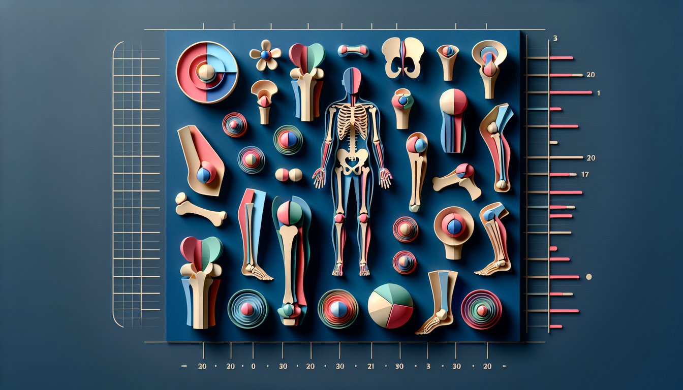
    Study the six synovial types: hinge (like the elbow), pivot (atlantoaxial joint), ball-and-socket (hip and shoulder), condyloid, saddle, and plane joints. Mnemonic "Some People Have Curly Ponytails Sometimes" helps you remember: Saddle, Plane, Hinge, Condyloid, Pivot, Sphere. Understanding these is key to mastering the types of joints quiz.

  3. Key Structural Components -

    Remember that a synovial joint comprises articular cartilage, a synovial membrane producing synovial fluid, fibrous capsule, and reinforcing ligaments. For example, the knee's menisci are two crescent-shaped fibrocartilage pads that improve joint congruence and distribute load. This structure-function link is a staple in any joints quiz anatomy challenge.

  4. Range of Motion & Biomechanics -

    Memorize basic movements - flexion/extension, abduction/adduction, rotation, and circumduction - and typical ROM degrees like ~150° flexion at the elbow. Use the formula: ROM_total = ROM_flexion + ROM_extension to calculate your expected joint mobility. This concept often appears in joint quiz questions to test applied understanding.

  5. Common Joint Disorders -

    Familiarize yourself with osteoarthritis (wear-and-tear), rheumatoid arthritis (autoimmune inflammation), and sprains (ligament injuries) to answer clinical-style joints quiz anatomy items. Recall "R-A-S" to spot Rheumatoid, Arthritis, Sprain fast. Being confident about these pathologies will boost your score on the joint quiz!

Powered by: Quiz Maker