Bone Tissue Labeling Quiz: Identify Osseous Components!
Think you can label the components of osseous tissue? Jump in!
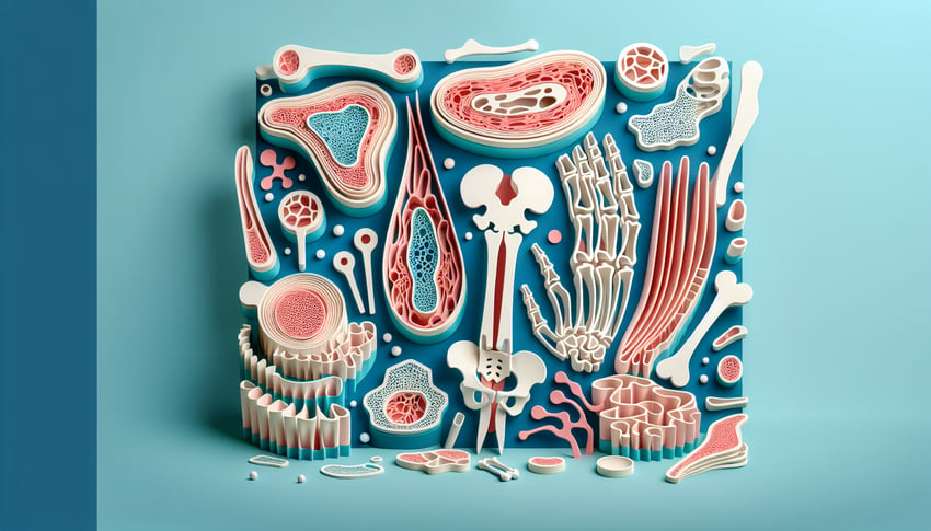
This Bone Tissue Labeling Quiz helps you practice naming key parts of compact bone - osteons, Haversian canals, and lamellae - and spot any gaps fast. Use it to review before an exam or between labs, then keep going with more bone tissue practice or try a broader skeletal quiz .
Study Outcomes
- Identify Osseous Tissue Components -
After completing the bone tissue labeling quiz, you will be able to accurately identify osteocytes, lamellae, Haversian canals, and other key parts of osseous tissue bone structures.
- Describe Structural Features -
Use anatomical terminology to describe the microarchitecture of osseous structure, including the arrangement of compact and spongy bone elements.
- Differentiate Bone Matrix Layers -
Distinguish between concentric, interstitial, and circumferential lamellae when interpreting labeled osseous tissue diagrams.
- Apply Labeling Techniques -
Practice accurate bone tissue labeling to label the components of osseous tissue in diagrams, reinforcing your grasp of skeletal anatomy.
- Analyze Functional Relationships -
Examine how osteon structures facilitate nutrient exchange and support biomechanical function within bone tissue.
- Evaluate Quiz Performance -
Self-assess your proficiency in bone tissue labeling and identify areas for further review to master osseous structure concepts.
Cheat Sheet
- Osteon Architecture -
The osteon or Haversian system is the fundamental functional unit of compact bone, featuring concentric lamellae surrounding a central Haversian canal that houses blood vessels and nerves. Think "tree rings" in bone - each lamella is a ring, and the central canal is the trunk, ensuring nutrient flow (Gray's Anatomy, University Histology resources). A handy mnemonic: "Osteon = Rings on a tree + Central canal."
- Lamellae and Bone Matrix -
Lamellae are layers of mineralized matrix rich in collagen fibers, alternating fiber orientations for tensile strength (American Society for Bone and Mineral Research). The inorganic component (hydroxyapatite Ca10(PO4)6(OH)2) provides rigidity, while the organic collagen offers flexibility, a "steel-rebar-in-concrete" model for bone resilience.
- Lacunae and Canaliculi Network -
Osteocytes reside in small cavities called lacunae and extend slender processes through canaliculi to communicate and exchange nutrients via gap junctions (Journal of Histochemistry & Cytochemistry). Remember "Osteocytes in Lacunae Love Canaliculi" to recall their interlinked survival system.
- Periosteum and Endosteum Layers -
The periosteum is a tough, vascularized outer sheath rich in osteoprogenitor cells for growth and repair, while the endosteum lines inner surfaces including marrow cavities (NIH Bone Health guidelines). Picture a sandwich: bone matrix is the filling, periosteum and endosteum are the tasty bread slices that nourish and regenerate bone.
- Spongy (Trabecular) Bone Organization -
Trabecular bone features a porous, lattice-like matrix that aligns along stress lines for weight distribution and houses red marrow (Bone Research International). Use the "scaffold in a building" analogy - trabeculae support loads and facilitate rapid metabolic exchange in the marrow spaces.