Unlock hundreds more features
Save your Quiz to the Dashboard
View and Export Results
Use AI to Create Quizzes and Analyse Results

Sign inSign in with Facebook
Sign inSign in with Google

Test Your Knee Anatomy Knowledge

Ready for a Knee Joint Anatomy Quiz? Dive In and Master Knee Ligaments!

Editorial: Review CompletedCreated By: Jenny GonzalezUpdated Aug 23, 2025
Difficulty: Moderate
2-5mins
Learning OutcomesCheat Sheet
Paper art illustration for knee anatomy quiz on a golden yellow background

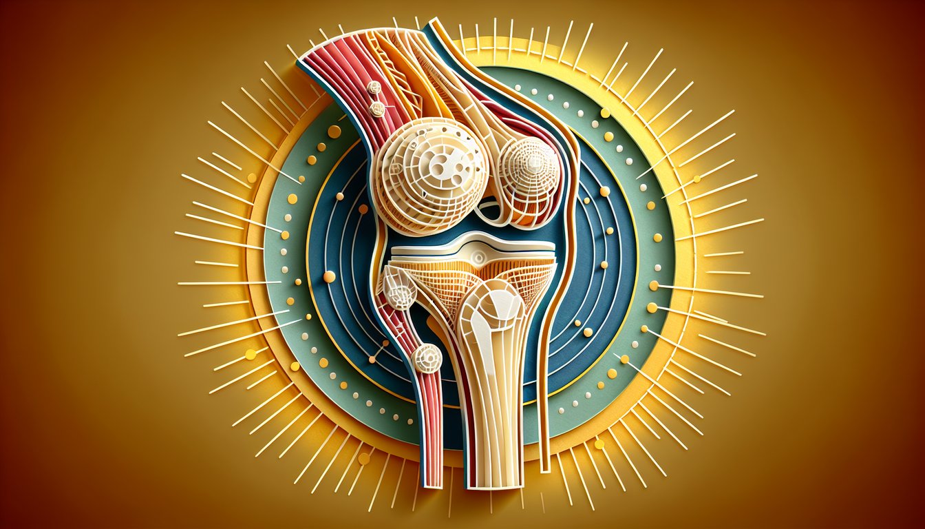
This knee anatomy quiz helps you practice identifying the knee joint's bones, menisci, and key ligaments. Use it to check gaps before an exam or a lab, and then try the companion quiz for more reps. You'll build recall and speed in a few focused minutes.

Which ligament is the primary restraint to anterior translation of the tibia on the femur?
Anterior cruciate ligament (resists anterior tibial translation)
Posterior cruciate ligament
Medial collateral ligament
Lateral collateral ligament
undefined
Which ligament runs from the posterior intercondylar area of the tibia to the medial femoral condyle?
Arcuate popliteal ligament
Posterior cruciate ligament (attaches posterior tibia to medial femoral condyle)
Medial patellofemoral ligament
Anterior cruciate ligament
undefined
Which meniscus is more firmly attached to the joint capsule and the deep fibers of the MCL?
Neither meniscus
Lateral meniscus
Medial meniscus (firm capsular and MCL attachments)
Both menisci equally
undefined
Which structure inserts on the tibial tuberosity?
Quadriceps tendon
Iliotibial band
Patellar ligament (distal continuation of the quadriceps mechanism)
Semimembranosus tendon
undefined
What forms the superior articular surface for the femoral condyles at the knee joint?
Patellar apex
Fibular head
Tibial plateau (medial and lateral tibial condyles)
Gerdy's tubercle
undefined
Which muscle unlocks the knee from full extension by laterally rotating the femur on the tibia?
Biceps femoris
Rectus femoris
Popliteus (initiates unlocking via lateral rotation of femur)
Gastrocnemius
undefined
Which structure inserts on Gerdy's tubercle of the tibia?
Popliteus tendon
Semitendinosus tendon
Biceps femoris tendon
Iliotibial band (lateral knee stabilizer inserting on Gerdy's)
undefined
Where is the suprapatellar bursa located?
Between patellar ligament and tibia
Between MCL and pes anserinus
Between skin and patella
Between the quadriceps tendon and the femur (extends the synovial cavity)
undefined
Which patellar facet is typically larger and bears more load during early flexion?
There are no distinct facets
Lateral facet (broader, engages trochlea early)
Odd facet
Medial facet
undefined
In the screw-home mechanism, what motion of the tibia occurs during the last degrees of open-chain knee extension?
Adduction of the tibia
External rotation of the tibia (locks the knee)
Internal rotation of the tibia
Abduction of the tibia
undefined
The oblique popliteal ligament is a reflected expansion of which tendon?
Biceps femoris
Semimembranosus (expands to reinforce posterior capsule)
Semitendinosus
Popliteus
undefined
The arcuate popliteal ligament attaches to which bony landmark?
Head of the fibula (reinforces posterolateral capsule)
Gerdy's tubercle
Adductor tubercle
Medial tibial plateau
undefined
Which meniscofemoral ligament passes anterior to the PCL from the lateral meniscus to the femur?
Coronary ligament
Transverse intermeniscal ligament
Ligament of Humphrey (anterior to PCL)
Ligament of Wrisberg
undefined
Which meniscofemoral ligament runs posterior to the PCL?
Ligament of Humphrey
Medial patellofemoral ligament
Ligament of Wrisberg (posterior to PCL)
Arcuate popliteal ligament
undefined
Which PCL bundle is most taut in knee flexion?
Posteromedial bundle
Anterolateral bundle (tight in flexion)
Anteromedial bundle of the ACL
Posterolateral bundle of the ACL
undefined
The anterolateral ligament (ALL) most commonly inserts on the tibia at which location?
On the posterior intercondylar area
On the medial tibial plateau
On the tibial tuberosity
Between Gerdy's tubercle and the fibular head (anterolateral tibia)
undefined
Which nerve predominantly supplies proprioceptive innervation to the cruciate ligaments?
Common fibular nerve
Anterior femoral cutaneous nerve
Saphenous nerve
Posterior articular branch of the tibial nerve (innervates cruciates)
undefined
A Segond fracture is an avulsion at which typical location?
Fibular styloid
Posterior medial tibial plateau
Anterolateral proximal tibia near the ALL insertion (lateral capsular region)
Tibial tuberosity
undefined
Which structure reinforces the posteromedial knee capsule and is a key stabilizer there?
Arcuate popliteal ligament
Medial patellofemoral ligament
Transverse intermeniscal ligament
Posterior oblique ligament (POL) (reinforces posteromedial corner)
undefined
Which artery gives rise to the anterior tibial recurrent branch that supplies the proximal tibiofibular joint and anterior knee region?
Medial genicular artery only
Posterior tibial artery recurrent branch
Popliteal artery directly
Anterior tibial artery via recurrent branch (anterior tibial recurrent)
undefined
0

Study Outcomes

  1. Identify Major Knee Joint Components -

    Recall and pinpoint the femur, tibia, fibula, and patella to build a solid foundation in knee anatomy quiz terminology.

  2. Distinguish Key Knee Ligaments -

    Differentiate between the anterior cruciate ligament, posterior cruciate ligament, medial collateral ligament, and lateral collateral ligament in the knee ligaments quiz.

  3. Locate Meniscal Structures -

    Recognize the medial and lateral menisci within the knee joint and understand their role in load distribution and joint stability.

  4. Analyze Ligament Attachments and Functions -

    Examine how each ligament's origin and insertion affect knee movement and stability during flexion and extension.

  5. Apply Knowledge to Injury Scenarios -

    Use your understanding of knee joint anatomy to predict which structures are most at risk in common sports and trauma-related injuries.

Cheat Sheet

  1. Osseous Framework of the Knee -

    Review the three primary knee bones: femur, tibia, and patella, noting the medial and lateral femoral condyles and tibial plateaus. Knowing these articulating surfaces is essential for any knee anatomy quiz or knee joint anatomy quiz and helps in understanding load distribution during movement.

  2. Menisci Morphology & Vascular Zones -

    The medial meniscus is C-shaped and the lateral is almost circular; remember their roles in shock absorption and joint stability. A handy mnemonic for their blood supply zones is "Red-Red, Red-White, White-White," drawn from orthopedic literature, to recall healing potential.

  3. Cruciate Ligaments: ACL & PCL -

    The ACL resists anterior tibial translation while the PCL checks posterior tibial translation; this is tested clinically with the Lachman and posterior drawer tests, respectively. Use the phrase "ACL: Always Checks Lateral" and "PCL: Prevents the Calcaneus Leaving" to nail your knee ligaments quiz.

  4. Collateral Ligaments: MCL & LCL -

    The MCL (medial) is broad and attaches to the medial meniscus, resisting valgus stress, whereas the LCL (lateral) is cord-like and resists varus forces. Referencing AAOS materials, remember they're extracapsular but key in maintaining side-to-side stability.

  5. Screw-Home Mechanism -

    In the final 10 - 20° of extension, the tibia externally rotates on the femur ("screw-home" lock), enhancing stability in full extension. This biomechanical feature, detailed in Gray's Anatomy, is a high-yield point for any ligaments of the knee quiz question on joint mechanics.

Powered by: Quiz Maker