Unlock hundreds more features
Save your Quiz to the Dashboard
View and Export Results
Use AI to Create Quizzes and Analyse Results

Sign inSign in with Facebook
Sign inSign in with Google

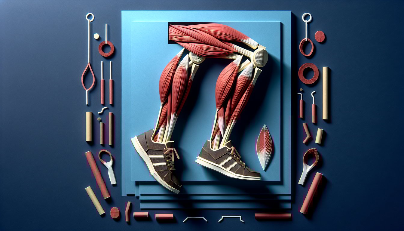
Muscles of the Leg Practice Quiz

Sharpen your leg muscle skills for exams

Difficulty: Moderate
Grade: Grade 10
Study OutcomesCheat Sheet
Paper art promoting Leg Muscle Showdown, a quiz for high school biology students on leg muscle anatomy.

What is the primary function of the quadriceps femoris group?
Hip abduction
Knee extension
Plantar flexion
Knee flexion
The quadriceps femoris group is responsible for extending the knee. This action is crucial for standing up, walking, and other fundamental movements.
Which muscle is located at the back of the thigh?
Rectus femoris
Vastus lateralis
Biceps femoris
Tibialis anterior
The biceps femoris is part of the hamstring group and is located at the back of the thigh. It plays an essential role in knee flexion.
Which muscle forms the most visible bulge of the calf?
Peroneus longus
Soleus
Gastrocnemius
Tibialis anterior
The gastrocnemius is the prominent superficial muscle of the calf that gives it its distinctive shape. It is also a key muscle in plantar flexion of the foot.
What is the primary action of the hamstring muscles at the knee?
Knee extension
Knee flexion
Hip abduction
Dorsiflexion of the foot
The hamstring muscles are primarily responsible for flexing the knee. They also contribute to hip extension during movement.
Where is the tibialis anterior muscle located?
Anterior compartment of the lower leg
Medial thigh
Posterior compartment of the lower leg
Lateral thigh
The tibialis anterior muscle is located in the anterior compartment of the lower leg. It plays an essential role in dorsiflexing the foot.
Which muscle of the quadriceps group crosses both the hip and knee joints?
Vastus intermedius
Rectus femoris
Vastus lateralis
Vastus medialis
The rectus femoris is unique among the quadriceps muscles because it originates from the pelvis and crosses both the hip and knee joints. This dual crossing allows it to assist in both hip flexion and knee extension.
Which muscle is the major contributor to plantar flexion of the foot?
Gastrocnemius
Peroneus longus
Hamstrings
Tibialis anterior
The gastrocnemius plays a central role in plantar flexion, particularly during activities like jumping and running. Its powerful contraction helps push the body forward.
Which muscle assists in stabilizing the patella by providing medial support?
Rectus femoris
Vastus medialis
Vastus lateralis
Biceps femoris
The vastus medialis, particularly its oblique fibers, helps stabilize the patella during knee movements. This medial support is essential for maintaining proper knee tracking.
Which of the following best describes the location and function of the sartorius muscle?
It is a deep muscle in the calf involved in plantar flexion
It is located on the lateral thigh and aids in knee extension only
It is on the posterior thigh and primarily flexes the knee
It runs diagonally across the thigh and assists in hip flexion and knee extension
The sartorius muscle is the longest muscle in the human body, running obliquely across the thigh. It assists in both hip flexion and knee extension, contributing to complex leg movements.
What is the primary role of the soleus muscle in leg movement?
Dorsiflexion of the foot
Plantar flexion of the foot
Hip abduction
Knee extension
The soleus muscle is a powerful muscle in the calf that is responsible for plantar flexion of the foot, especially during standing and walking. It works in coordination with the gastrocnemius to propel the body forward.
Which muscle is primarily responsible for the eversion of the foot?
Gastrocnemius
Tibialis anterior
Peroneus longus
Soleus
The peroneus longus is located on the lateral side of the leg and is the main muscle responsible for foot eversion. This action is critical for maintaining balance on uneven surfaces.
The hamstring muscles receive their nerve supply predominantly from which nerve?
Obturator nerve
Tibial nerve
Sciatic nerve
Femoral nerve
The hamstring muscles are primarily innervated by the sciatic nerve. This nerve supplies most of the posterior thigh and is essential for muscle contraction and coordination.
Which muscle is incorrectly paired with its primary function?
Biceps femoris - knee flexion
Vastus lateralis - knee flexion
Rectus femoris - hip flexion
Tibialis anterior - dorsiflexion
Vastus lateralis is part of the quadriceps group and is responsible for knee extension, not knee flexion. This question highlights a common misconception regarding quadriceps functions.
Which of these muscles is a component of the adductor group of the thigh?
Adductor longus
Vastus intermedius
Sartorius
Rectus femoris
The adductor longus is a primary muscle within the adductor group, located in the medial compartment of the thigh. It is responsible for drawing the thigh toward the body's midline.
The gastrocnemius muscle is composed of two heads. What are they called?
Inner head and outer head
Superior head and inferior head
Anterior head and posterior head
Medial head and lateral head
The gastrocnemius muscle has two distinct heads: a medial head and a lateral head. Together, they form the visible bulk of the calf and contribute to powerful plantar flexion.
Which muscle is often misclassified as a hamstring due to its minor role in knee flexion but actually belongs to the adductor group?
Sartorius
Semitendinosus
Gracilis
Biceps femoris
The gracilis muscle, although it can assist with knee flexion, is actually part of the adductor group of the thigh. Its origin from the pubic bone differentiates it from the true hamstring muscles.
Which major artery is responsible for supplying blood to most of the thigh muscles, including the quadriceps?
Popliteal artery
Deep circumflex iliac artery
Posterior tibial artery
Femoral artery
The femoral artery is the major blood vessel that supplies most of the thigh muscles, including the quadriceps. Its branches provide the essential oxygen and nutrients needed for muscle function.
Which structure separates the anterior compartment of the leg from the deep posterior compartment?
Achilles tendon
Interosseous membrane
Fascia lata
Plantar fascia
The interosseous membrane is a fibrous structure that divides the anterior compartment from the deep posterior compartment of the leg. It also provides attachment points for muscles and contributes to the stability of the leg.
Which nerve primarily innervates the muscles of the anterior compartment of the leg, such as the tibialis anterior?
Obturator nerve
Superficial fibular (peroneal) nerve
Saphenous nerve
Deep fibular (peroneal) nerve
The deep fibular (peroneal) nerve is responsible for innervating the muscles in the anterior compartment of the leg, including the tibialis anterior. This innervation is essential for dorsiflexion and toe extension, and damage to this nerve can lead to foot drop.
During dynamic movements, which leg muscle plays a critical role in stabilizing the knee and preventing patellar misalignment?
Adductor longus
Vastus medialis
Soleus
Gastrocnemius
The vastus medialis, particularly its oblique fibers, is crucial for stabilizing the knee joint by preventing lateral displacement of the patella. This stabilization is vital during high-impact activities such as landing from a jump.
0
{"name":"What is the primary function of the quadriceps femoris group?", "url":"https://www.quiz-maker.com/QPREVIEW","txt":"What is the primary function of the quadriceps femoris group?, Which muscle is located at the back of the thigh?, Which muscle forms the most visible bulge of the calf?","img":"https://www.quiz-maker.com/3012/images/ogquiz.png"}

Study Outcomes

  1. Identify and label the key muscles of the leg and their anatomical locations.
  2. Understand the functions and roles of major leg muscle groups during movement.
  3. Analyze the biomechanical interactions between leg muscles during various activities.
  4. Apply anatomical knowledge to assess muscle performance and potential injury risks.

Muscles of the Leg Game Cheat Sheet

  1. Major leg muscles - The quadriceps extend the knee for powerful strides, the hamstrings flex the knee to slow you down, and the calf muscles lift the heel for tiptoe moments. Picture them as teammates passing a baton in a relay to lock in their functions. Kenhub's Anatomy Mnemonics
  2. Origins and insertions - Knowing where a muscle starts and ends is like mapping its journey: for instance, the gastrocnemius begins at the femoral condyles and travels down to the calcaneus via the Achilles tendon. Crafting a catchy mnemonic can turn this detail into a memorable story. EpoMedicine Origin / Insertion Guide
  3. Anterior compartment mnemonic - "The Hospitals Are Not Dirty Places" helps you recall Tibialis anterior, extensor Hallucis longus, anterior tibial Artery, deep fibular Nerve, extensor Digitorum longus, and Peroneus tertius. Turning lists into silly sentences makes recall a breeze during exams. Anatomy Mnemonics Wiki
  4. Inversion versus eversion - Inversion (foot sole turns inward) is driven by tibialis anterior and posterior, while eversion (sole turns outward) is powered by peroneus longus and brevis. Remember "Little INtroVERted boys roll their feet in when talking to girls" for a fun mental cue. MedicStudent Mnemonics
  5. Nerve supply - The sciatic nerve handles the posterior thigh, the femoral nerve powers the anterior thigh, and the obturator nerve oversees the medial thigh. Visualize each nerve as a hotline connecting your spinal cord to its muscle "subscribers." Vivek Karn High-Yield Mnemonics
  6. Leg compartments - The leg's anterior, lateral, and posterior compartments each host unique muscles, nerves, and vessels. Think of each compartment as its own mini "neighborhood" with distinctive residents and utilities. Anatomy Compartments Wiki
  7. Role of the Achilles tendon - This mighty tendon is the common insertion of gastrocnemius and soleus, translating their contraction into powerful plantarflexion. Visualize it like a spring-loaded cable that propels you forward with each push-off. EpoMedicine on Achilles
  8. Tom, Dick, And Very Nervous Harry - This classic mnemonic lists Tibialis posterior, flexor Digitorum longus, posterior tibial Artery, posterior tibial Vein, tibial Nerve, and flexor Hallucis longus behind the medial malleolus. Turning anatomy into a catchy name makes testing feel like a game. Mnemonic List Wiki
  9. Popliteal fossa - The diamond-shaped hollow behind your knee houses crucial vessels and nerves, including the popliteal artery and tibial nerve. Understanding its borders and contents helps you navigate leg anatomy like a pro surgeon. MedicStudent Popliteal Tips
  10. Diagram practice - Regularly quiz yourself by labeling leg muscle diagrams or 3D models to reinforce spatial relationships. Active recall through drawing or pointing turns passive reading into long-term mastery. Kenhub Study Strategies
Powered by: Quiz Maker