Unlock hundreds more features
Save your Quiz to the Dashboard
View and Export Results
Use AI to Create Quizzes and Analyse Results

Sign inSign in with Facebook
Sign inSign in with Google

Respiratory System Labeling Practice Quiz

Master respiratory anatomy through interactive labeling practice

Difficulty: Moderate
Grade: Grade 8
Study OutcomesCheat Sheet
Paper art depicting a trivia quiz about labeling lung structures for high school biology students.

Which structure is known as the windpipe?
Trachea
Esophagus
Bronchiole
Larynx
The trachea is commonly referred to as the windpipe because it directs air to and from the lungs. Its role makes it an essential component of the respiratory system.
What is the main function of the diaphragm in respiration?
It contracts to allow inhalation
It produces sound
It filters the air
It pumps blood
The diaphragm contracts during inhalation, increasing the volume of the thoracic cavity and allowing the lungs to expand. This contraction is crucial for drawing air into the respiratory system.
Which of the following is NOT a part of the lower respiratory system?
Nasal cavity
Lungs
Trachea
Bronchi
The nasal cavity is part of the upper respiratory system where air is initially filtered and humidified. The lower respiratory system primarily includes the trachea, bronchi, and lungs.
How many lobes does the right lung have?
3
2
4
1
The right lung is divided into three lobes: upper, middle, and lower. This arrangement facilitates effective gas exchange and respiratory function.
Which structures are the primary sites of gas exchange in the lungs?
Alveoli
Bronchi
Trachea
Bronchioles
Alveoli are tiny air sacs where oxygen and carbon dioxide are exchanged with the blood. Their extensive surface area and thin walls make them ideal for rapid gas exchange.
What is the function of the pleura in the respiratory system?
It protects lung tissues and reduces friction
It produces mucus
It pumps blood
It aids in digestion
The pleura is a double-layered membrane that surrounds the lungs and lines the chest cavity. Its lubrication function reduces friction during breathing and protects the lung tissues.
Which structure branches into bronchioles within the lungs?
Bronchi
Trachea
Diaphragm
Alveoli
Bronchi further divide into smaller tubes called bronchioles, which then lead to the alveoli where gas exchange occurs. This branching ensures that air is distributed throughout the lungs.
The left lung is smaller than the right lung mainly due to its proximity to which organ?
Heart
Liver
Stomach
Kidney
The left lung is smaller because it has to accommodate the heart, which occupies space on the left side of the chest. This arrangement is necessary for proper organ placement and function.
Which of the following best describes the structure of the alveoli?
Thin-walled air sacs
Long, muscular tubes
Cartilaginous rings
Thick, fibrous layers
Alveoli are thin-walled, balloon-like structures designed to maximize gas exchange. Their delicate walls allow oxygen and carbon dioxide to diffuse rapidly between the air and blood.
What mechanism primarily allows the exchange of oxygen and carbon dioxide in the alveoli?
Diffusion
Osmosis
Active transport
Facilitated diffusion
Gas exchange in the alveoli occurs through diffusion, where oxygen and carbon dioxide move along their concentration gradients. This passive process does not require energy, making it highly efficient.
Which structure serves as the passageway for air to enter and exit the lungs?
Trachea
Larynx
Bronchi
Pharynx
The trachea, often called the windpipe, is the primary airway that conducts air into and out of the lungs. It functions as a critical passageway connecting the upper and lower respiratory systems.
What role do the cilia in the respiratory system play?
They filter and move mucus
They capture oxygen
They produce white blood cells
They generate sound
Cilia are hair-like structures that move rhythmically to sweep mucus and trapped particles out of the respiratory passages. This process helps keep the airways clear and reduces the risk of infection.
Which of the following is a primary function of the bronchial tree?
Conducting air to alveoli
Filtering blood supply
Producing lung surfactant
Housing respiratory muscles
The bronchial tree is made up of branching tubes that direct air throughout the lungs. Its primary function is the conduction of air to the alveolar sacs where gas exchange takes place.
What distinguishes bronchioles from bronchi?
Bronchioles lack cartilage
Bronchioles are larger in diameter
Bronchioles have more mucous glands
Bronchioles contain vocal cords
Bronchioles are smaller airways that do not possess cartilage in their walls, unlike the larger bronchi. This absence of cartilage allows for more flexible air distribution as the airways branch deeper into the lungs.
Which area of the lungs is most directly involved in the process of gas exchange?
Alveolar sacs
Intercostal muscles
Trachea
Bronchial walls
Alveolar sacs, composed of tightly clustered alveoli, are the primary sites where oxygen and carbon dioxide are exchanged with the blood. Their structure is optimally designed for efficient gas diffusion.
In a detailed lung diagram, which structure is labeled as the barrier between the lung tissue and the chest wall?
Pleura (visceral and parietal layers)
Mediastinum
Diaphragm
Alveolar septa
The pleura consists of two layers: the visceral pleura covering the lungs and the parietal pleura lining the chest wall. This double-layered membrane reduces friction and protects lung tissue during respiration.
When labeling lung structures, how is the right middle lobe distinguished in imaging?
It is located between the superior and inferior lobes on the right lung
It is the largest lobe
It is similar in size to the left lung
It is absent in standard lung anatomy
The right lung is divided into three lobes: superior, middle, and inferior. The middle lobe is uniquely positioned between the superior and inferior lobes, making it identifiable in lung imaging.
What structural feature of the alveoli facilitates rapid gas exchange?
Extensive capillary network and thin membrane walls
Their spiral shape
Large internal volume
Presence of smooth muscle
Alveoli benefit from an extensive network of capillaries and extremely thin walls. This configuration allows gases to diffuse quickly between the alveoli and the blood, making gas exchange highly efficient.
How do alveolar macrophages contribute to lung health beyond gas exchange?
They digest inhaled particles and pathogens
They produce surfactant
They contract to assist breathing
They secrete hormones
Alveolar macrophages are immune cells in the lungs that engulf and break down debris, pathogens, and particulate matter. This cleaning mechanism is essential for maintaining clear airways and preventing infection.
In anatomical labeling of the lungs, which structure serves as the central conducting airway leading into the branching bronchial network?
Trachea
Diaphragm
Esophagus
Mediastinum
The trachea is the main airway that channels air into the lungs, subsequently branching into the bronchi and bronchioles. It serves as the central conducting pathway crucial for proper ventilation.
0
{"name":"Which structure is known as the windpipe?", "url":"https://www.quiz-maker.com/QPREVIEW","txt":"Which structure is known as the windpipe?, What is the main function of the diaphragm in respiration?, Which of the following is NOT a part of the lower respiratory system?","img":"https://www.quiz-maker.com/3012/images/ogquiz.png"}

Study Outcomes

  1. Identify the key lung structures in a labeled diagram.
  2. Analyze the function of each respiratory system component.
  3. Synthesize visual information to accurately label lung anatomy.
  4. Apply conceptual knowledge to prepare for examination scenarios.

Respiratory System Labeling Cheat Sheet

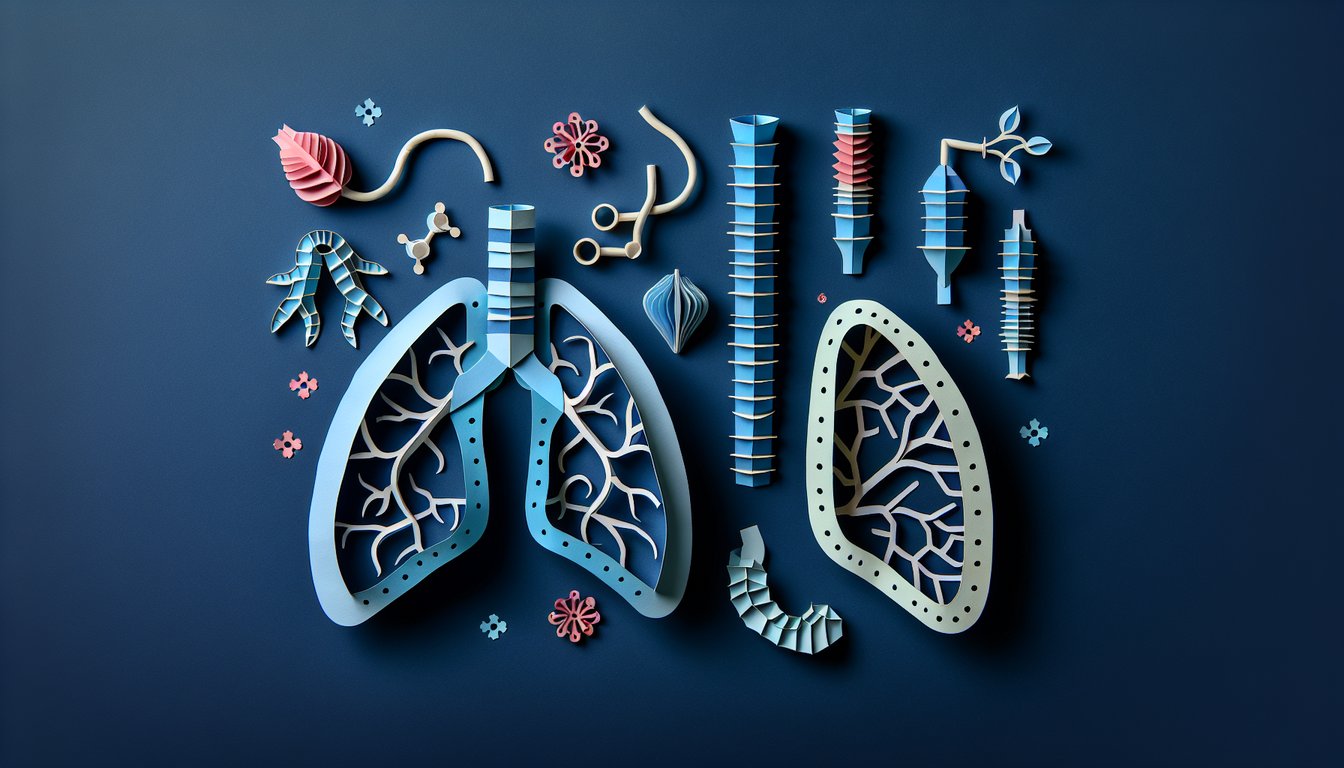
  1. Understand the basic structure of the lungs - Dive into the trachea, bronchi, bronchioles, and alveoli to see how air travels and gas exchange happens at the microscopic level. Grasping this roadmap makes it easier to picture how oxygen enters your blood and carbon dioxide exits. Wikipedia: Lung
  2. Recognize the differences between the right and left lungs - The right lung boasts three lobes (superior, middle, inferior), while the left lung has two lobes to make room for your heart. This clever asymmetry shows how anatomy maximizes space in your chest cavity. Wikipedia: Lung
  3. Learn the function of the diaphragm - This dome‑shaped muscle contracts downward to pull air into your lungs and relaxes upward to push air out. Understanding its piston‑like action helps you appreciate every breath you take. Wikipedia: Lung
  4. Familiarize yourself with the pleurae - These twin serous membranes envelop each lung, creating a slippery surface that reduces friction during breathing. Think of them as lubricated balloons that let your lungs expand and contract smoothly. Wikipedia: Lung
  5. Study the bronchial tree's branching pattern - Trace the airway journey from the trachea to primary bronchi, secondary (lobar) bronchi, tertiary (segmental) bronchi, and finally bronchioles. Mapping this network reveals how air gets distributed throughout your lungs. Wikipedia: Lung
  6. Identify the hilum of the lung - Spot the lung's "root" where blood vessels, nerves, and airways converge to enter or exit. Knowing this central hub is key to understanding lung connectivity and function. Wikipedia: Lung
  7. Recognize the alveoli - These tiny grape‑like air sacs are the true site of oxygen and carbon dioxide exchange between your lungs and blood. Appreciating their vast collective surface area (about the size of a tennis court!) highlights their efficiency. Wikipedia: Lung
  8. Understand the role of the intercostal muscles - Nestled between your ribs, these muscles assist the diaphragm by expanding and contracting the chest cavity. They fine‑tune your breathing, especially during deep inhales and powerful exhales. Wikipedia: Lung
  9. Learn about the cardiac notch - This slight indentation on the left lung accommodates the heart's shape, creating just enough room in your thorax. It's a neat example of how organs adapt to fit together. Wikipedia: Lung
  10. Practice labeling diagrams of the respiratory system - Actively drawing and labeling boosts memory retention far more than passive reading. Grab some blank schematics and challenge yourself to name every airway, muscle, and membrane. Kenhub: Respiratory System Quizzes and Labeled Diagrams
Powered by: Quiz Maker