Unlock hundreds more features
Save your Quiz to the Dashboard
View and Export Results
Use AI to Create Quizzes and Analyse Results

Sign inSign in with Facebook
Sign inSign in with Google

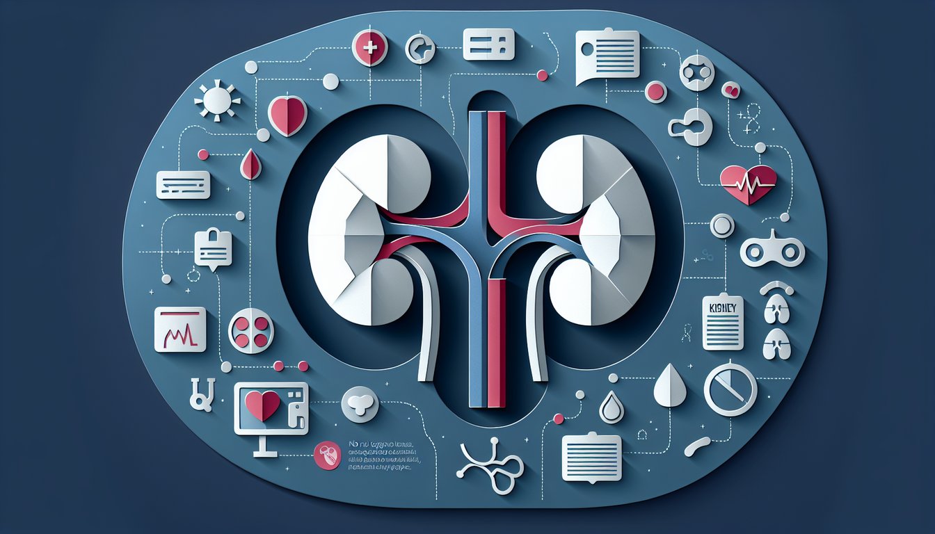
Label the Kidney Diagram: Practice Quiz

Improve your kidney labeling with guided practice

Difficulty: Moderate
Grade: Grade 10
Study OutcomesCheat Sheet
Interactive biology quiz art challenging high school students to label kidney structures.

In a kidney diagram, which outer region of the kidney is identified by its smooth and granular appearance?
Renal Pelvis
Renal Cortex
Ureter
Renal Medulla
The renal cortex is the outer layer of the kidney that appears smooth and granular. It contains most of the nephrons and is easily recognized in diagrams.
Which structure in the kidney acts as a funnel to collect urine before it flows into the ureter?
Renal Capsule
Renal Pelvis
Renal Hilum
Renal Medulla
The renal pelvis is a funnel-shaped structure that collects urine from the calyces before passing it to the ureter. It is a key landmark when labeling kidney diagrams.
Which structure in the kidney is the primary site for blood filtration in the nephron?
Distal Convoluted Tubule
Collecting Duct
Loop of Henle
Glomerulus
The glomerulus is a network of capillaries where blood filtration begins. It is enclosed within Bowman's capsule, forming the initial step of urine formation.
Which tube in the kidney diagram transports urine from the kidney to the urinary bladder?
Renal Artery
Nephron
Renal Vein
Ureter
The ureter is the duct responsible for carrying urine from the kidney to the bladder. Its identification is essential when labeling kidney structures.
What is the purpose of the renal capsule in a kidney diagram?
It transports urine to the bladder
It acts as a protective covering for the kidney
It collects urine from nephrons
It filters blood in the kidney
The renal capsule is a thin, fibrous layer that covers and protects the kidney. It helps maintain the organ's shape and provides a barrier against infections.
Which part of the nephron is primarily responsible for the reabsorption of water, ions, and nutrients?
Loop of Henle
Collecting Duct
Proximal Convoluted Tubule
Distal Convoluted Tubule
The proximal convoluted tubule reabsorbs a majority of water, electrolytes, and nutrients from the filtrate. This process is vital for maintaining the body's homeostasis.
Which structure in the kidney diagram collects urine from multiple nephrons before delivering it to the renal pelvis?
Bowman's Capsule
Collecting Duct
Renal Cortex
Loop of Henle
The collecting duct gathers urine from several nephrons and conveys it to the renal pelvis. Its role is crucial in the final concentration of urine, particularly under hormonal regulation.
What is the term for the notch on the kidney where blood vessels, nerves, and the ureter enter and exit?
Renal Medulla
Renal Hilum
Renal Capsule
Renal Pelvis
The renal hilum is the concave area of the kidney where major blood vessels, nerves, and the ureter are situated. Identifying this structure helps in understanding the kidney's anatomical layout.
Which component, enclosed within Bowman's capsule, is the network of capillaries where filtration occurs?
Distal Convoluted Tubule
Glomerulus
Loop of Henle
Proximal Convoluted Tubule
The glomerulus is a tuft of capillaries that facilitates the filtration of blood as the first step in urine production. It is a critical component of the nephron's filtration apparatus.
Which statement correctly differentiates the renal cortex from the renal medulla in a kidney diagram?
Renal cortex stores urine, while the renal medulla is primarily involved in blood filtration
Renal cortex is the inner region and contains the renal pelvis, while the renal medulla is the outer region with glomeruli
Both have identical functions and are indistinguishable in diagrams
Renal cortex is the outer layer housing most nephrons, while the renal medulla consists of pyramidal structures containing loops of Henle and collecting ducts
The renal cortex is the outer layer where most nephrons, including the glomeruli, are found, while the medulla contains the pyramidal structures that house the loops of Henle and collecting ducts. Understanding this contrast is essential for correctly interpreting kidney diagrams.
Which part of the nephron is involved in the countercurrent multiplication mechanism that concentrates urine?
Bowman's Capsule
Loop of Henle
Proximal Convoluted Tubule
Distal Convoluted Tubule
The loop of Henle establishes a concentration gradient in the kidney medulla through a countercurrent multiplication mechanism. This process is fundamental for the kidney's ability to produce concentrated urine.
What is considered the functional unit of the kidney responsible for filtering blood and forming urine?
Renal Medulla
Nephron
Renal Cortex
Renal Corpuscle
The nephron is the kidney's functional unit, comprising the glomerulus and the tubular system. It orchestrates the processes of filtration, reabsorption, and secretion to form urine.
Which segment of the nephron is primarily responsible for the secretion of additional waste products into the urine?
Glomerulus
Proximal Convoluted Tubule
Collecting Duct
Distal Convoluted Tubule
The distal convoluted tubule is the site where further secretion of ions and waste products is fine-tuned before final urine formation. This stage is crucial for regulating the chemical balance of the body.
How does the structure of the collecting duct contribute to its role in water reabsorption under the influence of antidiuretic hormone (ADH)?
Its structure has no role in water reabsorption
Its walls become highly permeable, facilitating water reabsorption
Its muscular contractions directly filter blood
Its rigid, impermeable walls prevent water loss
Under the influence of ADH, the collecting duct's permeability increases, allowing more water to be reabsorbed from the filtrate. This adaptation is key to maintaining the body's water balance.
What is the primary function of the renal artery as depicted in kidney diagrams?
To drain deoxygenated blood from the kidney
To transport urine away from the kidney
To envelop the kidney in a protective layer
To supply the kidney with oxygenated blood
The renal artery is responsible for delivering oxygen-rich blood to the kidney, providing the essential resources for filtration and metabolic activities. Its role is fundamental in supporting renal function.
When analyzing a detailed kidney diagram, what features are essential for accurately distinguishing between the cortex and the medulla?
Only the color differences in the diagram
The number of blood vessels present
The size of the kidney relative to other organs
The location, arrangement of nephrons, and presence of renal pyramids
Accurate differentiation between the cortex and medulla requires assessing their location and structural organization; the cortex is the outer layer rich in nephrons while the medulla features pyramidal structures. This understanding is vital for precise diagram labeling.
In a kidney diagram, how can renal pyramids in the medulla be distinguished from other structures?
They have a smooth, circular appearance
They are not visible in detailed diagrams
They are evenly distributed in the renal cortex
They appear as triangular or pyramid-shaped structures with striated zones
Renal pyramids are identifiable as triangular or pyramid-shaped regions with a striated appearance due to parallel arrangements of tubules. Recognizing these features is key for interpreting the kidney's medullary structure.
How can you differentiate between the proximal and distal convoluted tubules when labeling a kidney diagram?
The proximal tubule is located in the cortex and has a prominent brush border, unlike the distal tubule
Both tubules are identical in structure, so differentiation is not possible
The distal tubule is coiled, while the proximal tubule is straight
The proximal tubule is in the medulla, while the distal is in the cortex
The proximal convoluted tubule is distinguished by a dense brush border and its location in the cortex, which enhances reabsorption. In contrast, the distal convoluted tubule lacks this brush border and appears less elaborate.
Which kidney structure is most indicative of its role in acid-base balance as seen in detailed kidney diagrams?
The renal pelvis, which collects urine
The glomerulus, which only filters blood
The distal convoluted tubule, where hydrogen ions are secreted
The renal capsule, which provides structural support
The distal convoluted tubule plays a critical role in maintaining acid-base balance by secreting hydrogen ions. Correctly identifying this function on a diagram emphasizes its importance in renal physiology.
Why is it important to accurately label both the collecting duct and the loop of Henle in a detailed kidney diagram?
Because they are the same structure and can be used interchangeably
Because they have distinct roles in concentrating urine and errors can lead to misunderstandings of renal physiology
Because only the loop of Henle is involved in urine formation
Because labeling them is optional and does not affect comprehension
The collecting duct and the loop of Henle each contribute uniquely to urine concentration. Accurate labeling is crucial as it reflects their different functions, ensuring a correct understanding of renal physiology.
0
{"name":"In a kidney diagram, which outer region of the kidney is identified by its smooth and granular appearance?", "url":"https://www.quiz-maker.com/QPREVIEW","txt":"In a kidney diagram, which outer region of the kidney is identified by its smooth and granular appearance?, Which structure in the kidney acts as a funnel to collect urine before it flows into the ureter?, Which structure in the kidney is the primary site for blood filtration in the nephron?","img":"https://www.quiz-maker.com/3012/images/ogquiz.png"}

Study Outcomes

  1. Identify key kidney structures accurately.
  2. Label each part of the kidney diagram correctly.
  3. Analyze the functional importance of distinct kidney regions.
  4. Apply anatomical knowledge to answer exam questions.
  5. Evaluate the role of kidney structures in overall organ function.

Free: Label the Kidney Diagram Cheat Sheet

  1. External Kidney Anatomy - Discover the kidney's outer layers from the tough renal capsule to the cushioning perirenal fat, all working together like a superhero suit to protect this vital organ. Knowing these parts will help you appreciate how blood enters and exits in style. Kidney Anatomy Parts & Functions
  2. Internal Kidney Structures - Dive inside to explore the cortex, medulla, pyramids, columns, papillae, and pelvis - each area playing a unique role in the journey of urine. Imagine this as the kidney's theme park where filtration rides and reabsorption loops happen nonstop! Kidney Anatomy Parts & Functions
  3. The Mighty Nephron - Meet the nephron, your kidney's MVP, complete with a glomerulus, Bowman's capsule, and winding tubules that filter blood and craft urine drop by drop. Think of it as a tiny waterpark for plasma where every turn leads to cleaner blood! Kidney & Nephron Diagram
  4. Renal Artery & Vein Roles - The renal artery delivers a rush of blood for filtering, while the renal vein carries the cleansed results back to the circulation party. Understanding these highways clarifies how kidneys keep your fluids and electrolytes in check. Renal External Anatomy Details
  5. Ureter Function - Picture the ureter as a fast-moving slide carrying urine from the pelvis to your bladder's VIP lounge. Its muscular contractions ensure no backflow, making it a one-way ticket for detox duty! Kidney Function Quiz
  6. Renal Cortex Highlights - The outer cortex is packed with nephrons busy filtering blood and reclaiming crucial nutrients. It's like the initial checkpoint where all the magic of plasma purification begins! Renal External Anatomy Details
  7. Renal Medulla Marvels - Home to the pyramids and loops of Henle, the medulla concentrates urine like a pro, recovering water and salts with precision. Think of it as the kidney's in-house distillery for hydration control! Renal External Anatomy Details
  8. Renal Pelvis Function - This funnel-shaped chamber collects the freshly made urine before shipping it off through the ureter. It's the kidney's personal collection bucket, ensuring no drop spills out of place! Kidney Function Quiz
  9. Glomerulus Filtration - Inside each nephron, the glomerulus acts like a high-pressure sieve, letting water and small solutes pass while holding back cells and proteins. It's the ultimate bouncer for your blood! Kidney & Nephron Diagram
  10. Diagram Labeling Practice - Grab a kidney outline and start labeling to make these structures stick in your brain. Coloring and naming each part turns studying into an interactive adventure! Label & Color the Kidney Activity
Powered by: Quiz Maker