Unlock hundreds more features
Save your Quiz to the Dashboard
View and Export Results
Use AI to Create Quizzes and Analyse Results

Sign inSign in with Facebook
Sign inSign in with Google

Labeling the Heart Practice Quiz

Review essential heart anatomy for exam mastery

Difficulty: Moderate
Grade: Grade 7
Study OutcomesCheat Sheet
Paper art themed trivia quiz for high school biology students on heart anatomy labeling challenge.

Easy
Which chamber of the heart pumps oxygenated blood to the body?
Left Atrium
Right Atrium
Left Ventricle
Right Ventricle
The left ventricle is responsible for pumping oxygenated blood into the systemic circulation through the aorta. The other chambers serve different roles in circulating blood.
Which chamber receives deoxygenated blood from the body?
Left Ventricle
Right Ventricle
Right Atrium
Left Atrium
The right atrium collects deoxygenated blood from the body via the vena cava. The other chambers have roles related to pumping or receiving blood differently.
Which major blood vessel carries deoxygenated blood from the heart to the lungs?
Pulmonary Vein
Superior Vena Cava
Pulmonary Artery
Aorta
The pulmonary artery carries deoxygenated blood from the right ventricle to the lungs where it is oxygenated. The other vessels have distinct roles in the circulatory system.
What is the valve that controls blood flow between the left atrium and left ventricle?
Mitral Valve
Aortic Valve
Pulmonary Valve
Tricuspid Valve
The mitral valve, also known as the bicuspid valve, regulates blood flow from the left atrium to the left ventricle. The other valves are positioned on the right side or manage outflow from the ventricles.
Which valve is found on the right side of the heart, controlling blood flow from the right atrium to the right ventricle?
Aortic Valve
Mitral Valve
Tricuspid Valve
Pulmonary Valve
The tricuspid valve regulates the flow of blood from the right atrium to the right ventricle. It is distinct from other valves that manage flow in other regions of the heart.
Medium
Which structure is responsible for distributing oxygenated blood from the left ventricle to the body?
Pulmonary Artery
Aorta
Pulmonary Vein
Vena Cava
The aorta is the main artery that carries oxygenated blood from the left ventricle to the systemic circulation. The other vessels have different roles in blood transport.
Which component of the heart's conduction system delays the electrical impulse to allow proper ventricular filling?
Purkinje Fibers
Atrioventricular Node
Bundle of His
Sinoatrial Node
The atrioventricular (AV) node delays the electrical impulse from the atria to the ventricles, ensuring proper filling before contraction. The other structures have different roles in initiating or conducting electrical signals.
Which structure prevents backflow of blood into the left atrium during ventricular contraction?
Pulmonary Valve
Mitral Valve
Aortic Valve
Tricuspid Valve
The mitral valve prevents blood from flowing backward from the left ventricle into the left atrium during systole. The other valves regulate flow between other chambers or outflow tracts.
Which heart structure is the thick muscular layer that contracts to pump blood?
Pericardium
Myocardium
Epicardium
Endocardium
The myocardium is the thick, muscular layer that enables the heart to generate force for blood circulation. The other layers serve as inner lining or protective coverings.
The coronary arteries mainly supply blood to which part of the heart?
Pericardium
Myocardium
Endocardium
Atria
Coronary arteries deliver oxygen-rich blood directly to the myocardium, which is essential for the heart's continuous contraction. Other structures are not directly supplied by these arteries.
What is the primary function of the pulmonary veins in the heart's circulation?
Carry deoxygenated blood from the right ventricle to the lungs
Supply the myocardium with blood
Transport oxygenated blood from the lungs to the left atrium
Distribute blood from the left ventricle to the body
Pulmonary veins carry oxygenated blood from the lungs back to the left atrium. The other options describe functions of different vessels.
Which valve prevents the backflow of blood from the aorta into the left ventricle?
Mitral Valve
Aortic Valve
Tricuspid Valve
Pulmonary Valve
The aortic valve closes during ventricular diastole to prevent blood from flowing back into the left ventricle. The other valves control blood flow in other parts of the heart.
Which structure transmits the electrical impulse from the atrioventricular node to the ventricles?
Interventricular Septum
Purkinje Fibers
Bundle of His
Sinoatrial Node
The Bundle of His carries the impulse from the AV node to the bundle branches and then to the Purkinje fibers in the ventricles. The other structures play different roles in the conduction system.
During which phase of the cardiac cycle does ventricular contraction occur?
Atrial Systole
Isovolumetric Relaxation
Diastole
Systole
Systole is the phase when the ventricles contract to pump blood out of the heart. Diastole is the relaxation phase when the heart fills with blood.
Which structure serves as the protective sac enclosing the heart and reducing friction during its movement?
Pericardium
Epicardium
Myocardium
Endocardium
The pericardium is a double-layered sac that protects the heart and contains lubricating fluid to reduce friction. The other layers are involved in contraction or form the inner lining of the heart.
Hard
What is the correct sequence of blood flow through the heart starting from the right atrium?
Right atrium → Left atrium → Left ventricle → Aorta → Right ventricle → Pulmonary artery → Pulmonary veins
Right atrium → Right ventricle → Pulmonary vein → Lungs → Pulmonary artery → Left atrium → Left ventricle → Aorta
Right atrium → Right ventricle → Pulmonary artery → Lungs → Pulmonary veins → Left atrium → Left ventricle → Aorta
Right atrium → Right ventricle → Aorta → Left ventricle → Left atrium → Pulmonary veins → Pulmonary artery
The correct sequence starts with deoxygenated blood entering the right atrium, moving to the right ventricle, then to the pulmonary artery, reaching the lungs for oxygenation, and returning via the pulmonary veins to the left atrium before being pumped out through the left ventricle and aorta. The other sequences present incorrect orders.
When the atrioventricular node is dysfunctional, which structure typically acts as an emergency pacemaker for the ventricles?
Sinoatrial Node
Bundle of His
Tricuspid Valve
Purkinje Fibers
In cases of AV node failure, the Purkinje fibers can act as an escape pacemaker, maintaining a slow but steady ventricular rhythm. The other options do not serve as a backup pacemaker in such scenarios.
Which anatomical feature of the left ventricle allows it to generate the high pressure needed for systemic circulation?
A large atrial chamber
A compliant ventricular wall
A thick and muscular myocardium
A thin endocardial layer
The thick and muscular myocardium of the left ventricle is crucial for generating the high pressures required to pump blood throughout the systemic circulation. The other features do not contribute to high-pressure generation.
Which structure divides the left and right ventricles in the heart?
Aortic Arch
Interatrial Septum
Interventricular Septum
Pulmonary Trunk
The interventricular septum is the muscular wall that separates the left and right ventricles. The interatrial septum divides the atria, while the other structures are components of the great vessels.
What role do the papillary muscles play in heart function?
They anchor the chordae tendineae to prevent valve prolapse during systole
They form the heart's electrical conduction pathway
They initiate the heartbeat
They thicken the heart wall for high-pressure pumping
Papillary muscles contract during ventricular systole to tense the chordae tendineae, thereby preventing the atrioventricular valves from inverting. This function is critical for maintaining one-way blood flow in the heart.
0
{"name":"Which chamber of the heart pumps oxygenated blood to the body?", "url":"https://www.quiz-maker.com/QPREVIEW","txt":"Easy, Which chamber of the heart pumps oxygenated blood to the body?, Which chamber receives deoxygenated blood from the body?","img":"https://www.quiz-maker.com/3012/images/ogquiz.png"}

Study Outcomes

  1. Identify the major anatomical structures of the heart.
  2. Label each part of the heart accurately.
  3. Explain the functional role of each heart component.
  4. Analyze the relationship between heart chambers and valves.
  5. Assess anatomical knowledge through interactive quiz challenges.

Heart Labeling Cheat Sheet

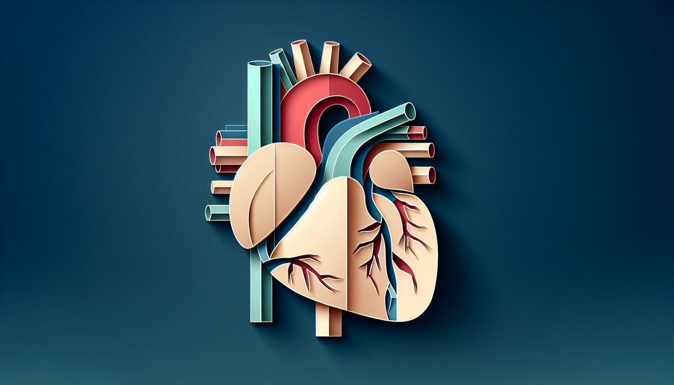
  1. Understand the four chambers of the heart - The right atrium, right ventricle, left atrium and left ventricle each play a unique role in circulating blood. Visualizing and sketching them in different colors turns memorization into a fun art project. Heart Anatomy Labeled Diagram
  2. Learn the major blood vessels - Major arteries and veins like the aorta, superior vena cava, and pulmonary vessels form highways for your blood cells. Understanding their routes helps you see how oxygenated and deoxygenated blood travel in a continuous loop. Heart Anatomy Labeled Diagram
  3. Familiarize yourself with heart valves - Heart valves act as one-way doors - tricuspid, pulmonary, mitral, and aortic - ensuring blood never backflows. Pinpointing their exact locations and matching them with their functions cements your recall. Heart Anatomy Labeled Diagram
  4. Practice with interactive labeling quizzes - Interactive quizzes let you label the heart's parts in real time, boosting retention through playful challenge. Gamifying your study turns review sessions into mini contests. Heart Labeling Quiz
  5. Use unlabeled heart diagrams - Grabbing an unlabeled diagram and filling in the blanks forces your brain to retrieve key structures. This "test yourself" strategy highlights weak spots for targeted review. Unlabeled Heart Diagrams
  6. Explore online flashcards - Flashcards are your pocket professors: quick to review and perfect for on-the-go cram sessions. Repetition with spaced intervals supercharges your long-term memory of key heart components. Heart Anatomy Flashcards
  7. Review the blood flow pathway - Tracing the journey of blood - from systemic veins, through the heart, then out via the aorta - builds a mental movie of circulation. Knowing this pathway inside out is the foundation for all cardiac physiology. Heart Anatomy Labeled Diagram
  8. Learn coronary arteries and veins - The coronary arteries and veins are the heart's own lifeline, delivering oxygen and nutrients to heart muscle. Mapping these vessels helps you grasp why blockages can spell trouble. Heart Anatomy Labeled Diagram
  9. Study the electrical conduction system - The SA node, AV node, and conduction fibers orchestrate each heartbeat like a natural pacemaker. Visualizing this electrical network deepens your understanding of rhythm disorders. Heart Anatomy Labeled Diagram
  10. Engage in interactive labeling games - Labeling games and virtual puzzles turn tedious memorization into a competitive sport. Chasing high scores on platforms like PurposeGames keeps you engaged and excited to learn more. Heart Anatomy Labeling Game
Powered by: Quiz Maker