Unlock hundreds more features
Save your Quiz to the Dashboard
View and Export Results
Use AI to Create Quizzes and Analyse Results

Sign inSign in with Facebook
Sign inSign in with Google

Ace the Brachial Plexus Practice Quiz

Enhance your nerve anatomy skills with practice questions

Difficulty: Moderate
Grade: Grade 12
Study OutcomesCheat Sheet
Colorful paper art promoting The Brachial Blitz Quiz, an anatomy-focused trivia for students.

What is the primary role of the brachial plexus?
Motor and sensory innervation of the upper limb
Regulation of heart rate
Control of digestive functions
Facilitation of respiratory movements
The brachial plexus is a network of nerves that supplies both motor and sensory innervation to the upper limb. Its function is essential for movement and sensation in the arm and hand.
Which spinal segments contribute to the formation of the brachial plexus?
C5 to T1
T2 to T6
C1 to C4
L1 to L5
The brachial plexus is formed by the ventral rami of spinal nerves C5, C6, C7, C8, and T1. Recognizing these segments is fundamental to understanding upper limb innervation.
Which of the following is NOT a component of the brachial plexus?
Divisions
Spleen
Roots
Trunks
The brachial plexus is organized into roots, trunks, divisions, cords, and terminal branches. The spleen, being an organ of the lymphatic system, is not a component of this neural network.
What type of nerve fibers does the brachial plexus contain?
Sensory only
Motor only
Both sensory and motor
Autonomic only
The brachial plexus carries both sensory fibers, which transmit sensory information from the skin, and motor fibers that control muscle movements of the upper limb. This dual role is central to its function.
Which of the following branches is a terminal branch of the brachial plexus?
Femoral nerve
Axillary nerve
Sciatic nerve
Obturator nerve
The axillary nerve is one of the terminal branches of the brachial plexus, supplying the deltoid and teres minor muscles and providing sensory input to the shoulder region. Identifying terminal branches helps in understanding upper limb function.
From which cord does the lateral root of the median nerve originate?
Lateral cord
Medial cord
Posterior cord
Superior cord
The lateral root of the median nerve originates from the lateral cord of the brachial plexus, carrying contributions from C5 to C7. This component is crucial in the formation of the median nerve.
Which nerve is primarily responsible for innervating the biceps brachii muscle?
Musculocutaneous nerve
Radial nerve
Median nerve
Axillary nerve
The musculocutaneous nerve innervates the biceps brachii, which is essential for elbow flexion and forearm supination. Its origin from the lateral cord highlights its significance within the brachial plexus.
What is the classic presentation of Erb-Duchenne palsy?
Arm abducted with a pronated forearm
Arm adducted with an extended and pronated forearm (waiter's tip position)
Arm elevated with a flexed elbow and wrist drop
Isolated hand and finger paralysis
Erb-Duchenne palsy typically results from injury to the upper trunk of the brachial plexus, leading to a 'waiter's tip' posture where the arm is adducted and internally rotated with an extended and pronated forearm. This clinical presentation is a hallmark in nerve injury assessments.
Which cord of the brachial plexus gives rise to the radial nerve?
Lateral cord
Medial cord
Posterior cord
Anterior cord
The radial nerve is formed from the posterior cord of the brachial plexus and plays a key role in extending the wrist and fingers. Understanding its origin is essential for localizing nerve injuries.
From which cord does the ulnar nerve primarily arise?
Lateral cord
Posterior cord
Medial cord
Superior cord
The ulnar nerve originates primarily from the medial cord, carrying fibers from C8 and T1. This nerve is vital for the motor and sensory functions of the medial side of the forearm and hand.
Which of the following muscles is primarily innervated by the radial nerve?
Biceps brachii
Triceps brachii
Deltoid
Pectoralis major
The radial nerve supplies the triceps brachii, which is essential for elbow extension. This relationship underscores the importance of the posterior cord's role in motor function.
What is the correct anatomical sequence of the brachial plexus structures from proximal to distal?
Trunks, roots, cords, divisions, branches
Roots, trunks, divisions, cords, branches
Roots, cords, trunks, divisions, branches
Divisions, trunks, roots, cords, branches
The brachial plexus is systematically organized as roots, trunks, divisions, cords, and finally terminal branches. This sequence is fundamental in mapping the origin and path of peripheral nerves.
Which nerve provides sensory innervation to the lateral aspect of the forearm?
Musculocutaneous nerve
Median nerve
Ulnar nerve
Radial nerve
The musculocutaneous nerve continues as the lateral antebrachial cutaneous nerve, which provides sensation to the lateral forearm. This sensory distribution is key in diagnosing specific nerve injuries.
Which nerve is most likely to be affected in a midshaft humerus fracture?
Radial nerve
Median nerve
Ulnar nerve
Axillary nerve
The radial nerve runs in close proximity to the midshaft of the humerus, making it particularly vulnerable during fractures. Injury to this nerve frequently results in wrist drop due to loss of extensor function.
Which structure in the brachial plexus splits into anterior and posterior divisions?
Roots
Trunks
Cords
Branches
Each trunk in the brachial plexus divides into an anterior and a posterior division. This division is an essential step in the organization of the plexus before the formation of cords and terminal branches.
A patient presents with wrist drop, characterized by loss of extension at the wrist and fingers. Which component of the brachial plexus is most likely injured?
Lateral cord
Medial cord
Posterior cord
Upper trunk
Wrist drop is most commonly associated with injury to the radial nerve, which arises from the posterior cord. Recognizing this correlation between clinical presentation and anatomical origin is vital for accurate diagnosis.
During surgical repair of a brachial plexus injury, which anatomical landmark is most useful for locating the lateral cord?
Axillary artery
Subclavian artery
Brachiocephalic vein
Clavicle
The lateral cord is situated lateral to the axillary artery in the infraclavicular region. This close anatomical relationship makes the axillary artery a key landmark during surgical procedures on the brachial plexus.
Which nerve is most commonly injured following an axillary dislocation, potentially leading to deltoid muscle paralysis?
Axillary nerve
Radial nerve
Ulnar nerve
Median nerve
The axillary nerve is vulnerable during shoulder dislocations because of its close proximity to the humeral head. Damage to this nerve can lead to deltoid muscle paralysis and impaired shoulder abduction.
In nerve conduction studies, an injury to the medial cord would most likely affect sensation over which area?
Lateral forearm
Ulnar aspect of the hand
Dorsal hand
Posterior arm
The medial cord gives rise primarily to the ulnar nerve, which provides sensory innervation to the ulnar aspect of the hand. An injury here typically results in sensory deficits in this specific distribution.
Which embryological structure is responsible for forming the neurons of the brachial plexus?
Sclerotome
Myotome
Neural crest cells
Endoderm
Neural crest cells are the embryological origin of peripheral neurons, including those that form the brachial plexus. This developmental process is essential for the formation of the peripheral nervous system.
0
{"name":"What is the primary role of the brachial plexus?", "url":"https://www.quiz-maker.com/QPREVIEW","txt":"What is the primary role of the brachial plexus?, Which spinal segments contribute to the formation of the brachial plexus?, Which of the following is NOT a component of the brachial plexus?","img":"https://www.quiz-maker.com/3012/images/ogquiz.png"}

Study Outcomes

  1. Analyze the anatomical structure of the brachial region.
  2. Apply knowledge of the brachial plexus to identify its key components.
  3. Explain the functional significance of nerves within the brachial region.
  4. Utilize anatomical terminology to accurately describe brachial pathways.

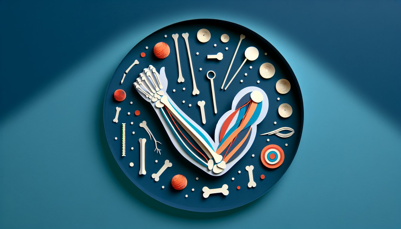
Brachial Plexus Cheat Sheet

  1. Five main components of the brachial plexus - The brachial plexus is built from five key parts: Roots, Trunks, Divisions, Cords, and Branches. Try the mnemonic "Rugby Teams Don't Cover Bruises" to lock it in your memory and make studying a bit more sporty. Sketching the plexus while you recite the phrase turns rote learning into a visual adventure! Kenhub Anatomy: Brachial Plexus
  2. Spinal nerve roots C5 - T1 - These roots fuse in various combinations to form the trunks of the brachial plexus and set the stage for all downstream pathways. Remembering "C5 to T1" is like knowing the starting lineup for a championship team. Seeing how the roots cascade into trunks helps you predict where each nerve will go. TeachMeAnatomy: Brachial Plexus Overview
  3. Three trunks: Superior, Middle, Inferior - Formed by root pairings (C5 - C6, C7, and C8 - T1), each trunk then splits into anterior and posterior divisions. Think of each trunk as a major highway exit ramp dividing into local roads - easy to follow on your mental map. Drawing these splits reinforces how signals route through your arm. TeachMeAnatomy: Trunks & Divisions
  4. Divisions recombine into three cords - Named by their position around the axillary artery - Lateral, Posterior, and Medial - these cords give rise to the major terminal nerves. Picture three vines twining around a central pole to remember their relative positions. Once you see the cords in relation to the artery, you'll never get lost in the plexus jungle! TeachMeAnatomy: Cords Explained
  5. Five terminal branches - Musculocutaneous, Axillary, Radial, Median, and Ulnar wrap up the plexus and dive into your arm. Use "My Aunt Rides My Unicorn" to recall their order and cast a spell on your memory. Visualizing each nerve's path off the "cord vines" cements their footprints in your mind! Kenhub Anatomy: Terminal Branches
  6. Motor and sensory roles - Each terminal nerve has its own muscle moves and skin zones, like the Radial nerve powering your extension and tickling your posterior arm. Quizzing yourself with flashcards - "Which nerve?" - turns study time into a mini-game. Active recall supercharges your retention engine! TeachMeAnatomy: Functions & Territories
  7. Common injuries like Erb's Palsy - Damage to the superior trunk (C5 - C6) can lead to the classic "waiter's tip" posture and teach you valuable clinical correlations. Turning injuries into case stories helps you empathize and remember. Imagine diagnosing a superhero with a plexus punch - studying has never been so heroic! TeachMeAnatomy: Injury Patterns
  8. Anatomical course from neck to arm - Follow the plexus from its neck origin, through the axilla, and into the upper limb like a treasure map. Tracing the pathway on a body chart makes the journey come alive. The more you "walk" the nerves, the more familiar territories become! TeachMeAnatomy: Pathway Map
  9. Use visual aids and diagrams - Sketching the brachial plexus in color codes - roots in red, trunks in blue, cords in green - cements patterns in your brain. Turning diagrams into doodles boosts neural connections and makes review sessions a breeze. Your future self will thank you for the colorful roadmap! TeachMeAnatomy: Diagrams & Charts
  10. Apply knowledge to clinical scenarios - Practice identifying injured plexus segments based on symptoms - are wrist drop and numb thumb pointing to a Radial nerve issue? Clinical vignettes transform abstract facts into practical skills. It's like solving medical mysteries before the real test! TeachMeAnatomy: Clinical Cases
Powered by: Quiz Maker