Unlock hundreds more features
Save your Quiz to the Dashboard
View and Export Results
Use AI to Create Quizzes and Analyse Results

Sign inSign in with Facebook
Sign inSign in with Google

Skeleton Labeling Practice Quiz

Boost learning with interactive skeleton label questions

Difficulty: Moderate
Grade: Grade 8
Study OutcomesCheat Sheet
Paper art illustrating a trivia quiz on skeletal structures for high school biology students.

Which bone is the longest in the human body?
Femur
Humerus
Tibia
Fibula
The femur is the longest and strongest bone in the human body. Its length and robust structure make it essential for supporting weight and facilitating movement.
Which bone protects the brain?
Skull
Ribcage
Pelvis
Vertebrae
The skull encases and protects the brain from physical damage. Composed of several fused bones, it forms a hard barrier against impacts.
Which bone is located in the upper arm?
Humerus
Scapula
Radius
Ulna
The humerus is the long bone in the upper arm, connecting the shoulder to the elbow. Its identification is fundamental in skeletal labeling.
What is the primary function of the ribcage?
Protects the heart and lungs
Supports leg movement
Facilitates hand motion
Aids in digestion
The ribcage creates a protective barrier around the heart and lungs. Its structure is critical for safeguarding vital organs within the thoracic cavity.
Which bone forms the lower jaw?
Mandible
Maxilla
Zygomatic
Temporal
The mandible is the sole movable bone of the skull and forms the lower jaw. Its distinct shape is key for oral functions such as chewing and speaking.
Which pair of bones forms the forearm?
Radius and Ulna
Femur and Tibia
Humerus and Scapula
Patella and Fibula
The forearm is composed of the radius and ulna, which allow for rotational movements such as pronation and supination. Their distinct locations and shapes make them easier to label when studied together.
What bone is commonly known as the collarbone?
Clavicle
Sternum
Scapula
Mandible
The clavicle, or collarbone, serves as a strut that connects the sternum to the scapula. Its horizontal orientation makes it a key landmark in skeletal labeling.
Which bone forms the back of the skull?
Occipital bone
Frontal bone
Parietal bone
Temporal bone
The occipital bone makes up the posterior portion of the skull and plays a crucial role in protecting the brain. Its broad, curved structure is distinctive in skeletal diagrams.
Which bone is commonly referred to as the kneecap?
Patella
Femur
Tibia
Fibula
The patella is a small, flat bone that protects the knee joint. Its prominent location and shape make it a frequent focus in labeling quizzes.
Which bone in the lower leg is larger and primarily weight-bearing?
Tibia
Fibula
Both are the same size
Femur
The tibia is the larger, weight-bearing bone of the lower leg, while the fibula is a slimmer bone that serves mainly for muscle attachment. Recognizing the tibia is essential in both anatomical and labeling studies.
Which bone primarily forms the eye socket?
Zygomatic bone
Maxilla
Vomer
Nasal bone
The zygomatic bone, or cheekbone, plays an integral role in forming the orbit of the eye. Its angular structure serves as a clear marker for correct labeling in craniofacial diagrams.
What is the primary function of the vertebral column?
Supports the body and protects the spinal cord
Facilitates hand movement
Helps with respiration
Assists in digestion
The vertebral column provides structural support for the body and encases the spinal cord within a series of protective vertebrae. Its segmented design is fundamental for both flexibility and protection.
Which bone connects the hip joint to the knee joint in the upper leg?
Femur
Tibia
Fibula
Pelvis
The femur is the only bone in the upper leg, extending from the hip to the knee. Its length and weight-bearing structure make it a key feature in labeling skeletal diagrams.
Which of the following is the uppermost and largest part of the hip bone?
Ilium
Ischium
Pubis
Sacrum
The ilium forms the broad, flaring portion of the hip bone and is the largest of the pelvic components. Its distinctive shape aids in identifying pelvic orientation during skeletal labeling.
Which of the following bones is part of the axial skeleton?
Ribcage
Femur
Humerus
Scapula
The axial skeleton includes the bones along the central axis of the body, such as the skull, spine, and ribcage. The ribcage plays a crucial role in protecting vital organs and serves as a clear marker in anatomical labeling.
Which bone in the lower leg is slender and often confused with its larger counterpart?
Fibula
Tibia
Femur
Patella
The fibula is a slender bone located alongside the tibia in the lower leg. Its subtle features can be easily overlooked, leading to frequent misidentification in skeletal labeling exercises.
Which cranial bone, known for its complex structure, is often challenging to label?
Sphenoid bone
Maxilla
Parietal bone
Occipital bone
The sphenoid bone has a butterfly-shaped, intricate structure that integrates with several other cranial bones. Its complexity makes it a common source of confusion during detailed labeling.
Which spinal region contains vertebrae that are most specialized for supporting significant body weight?
Lumbar vertebrae
Cervical vertebrae
Thoracic vertebrae
Sacral vertebrae
The lumbar vertebrae are the largest and most robust in the spinal column, designed specifically to support heavy loads. Their structure distinguishes them from the more flexible cervical region.
Which bone serves as a central landmark for shoulder movement due to its numerous muscle attachments?
Scapula
Clavicle
Sternum
Humerus
The scapula, or shoulder blade, is integral to shoulder mobility and has many muscle attachments that facilitate a range of movements. Its unique triangular shape and location make it a key landmark in advanced skeletal labeling.
Which bone's olecranon process forms a hinge joint at the elbow, making it essential for proper arm movement?
Ulna
Radius
Humerus
Scapula
The ulna features the olecranon process, a prominent hook-like projection that forms the elbow's hinge joint. This distinct feature is critical for understanding and labeling the mechanics of arm movement.
0
{"name":"Which bone is the longest in the human body?", "url":"https://www.quiz-maker.com/QPREVIEW","txt":"Which bone is the longest in the human body?, Which bone protects the brain?, Which bone is located in the upper arm?","img":"https://www.quiz-maker.com/3012/images/ogquiz.png"}

Study Outcomes

  1. Identify and label key skeletal structures accurately.
  2. Differentiates between various bones in the human skeleton.
  3. Apply anatomical terminology to describe skeletal features.
  4. Analyze skeletal diagrams to pinpoint specific bone landmarks.
  5. Evaluate and refine exam preparation through targeted practice.

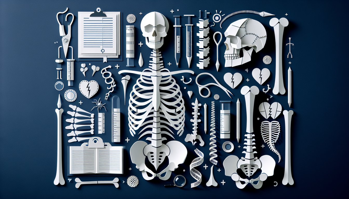
Skeleton Labeling Quiz: Study & Practice Cheat Sheet

  1. Major Bones of the Human Skeleton - Familiarize yourself with key bones like the skull, spine, ribs, and limbs so you can accurately label any skeleton. Think of your bones as the support crew for your body - once you know who's who, the rest falls into place effortlessly. Sciencing: Recognize Bones in Anatomy
  2. Cranial Bones Mnemonic - Use the quirky phrase "Old People From Texas Eat Spiders" to lock in the cranial bones: Occipital, Parietal, Frontal, Temporal, Ethmoid, and Sphenoid. Turning dry lists into fun sentences makes recall a breeze, especially when exam time rolls around. Synonym Classroom: Tricks for Remembering Bones
  3. Vertebral Column by Meal Times - Link the spine sections to your daily eats: 7 cervical (breakfast at 7 a.m.), 12 thoracic (lunch at noon), and 5 lumbar (dinner at 5 p.m.). This tasty timetable helps you order the vertebrae faster than you finish your cereal. Synonym Classroom: Spine Mnemonics
  4. Carpal Bones of the Wrist - Remember "Some Lovers Try Positions That They Can't Handle" for Scaphoid, Lunate, Triquetrum, Pisiform, Trapezium, Trapezoid, Capitate, and Hamate. Picture a wild dance crew to cement each name in your memory. Kenhub: Anatomy Mnemonics
  5. Tibia vs. Fibula - Distinguish the thicker inner bone (TIBia) from the fine, fluted lateral one (FIBuLa) by thinking "TIBia is Thick, FIBuLa is Fine." It's a quick mental check whenever you stare down a leg diagram. Sciencing: Bone Recognition Tips
  6. Rotator Cuff Muscles - Use "SITS" to recall Supraspinatus, Infraspinatus, Teres minor, and Subscapularis. Imagine a volleyball team (SITS) working together to keep your shoulder stable and strong. Kenhub: Muscle Mnemonics
  7. Lower Limb Bones - Lock in Hip, Femur, Patella, Tibia, Fibula, Tarsals, Metatarsals, Phalanges with "Help Five Police To Find Ten Missing Prisoners." Envision each officer on patrol to make the list stick. EUDesign: Bone Mnemonics
  8. Tarsal Bones of the Foot - Remember "The Circus Needs More Interesting Little Clowns" for Talus, Calcaneus, Navicular, Medial, Intermediate, Lateral cuneiforms, and Cuboid. A three-ring circus in your mind is hard to forget! FAC Medicine Forum: Clinical Anatomy Mnemonics
  9. Facial Bones Mnemonic - Use "Virgil Can Not Make My Pet Zebra Laugh" to nail down Vomer, Conchae, Nasal, Maxilla, Mandible, Palatine, Zygomatic, and Lacrimal. Picture a zebra in onlookers' shoes to solidify the sequence. Picmonic: Anatomy & Physiology Bones
  10. Regular Labeling Practice - Make flashcards, print blank skeletons, or use apps to drill names and locations. Consistent, short study bursts are your secret weapon for lightning-fast recall on test day. Kenhub: Study Strategies
Powered by: Quiz Maker