Unlock hundreds more features
Save your Quiz to the Dashboard
View and Export Results
Use AI to Create Quizzes and Analyse Results

Sign inSign in with Facebook
Sign inSign in with Google

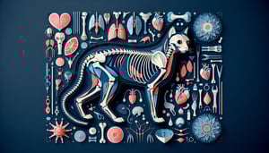
Anatomy & Physiology 1 Practice Quiz

Ace your A&P lab practical with confidence

Difficulty: Moderate
Grade: Other
Study OutcomesCheat Sheet
Colorful paper art promoting a trivia quiz on AP Lab Essentials for students.

What is the primary function of the stage on a light microscope?
To hold the specimen slide securely
To magnify the specimen
To provide illumination
To adjust focus
The stage serves to hold the slide in place so that the specimen can be observed under the microscope. It does not provide magnification, illumination, or focus adjustments, which are managed by other components.
What does the term 'binocular' indicate in relation to a microscope?
It has two eyepieces allowing stereo vision.
It contains two objective lenses.
It uses two light sources for better illumination.
It supports double the magnification capability.
Binocular microscopes are designed with two eyepieces, which provide a more comfortable viewing experience and enhance depth perception. This feature does not relate to having multiple objective lenses or light sources.
Why are safety goggles an essential part of lab safety?
They protect your eyes from chemical splashes and debris.
They help you see specimens more clearly.
They are used to prevent head injuries.
They serve as a fashion accessory in the lab.
Safety goggles are critical because they protect the eyes from harmful chemicals, splashes, or debris during experiments. They are not intended to improve vision clarity, prevent head injuries, or act as decorative items.
Which instrument is specifically designed to measure very small volumes of liquid in a lab?
Micropipette
Graduated cylinder
Beaker
Volumetric flask
A micropipette is engineered for dispensing very precise, small volumes of liquid, often in the microliter range. Alternative instruments like graduated cylinders and beakers are not designed for this level of precision.
What is considered a best practice when labeling specimens in a lab?
Using waterproof marker labels to ensure durability.
Writing labels on loose paper and taping them to specimens.
Marking specimens with a pencil directly on the tissue.
Not labeling to avoid interference with experiments.
Using waterproof markers helps ensure that labels remain legible and intact under various lab conditions. Proper labeling is crucial for accurate identification, and alternatives like loose paper or pencil marks can lead to errors.
What is the role of the coarse focus knob on a microscope?
To bring the specimen into rough focus.
To adjust the brightness of the light source.
To provide fine-tuned sharpness.
To change the magnification level.
The coarse focus knob is used to quickly move the stage and bring the specimen into rough focus. Fine adjustments for sharpness are then made using the fine focus knob, and brightness or magnification is controlled by other components.
Which component of the light microscope provides the primary magnification?
The objective lenses.
The eyepiece lenses.
The stage.
The diaphragm.
Objective lenses are the main source of magnification, capturing the detailed image of the specimen. While the eyepiece further magnifies the image, the primary magnification occurs through the objectives.
What is the function of immersion oil in microscopy?
It improves image resolution by reducing light refraction.
It cleans the microscope lenses.
It stains the specimen for better contrast.
It prevents the objective lens from overheating.
Immersion oil minimizes light refraction between the slide and the objective lens, thereby enhancing image resolution. It is not used for cleaning, staining, or cooling purposes in microscopy.
What does the term 'resolution' in microscopy refer to?
The ability to distinguish two closely spaced points as separate.
The total magnification power of the microscope.
The brightness of the observed image.
The depth of field of the microscope.
Resolution is the measure of a microscope's ability to differentiate between two points that are close together. It is distinct from magnification, brightness, or depth of field, focusing specifically on image clarity.
What is the primary purpose of using a hemocytometer in a lab?
To count cells in a given volume.
To measure the pH of a cell culture.
To weigh small samples precisely.
To separate cells from debris.
A hemocytometer is a specialized instrument used for accurately counting cells in a liquid suspension. Its grid system allows scientists to calculate cell concentrations based on the number of cells per defined volume.
What is the recommended procedure for handling broken glassware in the lab?
Wear gloves and use a brush to sweep up fragments into a designated container.
Use your hands to pick up the pieces quickly.
Leave the broken glassware on the bench without cleaning.
Dispose of it in regular trash immediately.
When handling broken glassware, safety is paramount. Wearing gloves and using appropriate tools like a brush ensures that sharp fragments are safely collected and disposed of in a designated container.
Why is tissue fixation a critical step in sample preparation for histology?
It preserves tissue morphology by preventing decay.
It enhances the color of the tissue naturally.
It enlarges the tissue for better viewing.
It dehydrates the tissue for long-term storage.
Tissue fixation stops enzymatic processes and decay, preserving the cellular structure for further analysis. This step ensures that the tissue's morphology remains intact during subsequent processing and staining.
Which safety measure is most important when working with hazardous chemical reagents in the lab?
Using a fume hood to prevent inhalation of toxic vapors.
Wearing open-toed shoes for better balance.
Dispensing reagents without a safety protocol to save time.
Working without goggles to maximize peripheral vision.
A fume hood is critical as it prevents exposure to harmful chemical vapors by venting them away from the user. Other options compromise safety and do not address the hazard presented by toxic reagents.
What is the primary purpose of a microtome in a histology laboratory?
To slice tissue specimens into uniformly thin sections.
To stain tissue sections for microscopic analysis.
To fix tissues to prevent decay.
To mount slides onto coverslips.
A microtome is used to cut very thin slices of tissue, allowing for detailed microscopic examination. Its function is distinct from staining, fixation, or mounting procedures.
What does the term 'cross-contamination' mean in a laboratory setting?
The accidental introduction of substances from one sample into another.
The deliberate mixing of different reagents for complex analyses.
The routine cleaning of equipment between experiments.
The use of multiple staining techniques on one specimen.
Cross-contamination involves the unintentional transfer of materials between samples, which can distort results and compromise experiments. It is an error to be rigorously avoided, unlike controlled procedures or proper cleaning.
Why are formalin-fixed specimens commonly used in dissection labs?
They preserve anatomical structures by preventing enzymatic degradation.
They enhance the coloration of tissues automatically.
They make the tissues more flexible for dissection.
They sterilize the specimen for consumption.
Formalin fixation stabilizes tissue by denaturing enzymes and halting decay, thereby preserving anatomical details. This method is used primarily for preservation rather than enhancing color, flexibility, or sterilization for consumption.
Why is regular calibration of lab instruments, such as pipettes, crucial in experimental procedures?
To ensure that measurements are accurate and experiments remain reproducible.
To improve the aesthetic appearance of the equipment.
Because calibration is required for equipment to start operating.
To reduce the frequency of cleaning the instruments.
Calibration verifies that instruments like pipettes dispense accurate volumes, which is vital for experimental precision and reproducibility. This process does not pertain to the appearance, startup, or cleaning frequency of the equipment.
If a microscopic image appears distorted at high magnification, what is a likely contributing factor?
Improper specimen preparation, such as uneven coverslip placement or fixation errors.
Excessive use of immersion oil beyond recommended amounts.
A malfunctioning coarse adjustment knob without any sample-related issues.
Ambient lighting variations in the room.
Distortion at high magnification is often linked to issues in specimen preparation, including uneven mounting or inadequate fixation. Addressing these preparation errors usually improves image clarity more effectively than adjustments to other factors.
What is the fundamental principle behind fluorescence microscopy in anatomical studies?
Specimens are labeled with fluorescent dyes that emit light when excited by specific wavelengths.
Specimens are heated so that they naturally emit light.
Chemical reactions in the specimen produce inherent luminescence without external dyes.
Magnification is solely achieved through the use of oil-immersion lenses.
Fluorescence microscopy relies on the use of fluorescent dyes which, when excited by specific wavelengths of light, emit light that can be detected for imaging. This technique is distinct from natural luminescence or conventional magnification methods.
Why is it important to segregate lab waste into biohazard, chemical, and sharps containers?
Because each type of waste requires specialized disposal methods to prevent contamination and injury.
Because it speeds up the disposal process regardless of waste type.
Because it makes all waste suitable for recycling.
Because it reduces the overall amount of waste produced.
Segregating lab waste is essential to ensure that each category is handled with the appropriate safety and disposal protocols. Different wastes pose unique hazards and require specific treatments to prevent contamination and injury.
0
{"name":"What is the primary function of the stage on a light microscope?", "url":"https://www.quiz-maker.com/QPREVIEW","txt":"What is the primary function of the stage on a light microscope?, What does the term 'binocular' indicate in relation to a microscope?, Why are safety goggles an essential part of lab safety?","img":"https://www.quiz-maker.com/3012/images/ogquiz.png"}

Study Outcomes

  1. Analyze anatomical specimens using key identification methods.
  2. Apply physiological concepts to interpret lab data effectively.
  3. Demonstrate proper lab safety and specimen handling protocols.
  4. Evaluate experimental procedures and outcomes in anatomy and physiology contexts.
  5. Integrate anatomical and physiological insights to solve practical lab problems.

A&P 1 Lab Practical 1 Cheat Sheet

  1. Master the Anatomical Position - Imagine yourself standing tall with feet shoulder-width apart, arms relaxed at your sides, and palms facing forward. This is the gold standard starting point for all anatomical descriptions - no more "left" or "right" confusion! Get this down, and you'll always speak the body's language like a champ. Quizlet Flashcards
  2. Familiarize with Directional Terms - Learn words like anterior (front), posterior (back), superior (above), and inferior (below) to nail pinpoint accuracy. These directional buddies help you describe where every organ, bone, or muscle calls home. Practice using them in sentences to make your study sessions pop! Quizlet Flashcards
  3. Understand Body Planes - Slice your mental body into sagittal (left/right), coronal (front/back), and transverse (top/bottom) planes. These imaginary cuts are your roadmap when looking at scans or dissections. Mastering them turns complex diagrams into clear snapshots! Quizlet Flashcards
  4. Learn the Four Primary Tissue Types - Break it down into epithelial (covering), connective (support), muscle (movement), and nervous (control). Spotting each under the microscope is like identifying characters in a superhero squad. Once you see their unique features, you'll breeze through labs! Quizlet Flashcards
  5. Identify Major Organ Systems - From the integumentary system's protective shield to the digestive system's food factory, each team has a vital role. Knowing their names and functions gives you a bird's-eye view of the human body's big picture. It's like assembling puzzle pieces for ultimate mastery! Cram Flashcards
  6. Recognize Common Epithelial Tissues - Spot simple squamous in lung air sacs, stratified squamous on your skin, simple cuboidal in kidney tubules, and simple columnar lining the digestive tract. Each type's shape and layers tell you exactly where it belongs. Picture them like different building blocks of a high-tech structure! Quizlet Flashcards
  7. Understand Connective Tissue Types - From the cushioning areolar to the fat-storing adipose, strong tendons' dense regular fibers, and the flexible cartilage trio - hyaline, elastic, and fibrocartilage - these tissues hold everything together. They're the body's scaffolding and shock absorbers all in one. Get familiar, and you'll ace those histology slides! Quizlet Flashcards
  8. Differentiate Muscle Tissues - Meet skeletal muscle for voluntary moves, cardiac muscle for your heartbeat, and smooth muscle lining hollow organs. Each type has its own structural quirks and functions - like heroes with specialized superpowers. Recognizing them will turn you into a muscle maven! Quizlet Flashcards
  9. Learn the Integumentary System Components - Dive into the epidermis, dermis, hypodermis, hair, nails, and glands that keep you protected and temperature-regulated. This system is your body's first line of defense and the ultimate thermostat. Knowing its parts is like unlocking your personal security system! Quizlet Flashcards
  10. Practice Hands-On Identification - Use models, slides, or virtual labs to point out bones, muscles, and tissues in real time. Touching, feeling, and seeing these structures cements your knowledge far better than just reading. Get your hands dirty - your practical exam will thank you! CliffsNotes Review
Powered by: Quiz Maker