Unlock hundreds more features
Save your Quiz to the Dashboard
View and Export Results
Use AI to Create Quizzes and Analyse Results

Sign inSign in with Facebook
Sign inSign in with Google

Test Your Skills: X-Ray Fracture Identification Quiz

Sharpen Your Radiology Fracture Diagnosis Skills

Difficulty: Moderate
Questions: 20
Learning OutcomesStudy Material
Colorful paper art depicting elements related to X-Ray Fracture Identification Quiz.

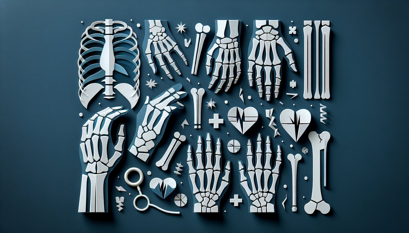
Ready to test your radiology prowess with a comprehensive X-ray fracture quiz? The X-Ray Fracture Identification Quiz challenges you with realistic imaging and questions designed to sharpen fracture diagnosis skills. Ideal for radiology students and educators seeking assessment, this fracture identification guide helps you master common bone injuries. Feel free to explore other identification exercises like our Animal Identification Quiz and Plant Identification Quiz . Plus, customize any free test in our quizzes editor.

Which fracture type is characterized by a break line that runs perpendicular to the bone's long axis?
Transverse fracture
Oblique fracture
Spiral fracture
Comminuted fracture
A transverse fracture has a break line that is perpendicular to the long axis of the bone. Oblique and spiral fractures run at an angle or corkscrew pattern, while comminuted fractures break into multiple fragments.
A fracture pattern where the bone is broken into three or more fragments is called:
Comminuted fracture
Greenstick fracture
Impacted fracture
Avulsion fracture
A comminuted fracture involves the bone shattering into three or more pieces. Greenstick fractures are incomplete breaks in children, impacted fractures involve bone ends driven into each other, and avulsion fractures occur when a fragment is pulled off by a tendon or ligament.
A break that occurs at an angle across the long axis of a bone describes which fracture type?
Oblique fracture
Spiral fracture
Transverse fracture
Segmental fracture
An oblique fracture runs at an angle across the bone's long axis. Spiral fractures twist around the bone, transverse fractures are perpendicular, and segmental fractures have two distinct fracture lines creating a segment.
Which type of fracture appears as a thin line and is often difficult to detect on X-ray images, commonly seen in overuse injuries?
Hairline (stress) fracture
Comminuted fracture
Greenstick fracture
Impacted fracture
Hairline or stress fractures are small cracks in bone often caused by repetitive stress and may not be immediately apparent on X-ray. Comminuted and greenstick fractures involve more obvious breaks.
In a systematic approach to reading X-rays, what is the first step to ensure accurate interpretation?
Confirm patient identity and correct side
Assess bone alignment
Evaluate soft tissues
Compare with prior images
The first step in any systematic X-ray review is to confirm patient identity and the correct side marker. This prevents misdiagnosis due to wrong-patient or wrong-side errors.
Which fracture type is most commonly seen in pediatric patients where one side of the bone bends and partially breaks?
Greenstick fracture
Comminuted fracture
Spiral fracture
Transverse fracture
Greenstick fractures occur in children because their bones are more flexible, causing one cortex to break while the other side bends. This pattern is not seen in adults.
A torus or buckle fracture is characterized by:
Buckling of the cortex without a complete break
Bone segmented into multiple pieces
A fracture line at a 45-degree angle
A complete circumferential break
A torus or buckle fracture results from compression that causes the cortex on one side of the bone to buckle without fully breaking. It is common in pediatric patients.
A segmental fracture is defined by:
Two separate fracture lines creating a distinct middle bone segment
A spiral break that encircles the bone
A single transverse break
Multiple small fragments around the site
In a segmental fracture, two parallel fracture lines isolate a segment of bone between them. Spiral fractures encircle bone, transverse fractures are single, and comminuted fractures have many fragments.
To minimize distortion on an X-ray image, how should the X-ray beam be oriented relative to the bone?
Perpendicular to the bone
Parallel to the bone
Tangential to the bone
At a 45-degree angle to the bone
Orienting the X-ray beam perpendicular to the long axis of the bone minimizes distortion and ensures accurate representation of the bone's true size and shape.
In fracture description, what does "angulation" refer to?
The angle formed between bone fragments at the fracture site
Soft tissue swelling around a break
Overlapping of bone ends
Length of the fracture line
Angulation describes the degree to which the distal fragment deviates in angle relative to the proximal fragment, measured in degrees at the fracture site.
What is a "butterfly fragment" in the context of long bone fractures?
A triangular wedge of bone at the fracture site between the main fragments
A small separate chip of bone
Complete shattering into multiple small pieces
A stress-related hairline crack
A butterfly fragment is the triangular piece of bone that is wedged between two larger fracture fragments, often seen in bending injuries.
According to the Salter-Harris classification, which type involves a fracture through the growth plate and metaphysis but not the epiphysis?
Type II
Type I
Type III
Type IV
Salter-Harris Type II fractures traverse the growth plate and extend into the metaphysis, but spare the epiphysis. Type I is only through the physis, III involves the epiphysis, and IV crosses both.
What term describes angulation of the distal fracture fragment toward the midline of the body?
Varus
Valgus
Dorsal
Volar
Varus describes inward angulation toward the midline. Valgus indicates outward angulation, while dorsal and volar refer to posterior and anterior orientations, respectively.
Which radiographic finding is most suggestive of an open (compound) fracture?
Gas or air in the surrounding soft tissues
Complete cortical disruption
A simple transverse break
Periosteal reaction
The presence of gas or air in soft tissues on X-ray strongly suggests communication with the external environment, indicating an open fracture.
In the ABCs of systematic X-ray evaluation, what does the "B" stand for?
Bones
Background
Borders
Brightness
In the ABC approach to reading X-rays, 'A' stands for Adequacy/Alignment, 'B' stands for Bones, 'C' for Cartilage, and 'S' for Soft tissues.
A patient presents with anatomical snuffbox tenderness after a fall, but the initial X-ray is normal. What is the most appropriate next step?
Immobilize the wrist and repeat X-ray in 10 - 14 days
Discharge without treatment
Obtain an immediate MRI
Proceed to CT scan at once
A normal initial X-ray cannot rule out a scaphoid fracture when clinical suspicion is high. Immobilization with repeat imaging in 10 - 14 days allows for fracture line visibility as early healing changes appear.
Which Salter-Harris fracture type extends through the growth plate and epiphysis?
Type III
Type II
Type I
Type IV
Salter-Harris Type III fractures cross the physis and extend into the epiphysis, potentially affecting the joint surface. Type II involves metaphysis, and Type IV crosses both physis and epiphysis.
When the bone is not in close contact with the image receptor during an X-ray, which artifact is most likely to occur?
Magnification
Underexposure
Motion blur
Foreshortening
When a bone is farther from the image receptor, the X-ray beam diverges, causing the image to appear larger or magnified. Close contact minimizes this effect.
A spiral fracture pattern is most commonly caused by what mechanism?
Twisting or rotational force applied to the bone
Direct blow perpendicular to the bone
Compression along the bone's axis
Tensile force pulling the bone apart
Spiral fractures result from rotational or twisting forces that encircle the bone in a corkscrew pattern. Direct blows and axial loads produce different fracture configurations.
At approximately how many days post-fracture does periosteal reaction first become visible on standard X-rays, indicating early healing?
7 - 10 days
1 - 2 days
3 - 5 weeks
Immediately after fracture
Periosteal reaction, a sign of new bone formation, typically becomes radiographically visible around 7 - 10 days after the initial fracture. Earlier imaging may not show this healing response.
0
{"name":"Which fracture type is characterized by a break line that runs perpendicular to the bone's long axis?", "url":"https://www.quiz-maker.com/QPREVIEW","txt":"Which fracture type is characterized by a break line that runs perpendicular to the bone's long axis?, A fracture pattern where the bone is broken into three or more fragments is called:, A break that occurs at an angle across the long axis of a bone describes which fracture type?","img":"https://www.quiz-maker.com/3012/images/ogquiz.png"}

Learning Outcomes

  1. Analyse radiographic images to distinguish different fracture types
  2. Identify common fracture patterns such as transverse and oblique
  3. Evaluate bone alignment and detect subtle hairline fractures
  4. Apply radiographic principles to interpret fracture severity
  5. Demonstrate proficiency in labeling fracture locations accurately
  6. Master the systematic approach to reading X-ray scans

Cheat Sheet

  1. Understand fracture types - Fractures fall into two big families: complete breaks that split the bone into separate pieces, and incomplete cracks that bend or nick the bone without full separation. Kids often get greenstick or torus (buckle) fractures, while adults face transverse, oblique, spiral, or comminuted types. Radiopaedia: Fracture Types Summary
  2. Recognize common fracture patterns - Each pattern tells a story: transverse fractures run straight across, oblique ones slice at an angle, and spiral fractures twist around the bone like a corkscrew. Spotting these shapes helps you imagine the injury mechanism - almost like detective work in the X-ray world! Radiology Masterclass: Trauma X-ray Patterns
  3. Assess bone alignment - After finding a fracture, check displacement (gap size), angulation (tilt between fragments), shortening (overlap), and rotation (twist). Quantifying these features lets you predict healing time and choose the right treatment - no guesswork! LearningRadiology: Recognizing Fractures
  4. Detect subtle fractures - Not all breaks scream "broken bone!" Look for soft tissue swelling, joint effusion or abnormal fat pad positions, which are like little breadcrumbs leading you to a hidden crack. These indirect signs keep you from missing sneaky injuries. PMC: Detecting Occult Fractures
  5. Apply radiographic principles - Always get at least two orthogonal views (think front and side) so you don't overlook a fracture hiding in one plane. Radiographic basics are your X-ray superpowers for a complete picture. Radiology Expert: Fracture Principles
  6. Label fracture locations accurately - Be specific: name the bone (e.g., distal radius) and note if it extends into a joint (intra-articular). Precise labels help your team know exactly where to focus. Radiology Expert: Fracture Classification
  7. Master systematic X-ray reading - Use the ABCS approach: Alignment, Bone density, Cartilage spaces, Soft tissues. A consistent checklist ensures you won't miss tiny clues that could change a diagnosis. LearningRadiology: ABCS Approach
  8. Understand pediatric fractures - Kids' bones are more flexible, producing unique breaks like greenstick or buckle fractures that rarely occur in adults. Treating these gently bent bones requires a special skill set. Radiology Expert: Pediatric Fractures
  9. Recognize fracture mimics - Normal anatomical quirks - like accessory ossicles or nutrient canals - can masquerade as cracks. Knowing the usual "imposters" helps you avoid false alarms. Radiology Expert: Fracture Mimics
  10. Learn common fracture eponyms - Named breaks like Colles' (dorsal tilt of the distal radius) and Smith's (palmar tilt) give quick clues about fracture geometry. Eponyms are your secret code for swift communication. LearningRadiology: Fracture Eponyms
Powered by: Quiz Maker