Unlock hundreds more features
Save your Quiz to the Dashboard
View and Export Results
Use AI to Create Quizzes and Analyse Results

Sign inSign in with Facebook
Sign inSign in with Google

Take the Ophthalmology Skills Assessment Quiz

Assess Core Ophthalmology Techniques and Concepts

Difficulty: Moderate
Questions: 20
Learning OutcomesStudy Material
Colorful paper art promoting Ophthalmology Skills Assessment Quiz.

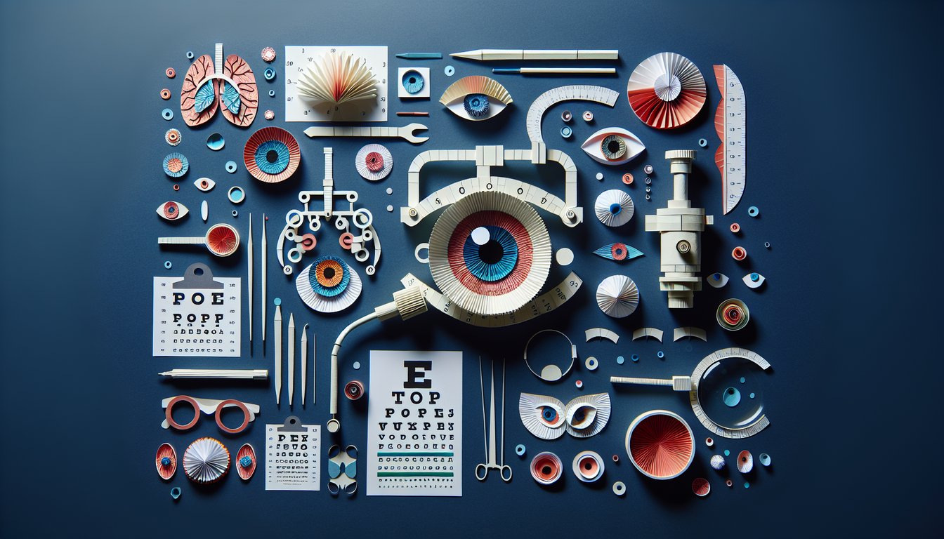
Ready to elevate your eye care expertise? This ophthalmology skills assessment quiz features 15 multiple-choice questions crafted for medical students and practitioners to sharpen diagnostic and treatment skills. Compare results with the Ophthalmology Knowledge Assessment Quiz or explore advanced cases in the Ophthalmology Clinical Knowledge Quiz. Personalize any question in our editor to suit your learning objectives, and adapt the quiz freely. Discover more quizzes spanning diverse medical topics.

What is the primary purpose of the Snellen chart in an eye examination?
Evaluate peripheral vision
Measure distance visual acuity
Measure intraocular pressure
Assess color vision
The Snellen chart is used to measure a patient's distance visual acuity by having them read letters at a fixed distance. It does not assess intraocular pressure, color vision, or peripheral fields.
Which structure is the transparent, anterior-most part of the eye that refracts light?
Lens
Iris
Cornea
Sclera
The cornea is the transparent front portion of the eye responsible for most of the eye's refractive power. The sclera is opaque, the iris controls the pupil, and the lens fine-tunes focus.
Which instrument is commonly used to examine the anterior chamber and lens under magnification?
Ophthalmoscope
Slit lamp biomicroscope
Tonometer
Phoropter
A slit lamp biomicroscope provides high-magnification, stereoscopic views of anterior segment structures. An ophthalmoscope is used for retinal exam, tonometers measure pressure, and phoropters assess refractive error.
What visual acuity is considered normal vision in adults?
20/10
6/12
20/40
20/20
A visual acuity of 20/20 is considered the standard for normal adult vision. Other values indicate below-normal (20/40) or better-than-normal (20/10) acuity.
During eye drop administration, the correct technique to minimize systemic absorption is to ask the patient to:
Press on the nasolacrimal duct
Tilt head forward
Look downward
Blink vigorously
Applying pressure over the nasolacrimal duct (punctal occlusion) reduces systemic absorption of the medication. Blinking, tilting forward, or looking downward do not effectively limit systemic uptake.
A patient presents with a red eye, purulent discharge, and eyelid crusting. What is the most likely diagnosis?
Viral conjunctivitis
Keratitis
Bacterial conjunctivitis
Blepharitis
Bacterial conjunctivitis often presents with purulent discharge and lid crusting. Viral conjunctivitis typically has watery discharge, keratitis involves corneal findings, and blepharitis affects the lids rather than causing copious discharge.
Which of the following tests assesses intraocular pressure?
Confrontation visual field
Slit lamp examination
Applanation tonometry
Fluorescein angiography
Applanation tonometry is the gold standard for measuring intraocular pressure. Slit lamp exams visualize structures, visual fields assess fields, and fluorescein angiography images retinal vasculature.
A relative afferent pupillary defect is best detected by performing which test?
Swinging flashlight test
Cover - uncover test
Confrontation visual field
Color vision test
The swinging flashlight test reveals a relative afferent pupillary defect by comparing direct and consensual responses. Cover - uncover tests assess strabismus, confrontation tests fields, and color tests assess color perception.
The Amsler grid is used primarily to detect dysfunction of which part of the eye?
Lens
Macula
Optic nerve
Cornea
The Amsler grid tests central visual field and reveals metamorphopsia due to macular disorders. It does not assess optic nerve function, corneal shape, or lens clarity.
Which condition is characterized by optic nerve cupping on fundus exam?
Uveitis
Glaucoma
Macular degeneration
Diabetic retinopathy
Glaucoma leads to increased cup-to-disc ratio due to retinal ganglion cell loss. Macular degeneration affects the macula, diabetic retinopathy shows hemorrhages, and uveitis involves inflammation.
Non-contact (air-puff) tonometry measures intraocular pressure by:
Using an optical scanner
Flattening the cornea with a puff of air
Measuring the return time of light
Indenting the cornea with a probe
Non-contact tonometry uses an air pulse to applanate (flatten) the cornea and gauge intraocular pressure. Indentation tonometry uses a probe, optical scanners image surfaces, and light-return time is not used here.
Metamorphopsia on an Amsler grid is most suggestive of:
Keratoconus
Age-related macular degeneration
Glaucoma
Cataract
Metamorphopsia (visual distortion) on an Amsler grid indicates macular pathology such as age-related macular degeneration. Glaucoma affects peripheral fields, cataracts cloud the lens, and keratoconus alters corneal shape.
Which imaging modality provides high-resolution cross-sectional images of the retina?
Ultrasound B-scan
Optical coherence tomography
Fluorescein angiography
Fundus photography
Optical coherence tomography (OCT) produces detailed cross-sectional images of retinal layers. Fundus photography captures surface images, ultrasound B-scan images deeper structures, and fluorescein angiography visualizes blood flow.
A 55-year-old patient has 20/20 distance acuity but complains of trouble reading. What is the most likely diagnosis?
Presbyopia
Myopia
Astigmatism
Hyperopia
Presbyopia is age-related loss of near focusing ability while distance acuity remains normal. Hyperopia and myopia affect distance vision, and astigmatism causes distorted vision at all distances.
When counseling a patient about pharmacologic pupil dilation, you should inform them that:
Color vision will be improved
They will be unable to open their eyes for hours
Near vision will be blurred and light sensitivity will increase
Blood pressure will significantly increase
Pharmacologic dilation impairs accommodation, blurring near vision, and causes photophobia. It does not affect blood pressure, eyelid opening, or color perception.
A patient has sudden, painless vision loss in one eye. Fundus exam shows retinal pallor with a cherry-red spot on the macula. What is the most likely diagnosis?
Central retinal vein occlusion
Optic neuritis
Macular degeneration
Central retinal artery occlusion
Central retinal artery occlusion presents with sudden painless vision loss and a cherry-red spot due to inner retinal pallor. Vein occlusion shows hemorrhages, macular degeneration is gradual, and optic neuritis is often painful.
Which lesion location most commonly produces a bitemporal hemianopia?
Lateral geniculate nucleus
Optic nerve
Optic tract
Optic chiasm
A lesion at the optic chiasm interrupts crossing nasal fibers from both eyes, causing bitemporal field loss. Lesions of the tract, nerve, or LGN produce different patterns of hemianopia.
A contact lens wearer presents with corneal ulcer and stromal infiltrate. What is the best first diagnostic step?
Order a CT scan of the orbit
Prescribe broad-spectrum antibiotics without testing
Start topical steroids
Perform corneal scraping for culture
Corneal scraping for culture identifies the causative organism and guides targeted therapy. Steroids can worsen infection, empirical treatment without culture may miss pathogens, and CT is unnecessary initially.
Which cranial nerve innervates the lateral rectus muscle?
Trochlear nerve (CN IV)
Abducens nerve (CN VI)
Trigeminal nerve (CN V)
Oculomotor nerve (CN III)
The abducens nerve (CN VI) innervates the lateral rectus muscle, responsible for eye abduction. The oculomotor nerve supplies most other extraocular muscles, the trochlear supplies superior oblique, and the trigeminal is sensory.
In retinal detachment repair, what does laser retinopexy accomplish?
Freezes the peripheral retina
Introduces a scleral buckle externally
Creates chorioretinal adhesion around the tear
Removes the vitreous gel
Laser retinopexy uses thermal burns to create a scar that seals the retina to the underlying tissue around a tear. Cryotherapy freezes tissue, scleral buckle indents the globe, and vitrectomy removes vitreous.
0
{"name":"What is the primary purpose of the Snellen chart in an eye examination?", "url":"https://www.quiz-maker.com/QPREVIEW","txt":"What is the primary purpose of the Snellen chart in an eye examination?, Which structure is the transparent, anterior-most part of the eye that refracts light?, Which instrument is commonly used to examine the anterior chamber and lens under magnification?","img":"https://www.quiz-maker.com/3012/images/ogquiz.png"}

Learning Outcomes

  1. Analyze patient eye examination data to diagnose common conditions.
  2. Identify key ocular anatomy structures and their functions.
  3. Demonstrate proper use of ophthalmic instruments and techniques.
  4. Apply clinical reasoning to select appropriate treatment options.
  5. Evaluate visual acuity results and interpret diagnostic tests.
  6. Master effective patient communication in ophthalmology care.

Cheat Sheet

  1. Understand the anatomy of the eye - Embark on a guided tour through the cornea, lens, retina, and optic nerve to see how these parts team up for perfect vision. This foundation is your backstage pass to diagnosing and treating all snazzy ocular conditions like a true eye expert! Anatomy and Clinical Exam - American Academy of Ophthalmology
  2. Master comprehensive eye exam techniques - From sharp visual acuity charts to pinpointing pupil reactions and dazzling slit-lamp views, you'll learn each move step by step. Nailing these skills ensures your patient assessments are spot-on every time. Ophthalmic Exam: Frequency, Procedure & More
  3. Familiarize yourself with common ocular pathologies - Get to know cataracts, glaucoma, and diabetic retinopathy, so you can spot their sneaky signs and symptoms quickly. Early recognition means faster treatment - which is great news for vision preservation! Eye Pathology - Optician Study Guide
  4. Practice interpreting visual acuity results - Decode Snellen and LogMAR charts like a boss to measure the crispness of someone's sight. Being fluent in these numbers helps you track vision changes and fine-tune patient care. Eye examination
  5. Gain proficiency with ophthalmic instruments - Shine your skills using the ophthalmoscope to peek at the retina and the slit lamp to inspect the front of the eye in dazzling detail. These trusty tools are your detective gadgets for uncovering hidden ocular clues. Anatomy and Clinical Exam - American Academy of Ophthalmology
  6. Understand OCT principles and applications - Dive into Optical Coherence Tomography's high-tech cross-sectional snapshots of the retina and optic nerve. This 3D imaging marvel is key for diagnosing diseases that hide beneath the surface. Optical coherence tomography -- principles, implementation, and applications in ophthalmology
  7. Perform a dilated fundus examination - Dilate pupils and peer deep into the back of the eye to inspect the retina and optic nerve head. It's a vital checkup for spotting diabetic retinopathy, glaucoma, and other vision villains early. Dilated fundus examination
  8. Develop effective communication skills - Hone your ability to explain diagnoses and treatment plans with clarity and empathy. Strong patient conversations build trust and ensure everyone's on the same page for better eye health. Board Prep Resources - American Academy of Ophthalmology
  9. Master the red reflex test - Shine a light into the eye to produce that familiar red-orange glow, helping you detect cataracts, retinal detachments, and more. A quick flick of the ophthalmoscope can be a life-saver for early pathology screening! Red reflex
  10. Review refraction techniques - Learn how lenses bend light to pinpoint the perfect prescription for glasses or contacts. Mastering refraction ensures your patients see their world in crystal clarity. Eye examination
Powered by: Quiz Maker