Unlock hundreds more features
Save your Quiz to the Dashboard
View and Export Results
Use AI to Create Quizzes and Analyse Results

Sign inSign in with Facebook
Sign inSign in with Google

Test Your Knowledge: MIR Neurosurgery Practice Quiz

Sharpen Neurosurgical Knowledge with Practice Questions

Difficulty: Moderate
Questions: 20
Learning OutcomesStudy Material
Colorful paper art promoting a fun MIR Neurosurgery Practice Quiz.

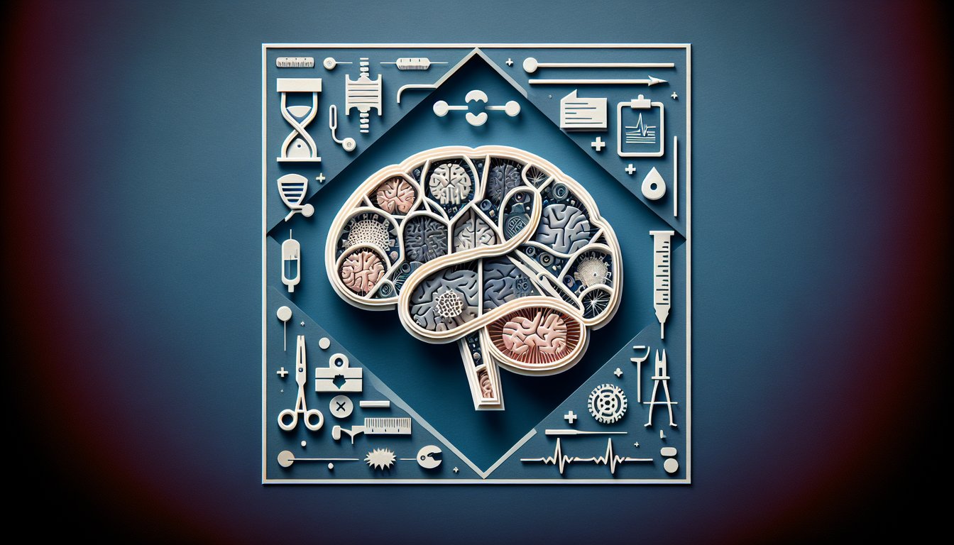
Ready to elevate neurosurgical knowledge and tackle challenging MIR exam questions? This MIR Neurosurgery Practice Quiz offers 15 targeted multiple-choice questions designed for medical students, residents, and aspiring neurosurgeons. Participants will sharpen diagnostic reasoning and surgical planning skills while reinforcing essential neuroanatomy principles. Each question is fully editable in our quiz editor, allowing customization of difficulty and focus areas. For foundational review, try the Medical Anatomy Practice Quiz , enhance overall test strategies with the Test Preparation Practice Quiz, or explore more quizzes today!

Which cranial nerve is responsible for motor innervation to the muscles of facial expression?
Trigeminal nerve (CN V)
Glossopharyngeal nerve (CN IX)
Oculomotor nerve (CN III)
Facial nerve (CN VII)
The facial nerve (CN VII) provides motor fibers to all muscles of facial expression. Other cranial nerves listed have different primary functions such as sensation or eye movement.
Which instrument is specifically designed to create burr holes in the skull during a craniotomy?
Rongeur
Cavitron Ultrasonic Surgical Aspirator
Craniotome
Kerrison punch
A craniotome is a specialized drill used to cut the skull for burr holes. Rongeurs and Kerrison punches remove bone but do not create initial bony openings.
What is the first-line imaging study for suspected acute subarachnoid hemorrhage?
CT angiography
MRI of the brain
Non-contrast head CT
Lumbar puncture
Non-contrast CT is highly sensitive for detecting acute subarachnoid blood within the first 24 hours. MRI is less sensitive acutely, and angiography or lumbar puncture are secondary steps.
The pterional (frontotemporal) approach is most commonly used to access lesions in which area?
Sellar region
Posterior fossa
Parietal lobe
Anterior circulation aneurysms
The pterional approach provides lateral access to the circle of Willis and anterior circulation aneurysms. Sellar and posterior fossa lesions require different approaches.
The ASA physical status classification system is used to assess what aspect of a surgical patient?
Postoperative care plan
Neurological function
Anesthesia risk
Tumor histologic grade
The ASA classification grades a patient's preoperative systemic health to predict anesthesia risk. It does not rate tumor characteristics or specific postoperative care.
Which preoperative imaging technique is best for mapping white matter tracts prior to neurosurgical resection?
Magnetic resonance angiography (MRA)
Positron emission tomography (PET)
functional MRI (fMRI)
Diffusion tensor imaging (DTI)
DTI visualizes diffusion of water along axonal pathways to map white matter tracts. fMRI maps cortical activation, while PET and MRA serve different purposes.
What is the primary use of a Cavitron Ultrasonic Surgical Aspirator (CUSA) in neurosurgery?
Hemostasis
Vessel clipping
Ultrasonic tissue aspiration
Bone drilling
CUSA uses ultrasonic vibrations to fragment and aspirate soft tissue selectively. It does not drill bone or apply clips, and hemostasis is achieved by other means.
In a patient with confirmed subarachnoid hemorrhage on CT scan, what is the next best step to identify the bleeding source?
Carotid Doppler ultrasound
Repeat CT after 24 hours
Lumbar puncture
Digital subtraction angiography (DSA)
DSA provides high-resolution vascular imaging to localize aneurysms or vascular malformations. Lumbar puncture is unnecessary after a positive CT, and ultrasound is not diagnostic for intracranial vessels.
Which surgical approach is most appropriate for resecting a pituitary macroadenoma extending into the sphenoid sinus?
Retrosigmoid approach
Suboccipital craniotomy
Pterional craniotomy
Endoscopic transsphenoidal
The endoscopic transsphenoidal approach provides direct access through the sphenoid sinus to the pituitary fossa, minimizing brain retraction. Craniotomies are more invasive for this lesion.
What is the main advantage of using neuronavigation during intracranial surgery?
Automatic hemostasis
Electrical stimulation mapping
Real-time anatomical guidance
Blood flow measurement
Neuronavigation integrates preoperative imaging to guide the surgeon's instruments in real time, improving accuracy. It does not provide automatic hemostasis or flow data.
What is the initial management strategy for a postoperative cerebrospinal fluid leak following skull base surgery?
High-dose steroids
Start broad-spectrum antibiotics
Lumbar drainage and bed rest
Immediate reoperation
Conservative management with bed rest and lumbar CSF drainage is first-line for low-output leaks. Reoperation and antibiotics may be considered if conservative measures fail.
Which imaging modality is considered the gold standard for preoperative vascular mapping in cerebral aneurysm cases?
Non-contrast MRI
Computed tomography (CT)
CT angiography (CTA)
Digital subtraction angiography (DSA)
DSA offers the highest spatial and temporal resolution for cerebral vasculature, making it the gold standard for aneurysm planning. CTA is less detailed.
During carotid endarterectomy, monitoring somatosensory evoked potentials (SSEPs) primarily helps detect what?
Peripheral nerve injury
Spinal cord function
Cardiac arrhythmias
Cortical ischemia
SSEPs measure cortical responses to peripheral stimulation, providing real-time feedback on cerebral perfusion during cross-clamping. They do not monitor cardiac or spinal function.
In planning a frontal craniotomy, which external landmark is most commonly used to locate the burr hole?
Mastoid tip
Superior nuchal line
Inion
Coronal suture
The coronal suture demarcates the anterior limit of the parietal bone and is used to align frontal burr holes. Other landmarks relate to posterior or lateral skull anatomy.
What intraoperative technique helps maximize resection of a glioma while preserving motor function?
Cortical mapping with awake craniotomy
Frozen section pathology
Intraoperative angiography
Intraoperative ultrasound
Awake craniotomy with cortical mapping identifies and preserves motor cortex and pathways in real time, optimizing tumor resection and function. Other tools do not provide functional mapping.
Which structure is the origin of vestibular schwannomas in the cerebellopontine angle?
Dura mater cells
Arachnoid cap cells
Schwann cells of the cochlear nerve
Schwann cells of the vestibular nerve
Vestibular schwannomas arise from Schwann cells of the vestibular portion of cranial nerve VIII in the cerebellopontine angle. They seldom originate from cochlear Schwann cells.
For a butterfly glioma crossing the corpus callosum, which surgical approach provides optimal midline access?
Bifrontal interhemispheric approach
Pterional craniotomy
Subtemporal approach
Retrosigmoid approach
The bifrontal interhemispheric approach provides direct midline exposure to tumors involving the corpus callosum bilaterally. Other approaches are more lateral or posterior.
During arteriovenous malformation surgery, which intraoperative tool assists in identifying arterial feeders and confirming resection?
Electrophysiological monitoring
Intraoperative ultrasonography
Neuronavigation
Indocyanine green angiography
Indocyanine green angiography visualizes blood flow in vessels intraoperatively, allowing surgeons to identify feeders and verify AVM removal. Other tools do not directly image flow dynamics.
What is the most likely complication following a suboccipital craniotomy for cerebellar tumor resection?
Diabetes insipidus
Aphasia
Homonymous hemianopia
Hydrocephalus
Suboccipital craniotomy can disrupt CSF pathways behind the foramen magnum, leading to postoperative hydrocephalus. Visual field defects and endocrine complications are less common in this region.
Preoperative embolization of meningioma feeders most commonly targets which artery to reduce intraoperative bleeding?
Ophthalmic artery
Middle meningeal artery
Posterior inferior cerebellar artery
Anterior cerebral artery
The middle meningeal artery supplies the dural attachment of many convexity meningiomas and is routinely embolized to decrease blood loss. Other arteries serve different brain regions.
0
{"name":"Which cranial nerve is responsible for motor innervation to the muscles of facial expression?", "url":"https://www.quiz-maker.com/QPREVIEW","txt":"Which cranial nerve is responsible for motor innervation to the muscles of facial expression?, Which instrument is specifically designed to create burr holes in the skull during a craniotomy?, What is the first-line imaging study for suspected acute subarachnoid hemorrhage?","img":"https://www.quiz-maker.com/3012/images/ogquiz.png"}

Learning Outcomes

  1. Analyze cranial neuroanatomy and its surgical relevance
  2. Identify critical neurosurgical instruments and applications
  3. Apply diagnostic reasoning to common neurosurgical case scenarios
  4. Evaluate surgical approaches for intracranial pathologies
  5. Master preoperative planning and operative workflow in neurosurgery
  6. Demonstrate postoperative care strategies and complication management

Cheat Sheet

  1. Circle of Willis Anatomy - Dive into the life-saving arterial ring at the brain's base and discover how its circular shape provides backup blood flow when one pathway is blocked. Mastering its arteries - like the anterior and posterior cerebral branches - gives you a head start in assessing stroke risks and planning interventions. Operative Review: Cerebral Circulation
  2. Essential Neurosurgical Instruments - Get hands-on with cranial perforators, rongeurs, and microsurgical tools and learn which instrument is your hero in each step of surgery. From drilling precise burr holes to delicately teasing tissue apart, knowing their functions boosts both your confidence and skill. Profile Surgical: Instrument Guide
  3. 12 Cranial Nerves Mnemonics - Turn a list of twelve nerves into a memorable chant and never blank on nerve order again. Fun phrases lock in their names and functions, making clinical exams feel like a game rather than a quiz. Operative Review: Cranial Nerves Mnemonics
  4. Image-Guided Surgery Basics - Think of this as GPS for your scalp incision: it fuses real-time imaging with navigation tools to steer you through delicate terrain. The result? Pinpoint accuracy, less trial-and-error, and reduced damage to healthy tissue. Wikipedia: Image-Guided Surgery
  5. Endoscopic Endonasal Approach Overview - Slide an endoscope through the nasal passages to reach the skull base without large scalp flaps - talk about a nose-to-brain shortcut! This minimally invasive doorway demands keen anatomical insight and steady hands. PubMed: Endoscopic Endonasal Approach
  6. Mayfield Headrest & Skull Clamp - Stabilize like a pro with devices that lock the patient's head in perfect position, giving you a rock-steady field. Reducing movement risk means smoother operations and fewer surprises mid-procedure. Wikipedia: Mayfield Headrest
  7. Minimally Invasive Neurosurgery Advances - Explore how tiny incisions, endoscopic cameras, and stereotactic frames work together to shrink surgical footprints. Faster recoveries and happier patients are the real perks of these sleek techniques. PubMed: Minimally Invasive Neurosurgery
  8. Neuro-Navigation Systems - Imagine performing brain surgery guided by 3D maps that update in real time - these systems are the ultimate sidekick. Precision plotting and immediate feedback keep you on course and out of trouble. Vaia: Neurosurgical Technology
  9. Endoscopic Endonasal Anatomy Deep Dive - Beyond the basics, learn each bony landmark, vascular corridor, and mucosal layer you'll encounter en route to the anterior cranial fossa. This level of detail turns a tricky approach into familiar territory. PubMed: Surgical Anatomy Details
  10. Comprehensive Instruments Reference - Bookmark this ultimate instrument compendium covering forceps, dissectors, aspirators, and beyond - each entry explains the "why" and "how" of use. Armed with this knowledge, you'll choose tools like a neurosurgical ninja. Surgical Supplies: Instruments Guide
Powered by: Quiz Maker