Microscopy and Genetics Knowledge Test Challenge
Test your cell imaging and heredity knowledge
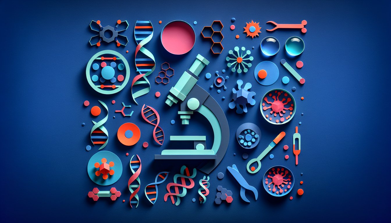
Ready to gauge your mastery of microscopy and genetics concepts? This Genetics Knowledge Assessment Quiz challenges learners with clear explanations, ideal for students and educators alike. Whether you're reviewing cell imaging techniques or heredity fundamentals in our Basic Genetics Quiz, you'll discover where your strengths shine and which areas need a refresher. All questions are fully editable in our quiz editor, so you can tailor the quizzes to suit your classroom or personal study goals.
Learning Outcomes
- Identify key components of light and electron microscopy
- Analyse chromosomal structures and genetic markers
- Demonstrate microscopy techniques in genetic research
- Evaluate the role of microscopy in observing mutations
- Apply staining and imaging methods to visualize gene expression
- Master core concepts of cell structure and inheritance patterns
Cheat Sheet
- Key Components of Light and Electron Microscopes - Explore how lenses, mirrors, and electron beams team up to reveal the tiniest details of cells and tissues. Uncover the secret sauce behind resolution and magnification that makes the invisible visible. Electron Microscope
- Chromosomal Structures and Genetic Markers - Get hands-on with FISH (Fluorescence In Situ Hybridization) to tag DNA sequences in glowing colors. Learn how these fluorescent probes light up specific genes on chromosomes, helping you spot genetic markers in a flash. Molecular Cytogenetics
- Demonstrating Fluorescence Microscopy - Shine a light (literally) on gene expression by using fluorescent dyes and antibodies to visualize active genes in living cells. Practice capturing stunning images that reveal which proteins are doing the most work under the microscope. Fluorescence Microscope
- Observing Mutations with Comparative Genomic Hybridization - Use CGH to compare normal and mutated DNA side by side for a detailed look at gene gains or losses. This technique highlights chromosomal abnormalities in brilliant color, making mutation detection a breeze. Comparative Genomic Hybridization
- Staining and Imaging Gene Expression - Dive into the world of fluorescent proteins and dyes that bind to specific cellular components. Master these staining tricks to illuminate the inner workings of cells and track gene activity over time. Fluorescence Techniques
- Core Concepts of Cell Structure and Inheritance - Brush up on chromosome organization, nucleus architecture, and the foundational rules of Mendelian genetics. Connect these basics to modern cytogenetic analyses for a well-rounded understanding. Genetic Foundations
- TEM vs. SEM: What's the Difference? - Compare Transmission Electron Microscopy's high-resolution slices with Scanning Electron Microscopy's 3D surface scans. Discover how sample prep, vacuum conditions, and detectors change the way you see your specimen. Electron Microscopy Types
- Applications of Fluorescence Microscopy - Detect specific proteins and nucleic acids by tagging them with fluorescent markers. Use live-cell imaging to watch molecular interactions unfold in real time, turning your slides into a live science show. Fluorescence Applications
- Principles of Molecular Cytogenetics - Learn how chromosomal analysis uncovers genetic disorders and guides clinical diagnoses. From karyotyping to high-throughput sequencing, see how modern tools map the human genome's twists and turns. Chromosomal Analysis
- Sample Preparation for Electron Microscopy - Master fixation, sectioning, and staining methods to protect delicate structures under the electron beam. Perfect your protocol to achieve crisp images that reveal ultrastructural details. EM Sample Prep