Unlock hundreds more features
Save your Quiz to the Dashboard
View and Export Results
Use AI to Create Quizzes and Analyse Results

Sign inSign in with Facebook
Sign inSign in with Google

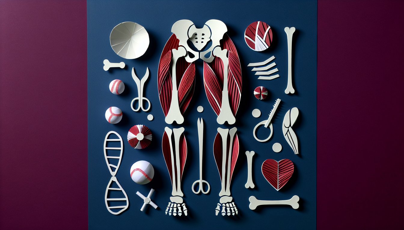
Take the Lower Limb Muscle Anatomy Quiz

Test Your Knowledge of Leg Muscle Structure

Difficulty: Moderate
Questions: 20
Learning OutcomesStudy Material
Colorful paper art featuring a quiz on lower limb muscle anatomy.

Are you ready to test your mastery of lower limb muscle anatomy? This lower limb muscle anatomy quiz challenges your grasp of muscle origins, insertions, and functions in everyday movement. Perfect for anatomy students and fitness professionals seeking to strengthen their understanding, it complements the Muscle Anatomy Knowledge Quiz and the Lower Extremity Assessment Quiz. Answers and explanations can be freely adjusted in our editor, making it a versatile tool for personalized study plans. Explore more quizzes and advance your anatomy skills today.

What is the primary action of the quadriceps femoris muscle group?
Knee extension
Knee flexion
Hip abduction
Plantarflexion
The quadriceps femoris group crosses the knee joint anteriorly and its main function is to extend the knee. Other actions listed are not primary functions of this muscle group.
Which muscle is superficial in the posterior compartment of the leg?
Flexor digitorum longus
Gastrocnemius
Soleus
Tibialis posterior
The gastrocnemius is the most superficial muscle of the posterior leg, forming the visible calf fullness. The other muscles lie deeper beneath it.
Which muscle is responsible for dorsiflexing the foot at the ankle joint?
Flexor hallucis longus
Peroneus longus
Tibialis anterior
Gastrocnemius
Tibialis anterior is the primary dorsiflexor of the foot, lifting the dorsum towards the shin. The other muscles either plantarflex or evert/invert the foot.
Which nerve innervates the hamstring muscles?
Femoral nerve
Superior gluteal nerve
Obturator nerve
Sciatic nerve
The hamstrings are innervated by the sciatic nerve (tibial portion), which supplies motor fibers to these posterior thigh muscles. The other nerves serve different compartments.
Which muscle primarily abducts the thigh at the hip joint?
Iliopsoas
Gluteus medius
Adductor longus
Gluteus maximus
Gluteus medius is the prime abductor of the hip, positioned laterally on the pelvis. Gluteus maximus extends the hip; the adductors and iliopsoas flex or adduct.
Which muscle originates from the anterior superior iliac spine and flexes, abducts, and laterally rotates the hip?
Iliopsoas
Tensor fasciae latae
Sartorius
Rectus femoris
Sartorius originates at the ASIS and crosses the hip and knee, performing hip flexion, abduction, and lateral rotation. The other muscles have different origins or actions.
The pes anserinus on the medial tibia is formed by the tendons of which set of muscles?
Biceps femoris, semimembranosus, semitendinosus
Vastus medialis, adductor longus, gracilis
Sartorius, gracilis, semitendinosus
Tibialis posterior, flexor digitorum longus, flexor hallucis longus
The pes anserinus is the conjoined insertion of sartorius, gracilis, and semitendinosus on the anteromedial tibia. The other groupings insert elsewhere.
The tibialis posterior primarily inserts on which bone in the foot?
Navicular
Cuboid
Calcaneus
Talus
Tibialis posterior inserts mainly on the navicular tuberosity and adjacent tarsal bones, supporting the medial arch. It does not attach to the calcaneus, cuboid, or talus directly.
Which muscle lies deep to the gastrocnemius and also contributes to plantarflexion?
Tibialis posterior
Plantaris
Soleus
Flexor hallucis longus
The soleus lies deep to the gastrocnemius in the posterior compartment and is a powerful plantarflexor. Plantaris is more superficial and smaller; the others have different primary actions.
Which muscle abducts the thigh and stabilizes the pelvis during gait?
Gluteus medius
Gluteus maximus
Piriformis
Tensor fasciae latae
Gluteus medius prevents contralateral pelvic drop during stance by abducting the hip. Gluteus maximus extends the hip; piriformis laterally rotates; TFL assists abduction but is less dominant.
What is the primary innervation of the iliopsoas muscle?
Femoral nerve
Sciatic nerve
Obturator nerve
Inferior gluteal nerve
The iliopsoas (psoas major and iliacus) is innervated mainly by the femoral nerve (L2 - L4). The other nerves supply different compartments of the lower limb.
Which muscle is dorsiflexed and everted by the deep fibular nerve?
Tibialis anterior
Fibularis longus
Gastrocnemius
Tibialis posterior
Tibialis anterior dorsiflexes and inverts (not everts) the foot and is innervated by the deep fibular nerve. Fibularis longus everts the foot and is innervated by the superficial fibular nerve.
During the toe-off phase of gait, which muscle is most active?
Tibialis anterior
Gastrocnemius
Gluteus medius
Rectus femoris
Gastrocnemius generates powerful plantarflexion during toe-off to propel the body forward. Tibialis anterior controls dorsiflexion; gluteus medius stabilizes pelvis; rectus femoris flexes hip/extending knee.
When the hip is flexed beyond 90 degrees, the piriformis changes its function to:
Medially rotate the thigh
Extend the thigh
Abduct the thigh
Flex the knee
When the hip is flexed past 90°, piriformis shifts from being a lateral rotator to acting as a hip abductor. It does not medially rotate or flex knee/extend thigh.
The hamstring portion of adductor magnus is innervated by which nerve?
Femoral nerve
Tibial division of the sciatic nerve
Obturator nerve
Superior gluteal nerve
The adductor (ischiocondylar) portion of adductor magnus is innervated by the tibial division of the sciatic nerve; the adductor portion is supplied by the obturator nerve.
Which muscle unlocks the knee by medially rotating the tibia on the femur at the initiation of flexion?
Popliteus
Plantaris
Soleus
Gastrocnemius
Popliteus unlocks the knee by medially rotating the tibia (or laterally rotating the femur) to initiate knee flexion. The other muscles do not play this specific role.
The plantaris muscle originates from which part of the femur?
Greater trochanter
Lateral supracondylar ridge
Linea aspera
Medial epicondyle
The plantaris originates on the lateral supracondylar ridge of the femur above the lateral head of gastrocnemius. The other landmarks belong to different muscles.
The fibularis longus tendon passes under the foot to insert on which structures?
Navicular and talus
Calcaneus and cuboid
Cuboid and fifth metatarsal
Medial cuneiform and base of first metatarsal
Fibularis longus passes beneath the foot to insert on the medial cuneiform and base of the first metatarsal, supporting the transverse arch. The other sites are incorrect.
Which muscle of the medial thigh has both an adductor part and a hamstring part with dual innervation?
Adductor longus
Pectineus
Gracilis
Adductor magnus
Adductor magnus has two parts: the adductor portion (obturator nerve) and the hamstring portion (tibial division of sciatic nerve). The others have single innervation.
Which muscle is most active in maintaining upright posture due to its high proportion of slow-twitch fibers?
Peroneus brevis
Gastrocnemius
Tibialis anterior
Soleus
Soleus has a high proportion of type I (slow-twitch) fibers and remains active during standing to maintain posture. Gastrocnemius has more fast-twitch fibers.
0
{"name":"What is the primary action of the quadriceps femoris muscle group?", "url":"https://www.quiz-maker.com/QPREVIEW","txt":"What is the primary action of the quadriceps femoris muscle group?, Which muscle is superficial in the posterior compartment of the leg?, Which muscle is responsible for dorsiflexing the foot at the ankle joint?","img":"https://www.quiz-maker.com/3012/images/ogquiz.png"}

Learning Outcomes

  1. Identify major muscles of the lower limb and their anatomical positions
  2. Analyze muscle functions during common lower limb movements
  3. Apply knowledge of muscle origins, insertions, and actions
  4. Differentiate between superficial and deep muscles in the leg
  5. Evaluate the role of key muscles in gait and posture
  6. Demonstrate understanding of innervation patterns for lower limb muscles

Cheat Sheet

  1. Major lower limb muscles - Get to know the gluteal group, quadriceps, hamstrings, and calf muscles by name and location so you can picture how they work together when you move. Visualizing these powerhouse muscles makes memorization a breeze and sets the stage for deeper study. Muscles of the Lower Limb
  2. Primary muscle functions - Discover which muscles extend, flex, abduct, or rotate key joints by linking names to actions - like how your quadriceps straighten the knee and your hamstrings bend it. This understanding is essential for analyzing movement mechanics in sports, rehab, or everyday life. Muscles of the Lower Limb - Listed Alphabetically
  3. Origins and insertions - Learn where each muscle starts and ends, such as the gluteus maximus popping off the ilium and sacrum and inserting on the femur to power hip extension. Knowing these anchor points explains how muscles create leverage and torque during movement. Muscles of the Lower Limb - Listed Alphabetically
  4. Superficial vs. deep layers - Distinguish between skin-level champs like the gastrocnemius and hidden heroes like the tibialis posterior to map out muscle compartments. Understanding layering helps you predict which muscles activate first and where injuries are most likely. Lower limb anatomy: Bones, muscles, nerves, vessels
  5. Gait and posture stabilizers - Explore how muscles such as the gluteus medius steady your pelvis when you walk, preventing that wobbly hip drop. Recognizing these stabilizers is key to both performance training and injury prevention. Muscles of the Lower Limb
  6. Innervation patterns - Map nerves to muscles - like the sciatic nerve firing your hamstrings and the femoral nerve powering your quads - to grasp how signals travel from brain to biceps femoris. This knowledge is vital for understanding neurological assessments and treating nerve injuries. Lower limb anatomy: Bones, muscles, nerves, vessels
  7. Mnemonic tricks - Use memorable phrases like "Tom, Dick, and Harry" to recall the tibialis posterior, flexor Digitorum longus, and flexor Hallucis longus tendons. Clever mnemonics turn complex lists into fun mental games and speed up recall. Muscles of the Lower Limb
  8. Diagram drills - Practice labeling muscles on diagrams or cadaver shots to cement their shapes, positions, and relationships in your mind. Visual repetition supercharges your learning and makes exam prep more engaging. Lower limb anatomy: Bones, muscles, nerves, vessels
  9. Blood supply essentials - Track arteries like the femoral artery supplying your quads and the posterior tibial artery nourishing your calf muscles to appreciate how oxygen reaches tissues. Understanding vascular routes is crucial for grasping injury healing and surgical approaches. Lower limb anatomy: Bones, muscles, nerves, vessels
  10. Common muscle injuries - Study typical ailments such as hamstring strains or Achilles tendonitis to link anatomy to real-world clinical cases. Knowing how and why these injuries occur guides prevention and effective rehab strategies. Muscles of the Lower Limb
Powered by: Quiz Maker