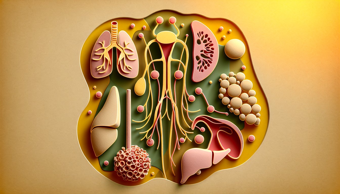
Jump into our ultimate lymphatic system quiz to see if you can pinpoint every organ's role! Perfect for students, nursing hopefuls, and curious biology buffs, this free quiz about lymphatic system invites you to match the lymphatic organ with its description and tackle targeted lymphatic system practice problems. You'll explore everything from lymphatic capillaries and nodes trivia to fluid filtration, immune defense, and overall anatomy and function. As you progress, you'll reinforce key concepts found in any lymphatic system function quiz and gain confidence with medical terminology lymphatic system. Ready to challenge yourself? Start the adventure now or begin your quiz here - let's get learning!
What is the primary function of the lymphatic system?
Maintaining fluid balance by returning interstitial fluid to the bloodstream
Facilitating gas exchange in the lungs
Transporting hormones throughout the body
Eliminating metabolic waste through urine
The lymphatic system collects excess interstitial fluid and returns it to the circulatory system, preventing edema and maintaining fluid homeostasis. It also plays roles in dietary fat absorption and immune defense. Unlike the cardiovascular system, it does not pump fluid via a central pump but relies on pressure gradients and valves. For more details, see NCBI Bookshelf.
Which structure filters lymph to remove pathogens and debris?
Lymph nodes
Spleen
Thymus
Tonsils
Lymph nodes are small, bean-shaped structures located along lymphatic vessels that filter lymph fluid, trapping bacteria, viruses, and foreign particles. They contain immune cells such as lymphocytes and macrophages to destroy pathogens. The spleen filters blood rather than lymph, and the thymus is primarily for T cell maturation. For further reading, see NCI.
The largest lymphatic organ in the body is the:
Spleen
Thymus
Lymph node
Tonsil
The spleen is the largest lymphatic organ and is located in the upper left quadrant of the abdomen. It filters blood, recycles aged red blood cells, and helps mount immune responses to blood-borne pathogens. The thymus, lymph nodes, and tonsils are smaller and have more specialized roles. More information is available at Mayo Clinic.
The thoracic duct drains lymph from which regions of the body?
Entire body except the right upper quadrant
Only the right upper limb and right side of head
Both lower limbs only
Left upper limb and left side of head only
The thoracic duct is the largest lymphatic vessel and drains lymph from both lower limbs, the abdomen, the left thorax, and the left upper limb and head. It empties into the junction of the left subclavian and internal jugular veins. The right lymphatic duct handles the right upper quadrant. See TeachMeAnatomy for details.
What is the name of the fluid transported within lymphatic vessels?
Lymph
Blood
Chyle
Interstitial fluid
Lymph is the clear to slightly yellow fluid that circulates through the lymphatic vessels. It originates from interstitial fluid that enters lymph capillaries and contains immune cells, proteins, and sometimes chyle from the digestive tract. Chyle is a lipid-rich form of lymph found specifically in lacteals after fat absorption. More on lymph at National Kidney Foundation.
Which lymphatic tissue is found in the pharynx and aids in defending against inhaled pathogens?
Tonsils
Peyer’s patches
Thymus
Spleen
The tonsils are collections of lymphoid tissue located in the pharynx that trap and destroy pathogens entering through the mouth or nose. They contain germinal centers full of B cells and are part of the mucosa-associated lymphoid tissue (MALT). Peyer’s patches are found in the small intestine, and the thymus and spleen have different functions. More information at NCBI Bookshelf.
The thymus is the primary site for the maturation of which cells?
T lymphocytes (T cells)
B lymphocytes (B cells)
Natural killer cells
Macrophages
The thymus is a primary lymphoid organ where precursor T cells complete their maturation and selection processes. It ensures self-tolerance by eliminating self-reactive T cells during negative selection. B cells mature primarily in the bone marrow. Learn more at NCBI Bookshelf.
Which duct drains lymph from the right upper limb, right side of the thorax, and right halves of the head and neck?
Right lymphatic duct
Thoracic duct
Cisterna chyli
Left lymphatic duct
The right lymphatic duct drains lymph from the right upper limb, right side of the thorax, and right halves of the head and neck, emptying into the right subclavian vein. The thoracic duct handles the remainder of the body’s lymph drainage. The cisterna chyli is a dilated sac at the start of the thoracic duct. See TeachMeAnatomy.
Peyer’s patches are specialized lymphoid nodules located in which part of the gastrointestinal tract?
Ileum of the small intestine
Jejunum
Duodenum
Colon
Peyer’s patches are clusters of lymphoid follicles found predominantly in the ileum, the distal part of the small intestine. They monitor intestinal bacteria and generate immune responses to pathogens entering through the gut. Jejunum and duodenum contain fewer or no Peyer’s patches, and the colon has distinct lymphoid aggregates. Read more at NCBI Bookshelf.
What type of lymphatic capillaries absorb dietary fats in the small intestine?
Lacteals
Blood capillaries
Pulmonary capillaries
Glomerular capillaries
Lacteals are specialized lymphatic capillaries present in the villi of the small intestine that absorb dietary lipids and transport them as chyle. Blood capillaries handle nutrient absorption into the bloodstream, while pulmonary and glomerular capillaries serve different organs. For further details, see NCBI Bookshelf.
The cisterna chyli is best described as:
An enlarged sac at the beginning of the thoracic duct
A cluster of lymph nodes in the neck
A lymphoid organ in the spleen
A duct draining the right upper quadrant
The cisterna chyli is a dilated, sac-like structure in the abdomen that serves as a reservoir for lymph from the lower body and intestinal trunk before it enters the thoracic duct. It is located near the L1 and L2 vertebrae. It is not a cluster of lymph nodes or part of the spleen. More at Visible Body.
Which of the following is NOT considered a primary lymphoid organ?
Spleen
Bone marrow
Thymus
None of the above
Primary lymphoid organs are where lymphocytes develop and mature: the bone marrow and thymus. The spleen is a secondary lymphoid organ where immune responses are coordinated but not lymphocyte development. Thus, the spleen is not a primary lymphoid organ. See NCBI Bookshelf for more.
In the formation of lymph, which force drives fluid out of blood capillaries into the interstitial space?
Capillary hydrostatic pressure
Blood colloid osmotic pressure
Interstitial oncotic pressure
Venous pressure
Capillary hydrostatic pressure pushes plasma out of blood capillaries into the interstitial space, forming interstitial fluid that enters lymphatic capillaries. Blood colloid osmotic pressure opposes this movement. Once in lymphatic vessels, this fluid is called lymph. For more information, visit OpenStax.
Which mechanism is most responsible for propelling lymph through lymphatic vessels?
Contraction of surrounding skeletal muscles
Cardiac pumping action
Peristalsis of smooth muscle in vessels
Respiratory pump only
Lymph is propelled largely by the contraction of surrounding skeletal muscles during movement, aided by one-way valves in lymphatic vessels. The respiratory pump also contributes, but there is no central pump like the heart. Smooth muscle peristalsis plays a minor role. For more details, see NCI.
Which cells in lymph nodes are primarily responsible for presenting antigens to T lymphocytes?
Dendritic cells
B cells
Reticular cells
Natural killer cells
Dendritic cells are potent antigen-presenting cells that capture antigens in peripheral tissues and migrate to lymph nodes to activate naïve T cells. B cells and macrophages can present antigen but are not the primary presenters for T cell activation. Reticular cells form the structural scaffold. Learn more at NCBI Bookshelf.
MALT is an acronym for:
Mucosa-Associated Lymphoid Tissue
Major Antigen-Linked Tissues
Memory-Activated Lymphocyte Transport
Macrophage-Antigen Lymphatic Trap
MALT stands for Mucosa-Associated Lymphoid Tissue, which includes lymphoid tissues in the gastrointestinal, respiratory, and genitourinary tracts. It provides immune surveillance and response at mucosal surfaces. Peyer’s patches and tonsils are part of MALT. More info at NCBI Bookshelf.
Which immunoglobulin is most abundant in lymph fluid?
IgG
IgA
IgM
IgE
IgG is the most abundant immunoglobulin in both serum and lymph fluid due to its small size and ability to diffuse through capillary walls. IgA is abundant in mucosal secretions but less so in lymph. IgM and IgE are present in lower concentrations. See NCBI Bookshelf.
The red pulp of the spleen is primarily involved in:
Filtering and removal of aged red blood cells
Maturation of T lymphocytes
Production of antibodies
Lymphocyte proliferation
The red pulp of the spleen contains sinusoids and macrophages that filter blood, removing aged or damaged red blood cells and pathogens. The white pulp is involved in immune functions such as antibody production and lymphocyte activation. T cell maturation occurs in the thymus. More at NCBI Bookshelf.
The paracortex region of a lymph node primarily contains which cell type?
T lymphocytes
B lymphocytes
Macrophages
Plasma cells
The paracortex of a lymph node is rich in T lymphocytes and dendritic cells that present antigens to these T cells. B lymphocytes are concentrated in the cortex and follicles. Macrophages and plasma cells reside mostly in the medullary cords. More details at NCBI Bookshelf.
Which chemokine receptor guides naïve lymphocytes into lymph nodes via high endothelial venules?
CCR7
CXCR4
CCR5
CXCR5
CCR7 is expressed on naïve T and B lymphocytes and directs them to lymph nodes by recognizing CCL19 and CCL21 on high endothelial venules. CXCR5 guides B cells to follicles, while CXCR4 and CCR5 have roles in other homing processes. For a full review, see PMC.
Lymphatic vessels are characterized by the presence of:
One-way valves to prevent backflow
Continuous basal lamina with tight junctions
Fenestrated endothelium like in kidneys
Thick smooth muscle layers
Lymphatic vessels have one-way valves that prevent the backflow of lymph and ensure unidirectional flow toward the venous circulation. Their endothelium is more permeable and lacks a continuous basal lamina. They do not have thick smooth muscle layers like arteries. See National Kidney Foundation.
Lymphangiogenesis refers to:
Formation of new lymphatic vessels
Destruction of lymphatic vessels
Inflammation of lymph nodes
Migration of lymphocytes
Lymphangiogenesis is the process by which new lymphatic vessels form from pre-existing lymphatics, often in development, wound healing, or tumor metastasis. It is distinct from angiogenesis, which refers to blood vessels. For more information, see PMC.
Which transcription factor is critical for T-cell lineage commitment within the thymus?
Notch1
AIRE
FOXP3
GATA3
Notch1 signaling is essential for early T-cell lineage commitment and differentiation in the thymus. It directs progenitor cells away from the B-cell lineage. AIRE is involved in negative selection in medullary thymic epithelial cells. For a deeper dive, see PMC.
Which cytokine produced by follicular dendritic cells is essential for germinal center formation?
BAFF (B-cell activating factor)
IL-2
TNF-alpha
Interferon-gamma
BAFF (B-cell activating factor) produced by follicular dendritic cells and stromal cells is crucial for B-cell survival and germinal center formation. It helps maintain B-cell niches and promotes class-switch recombination. IL-2 and IFN-gamma have different roles in T-cell responses. For more, see Nature Reviews Immunology.
The specialized medullary thymic epithelial cells that present self-antigens for negative selection express:
AIRE
MHC class I only
FOXP3
CCR7
AIRE (Autoimmune Regulator) is expressed by medullary thymic epithelial cells and promotes the expression of tissue-restricted antigens, enabling negative selection of self-reactive T cells. MHC molecules present these antigens, but AIRE is the transcription factor that drives their expression. For more, see NCBI Bookshelf.
0
{"name":"What is the primary function of the lymphatic system?", "url":"https://www.quiz-maker.com/QPREVIEW","txt":"What is the primary function of the lymphatic system?, Which structure filters lymph to remove pathogens and debris?, The largest lymphatic organ in the body is the:","img":"https://www.quiz-maker.com/3012/images/ogquiz.png"}
Study Outcomes
Identify key lymphatic structures -
Pinpoint major organs and tissues in the lymphatic system quiz, including lymph nodes, spleen, thymus, and tonsils.
Describe lymphatic organ functions -
Explain how each lymphatic organ contributes to fluid balance, immune defense, and fat absorption.
Match the lymphatic organ with its description -
Accurately pair organs with their descriptions to sharpen your skills in our match the lymphatic organ with its description activity.
Apply lymphatic system knowledge -
Tackle lymphatic system practice problems to reinforce concepts and improve recall during the quiz about lymphatic system structures.
Analyze fluid and immune pathways -
Trace lymph flow through vessels and nodes to understand how the lymphatic network supports immune surveillance.
Evaluate your mastery -
Use the lymph system quiz results to gauge your understanding and identify areas for further study.
Cheat Sheet
Lymph Formation & Starling Forces -
Lymph originates when interstitial fluid is pushed into lymphatic capillaries by net filtration from blood using the Starling equation: Jv = Kf [(Pc - Pi) - σ(πc - πi)]. Remember "pressure in, proteins stay out" to keep fluid moving toward the lymph nodes. Reviewing this formula on your lymphatic system practice problems will solidify how lymphatics maintain fluid balance.
Lymphatic Vessel Structure & Valves -
Lymphatic vessels are blind-ended, thin-walled capillaries with overlapping endothelial cells that act as one-way valves. These valves prevent backflow, much like venous valves, ensuring unidirectional lymph transport. A quick mnemonic: "Bale Every Vessel" helps you recall Blind-ended, Endothelial overlaps, Valves for your lymph system quiz.
Primary Lymphoid Organs: Bone Marrow & Thymus -
The bone marrow generates B cells while the thymus is where T cells mature and differentiate (B for bone, T for thymus). Recall the phrase "Be There": B in Bone, T in Thymus to ace any question that asks you to match the lymphatic organ with its description. Solidify this by checking university anatomy resources like Stanford Medicine or NIH outlines.
Lymph nodes filter lymph via afferent and efferent vessels (multiple afferents, one efferent) while the spleen filters blood; MALT (e.g., tonsils, Peyer's patches) protects mucosal surfaces. A handy acronym is "SLiM" (Spleen, Lymph nodes, MALT) to recall these organs on a quiz about lymphatic system. Practicing diagram labeling on these organs will boost your confidence on match-the-description items.
Thoracic & Right Lymphatic Duct Drainage Regions -
The thoracic duct drains ~75% of body lymph into the left subclavian vein, whereas the right lymphatic duct drains the upper right quadrant into the right subclavian vein. Remember "Left Large, Right Right" to recall that the thoracic duct collects more territory. Try drawing the drainage map - visual practice is key for quick recall on lymphatic system practice problems.