Unlock hundreds more features
Save your Quiz to the Dashboard
View and Export Results
Use AI to Create Quizzes and Analyse Results

Sign inSign in with Facebook
Sign inSign in with Google

Embryology of the Genitourinary System Quiz

Think you can ace this genitourinary system development quiz? Let's begin!

Difficulty: Moderate
2-5mins
Learning OutcomesCheat Sheet
Paper cut embryo kidney and reproductive tract illustration on coral background

Feeling curious about how kidneys and reproductive tracts form? Our genitourinary embryology quiz invites you to evaluate your knowledge of these critical developmental journeys. Whether you're brushing up on embryology of genitourinary system quiz topics, exploring a genitourinary system development quiz segment, or tackling our urinary system quiz for extra practice, this medical embryology trivia will keep you motivated. You'll dive into embryology anatomy quiz questions, uncover key organogenesis milestones, and compare your score with peers. Take our embryology test now and level up your expertise!

Which embryonic structure gives rise to the definitive kidney?
Metanephric blastema
Pronephros
Mesonephros
Urorectal septum
The definitive kidney, or metanephros, arises from the metanephric blastema, which interacts with the ureteric bud to form nephrons and collecting ducts. Early in development, the pronephros is transient and nonfunctional, and the mesonephros serves a temporary excretory role. The metanephric blastema persists to form the adult kidney. NCBI: Kidney Development
From which duct does the ureteric bud originate?
Mesonephric (Wolffian) duct
Paramesonephric (Müllerian) duct
Allantois
Neural crest
The ureteric bud sprouts from the mesonephric (Wolffian) duct and invades the metanephric blastema, where it undergoes branching morphogenesis to form the collecting system. The paramesonephric duct develops into female reproductive structures, not the ureteric bud. Proper induction of the ureteric bud is essential for nephron formation. Embryology.ch: Kidney Development
During which week of gestation does the metanephric kidney begin to produce urine?
Week 9
Week 4
Week 20
Week 30
Urine production by the metanephric kidney typically begins around week 9 of gestation, contributing to amniotic fluid volume. Before this, the mesonephros provides limited excretory function from weeks 4 to 10 but does not produce significant urine. Proper timing of kidney function is crucial for normal amniotic fluid homeostasis. NCBI PMC: Fetal Renal Development
What does the allantois contribute to in urinary tract development?
Urachus
Urinary bladder trigone
Collecting ducts
Glomerulus
The allantois forms the urachus, a fibrous remnant connecting the bladder apex to the umbilicus, which becomes the median umbilical ligament after birth. The urinary bladder trigone arises from incorporated mesonephric ducts, not the allantois. The collecting ducts derive from the ureteric bud, and glomeruli arise from metanephric mesenchyme. NCBI: Urinary Development
Which part of the nephron is derived from the ureteric bud?
Collecting duct
Proximal convoluted tubule
Glomerular capsule
Loop of Henle thick ascending limb
The ureteric bud gives rise to the collecting ducts, calyces, renal pelvis, and ureter through successive branching events. The proximal convoluted tubule, glomerular capsule, and most of the loop of Henle originate from the metanephric mesenchyme. Coordination between bud and blastema is critical for complete nephron formation. National Kidney Foundation
What is the fate of the pronephros in human development?
Degenerates early in the cervical region
Forms the permanent kidney
Becomes the ureter
Develops into the bladder
The pronephros appears in the cervical region around week 4 and rapidly degenerates without forming functional nephrons in humans. It is a vestigial structure with no permanent contribution to the urinary system. The mesonephros then temporarily functions as an excretory organ until the metanephros takes over. UNSW Embryology: Pronephros
Which embryonic duct gives rise to the female internal genital tract?
Paramesonephric (Müllerian) duct
Mesonephric (Wolffian) duct
Urachus
Allantois
The paramesonephric (Müllerian) ducts form the fallopian tubes, uterus, cervix, and upper vagina in females when they persist and differentiate. The mesonephric ducts regress in the absence of androgens. Proper signaling and absence of anti-Müllerian hormone allow these structures to develop. NCBI: Development of the Female Reproductive System
Which structure arises from the urorectal septum during cloacal division?
Urogenital sinus
Anal canal
Nephric tubule
Allantois
The urorectal septum divides the cloaca into the urogenital sinus anteriorly and the anorectal canal posteriorly. The urogenital sinus later gives rise to the bladder, proximal urethra, and in males parts of the prostate. Failure of this division can lead to cloacal anomalies. Embryology.ch: Bladder and Cloaca
The trigone of the bladder is derived from which embryonic component?
Mesonephric duct
Metanephric blastema
Paramesonephric duct
Pronephros
The trigone of the bladder forms from the incorporated caudal portions of the mesonephric ducts as the bladder develops from the urogenital sinus. The rest of the bladder wall is of endodermal origin. This distinct origin explains the different innervation patterns in the trigone. NCBI: Bladder Development
Which germ layer gives rise to the urogenital sinus epithelium?
Endoderm
Ectoderm
Mesoderm
Neural crest
The epithelial lining of the urogenital sinus is derived from endoderm. Surrounding connective tissue and smooth muscle arise from splanchnic mesoderm. Proper endodermal differentiation is required for normal bladder and urethral development. NCBI: Urogenital Sinus Development
Which embryonic structure evolves into the external genital tubercle?
Genital tubercle
Urogenital fold
Labioscrotal swelling
Cloacal membrane
The genital tubercle is the primordium that elongates to form the penis in males or the clitoris in females. It arises anterior to the cloacal membrane early in development. Urogenital folds and labioscrotal swellings form adjacent structures of the genitalia. Embryology.ch: External Genitalia
What do the labioscrotal swellings become in males?
Scrotum
Labia minora
Glans penis
Urethral meatus
In male embryos, the labioscrotal swellings fuse in the midline to form the scrotum. In females, they remain unfused to become the labia majora. Fusion depends on dihydrotestosterone signaling. NCBI: External Genital Development
Which hormone induces regression of the paramesonephric ducts in males?
Anti-Müllerian hormone
Testosterone
Progesterone
Estrogen
Sertoli cells of the developing testes secrete anti-Müllerian hormone, causing paramesonephric ducts to regress in male embryos. Testosterone supports differentiation of mesonephric ducts into male internal genitalia. Without AMH, Müllerian structures persist as seen in persistent Müllerian duct syndrome. NCBI: Sexual Differentiation
What structure develops into the epididymis in males?
Mesonephric duct
Paramesonephric duct
Ureteric bud
Metanephric blastema
The mesonephric (Wolffian) duct differentiates into the epididymis, vas deferens, seminal vesicles, and ejaculatory duct under the influence of testosterone. The paramesonephric duct regresses in males. Proper testosterone signaling ensures male internal duct formation. NCBI: Wolffian Duct Derivatives
From which embryonic tissue do the glomeruli originate?
Metanephric mesenchyme
Ureteric bud
Endoderm of urogenital sinus
Ectoderm
Glomeruli and most nephrons arise from the metanephric mesenchyme, which differentiates into podocytes, Bowman’s capsule, and tubules. The ureteric bud contributes only to the collecting system. Endoderm and ectoderm do not form nephron structures. National Kidney Foundation
Which structure forms the neck of the bladder?
Pelvic part of urogenital sinus
Vesical part of urogenital sinus
Allantois
Mesonephric duct
The urogenital sinus is divided into vesical, pelvic, and phallic parts. The pelvic part forms the urethra and bladder neck, while the vesical part becomes the bladder body. The allantois forms the urachus. NCBI: Bladder and Urethra
Which growth factor secreted by the metanephric mesenchyme induces ureteric bud branching?
GDNF
FGF8
BMP4
TGF-?
Glial cell line–derived neurotrophic factor (GDNF) is secreted by the metanephric mesenchyme and binds to RET receptors on the ureteric bud, stimulating branching morphogenesis. Mutations in GDNF or RET can cause renal agenesis. Other factors modulate but do not primarily initiate branching. NCBI: Renal Branching Morphogenesis
Mutation of which gene is most commonly associated with Wilms tumor?
WT1
RET
GDNF
PAX2
WT1 is a tumor suppressor gene expressed in the developing kidney, and its mutation is found in about 10–20% of Wilms tumors. Loss of WT1 function leads to unchecked proliferation of nephrogenic blastema. Other genes contribute, but WT1 mutation is most characteristic. NCBI: Wilms Tumor
Which transcription factor is essential for early urogenital ridge formation?
PAX2
WT1
SOX9
SRY
PAX2 is expressed in the intermediate mesoderm and is critical for urogenital ridge formation, kidney development, and ureteric bud outgrowth. Mutations in PAX2 can cause renal-coloboma syndrome. WT1 acts later in nephron differentiation, while SOX9 and SRY are involved in sex determination. NCBI: PAX Genes
Failure of which enzyme leads to 5?-reductase deficiency in sexual differentiation?
Type 2 5?-reductase
Aromatase
17?-hydroxylase
3?-HSD
Type 2 5?-reductase converts testosterone to dihydrotestosterone (DHT), which is crucial for masculinization of external genitalia. Deficiency results in undervirilized male external genitalia despite normal internal ducts. Aromatase converts androgens to estrogens, not DHT. NCBI: 5?-Reductase Deficiency
Which malformation results from a patent urachus?
Urine leaking from the umbilicus
Bladder exstrophy
Hydrocele
Ureterocele
A patent urachus results when the allantois fails to obliterate, leaving a channel between the bladder and umbilicus, allowing urine to leak externally. Bladder exstrophy is due to abdominal wall closure defects, hydrocele is a tunica vaginalis issue, and ureterocele is a ureteral cyst. Management involves surgical closure. NCBI: Urachal Anomalies
Potter sequence is most directly caused by which embryologic defect?
Bilateral renal agenesis
Unilateral renal agenesis
Hydronephrosis
Horseshoe kidney
Potter sequence arises from oligohydramnios due to bilateral renal agenesis, leading to pulmonary hypoplasia, limb deformities, and characteristic facies. Unilateral agenesis does not severely reduce amniotic fluid. Hydronephrosis and horseshoe kidney have variable fluid impacts. NCBI: Potter Sequence
Which molecular signal restricts ureteric bud outgrowth to a single location on the mesonephric duct?
BMP4
GDNF
FGF10
Wnt2b
BMP4 in the surrounding mesenchyme inhibits ectopic ureteric bud formation, ensuring outgrowth occurs only where GDNF expression is highest. GDNF promotes budding but BMP4 confines it spatially. Disruption leads to duplex ureters or agenesis. NCBI: BMP4 in Kidney
Mayer-Rokitansky-Küster-Hauser syndrome results from agenesis of which structure?
Paramesonephric ducts
Mesonephric ducts
Ureteric buds
Urogenital sinus
Müllerian agenesis (MRKH syndrome) involves failure of paramesonephric ducts to develop, resulting in absence of the uterus and upper vagina with normal ovaries. Mesonephric duct derivatives and external genitalia are normal. It is not related to ureteric bud anomalies. NCBI: MRKH Syndrome
Hypospadias is due to defective fusion of which embryonic structures?
Urogenital folds
Labioscrotal swellings
Genital tubercle
Cloacal membrane
Hypospadias results from incomplete fusion of the urogenital folds along the ventral side of the penis, creating an abnormal urethral opening. Labioscrotal swellings form the scrotum, and the genital tubercle forms the glans. Normal androgen signaling is crucial for fold fusion. NCBI: Hypospadias
Which gene mutation is commonly implicated in autosomal dominant polycystic kidney disease?
PKD1
WT1
GDNF
PAX2
PKD1 encodes polycystin-1 and is mutated in about 85% of autosomal dominant polycystic kidney disease cases. These mutations disrupt tubular differentiation leading to cyst formation. PKD2 mutations account for most remaining cases. NCBI: ADPKD
Embryologic origin of the internal urethral sphincter is from which tissue?
Smooth muscle of urogenital sinus
Somatic mesoderm
Neural crest
Endoderm of bladder
The internal urethral sphincter consists of smooth muscle derived from the pelvic part of the urogenital sinus mesenchyme. It is under involuntary control. Somatic mesoderm forms skeletal muscle, not smooth sphincter muscle. NCBI: Urethral Sphincters
Duplication of the ureter is most often due to abnormal budding of which structure?
Ureteric bud
Metanephric mesenchyme
Mesonephric duct
Allantois
Ureteral duplication arises when two ureteric buds sprout from the mesonephric duct, leading to double ureters on one side. Timing and position of budding determine drainage patterns. Metanephric mesenchyme does not produce ureter primordia. NCBI: Ureteral Duplication
Which anomaly results from fusion of the inferior poles of the kidneys during ascent?
Horseshoe kidney
Crossed fused ectopia
Pelvic kidney
Duplicated collecting system
A horseshoe kidney forms when the inferior poles of the metanephric kidneys fuse during ascent, becoming trapped by the inferior mesenteric artery. This anomaly alters the location and rotation of the kidneys. Crossed fused ectopia involves one kidney crossing midline and fusing with the other. NCBI: Horseshoe Kidney
Posterior urethral valves in males arise from abnormal development of which structure?
Membranous folds in the prostatic urethra
Urethral meatus
Seminal vesicles
Ejaculatory ducts
Posterior urethral valves are obstructive membranous folds in the prostatic urethra, thought to result from anomalous embryologic development of the urogenital sinus. They can cause significant obstruction of urinary outflow in boys. Early diagnosis and valve ablation prevent renal damage. NCBI: Posterior Urethral Valves
Which syndrome involves mutation of the WT2 region and leads to nephroblastomatosis and organomegaly?
Beckwith-Wiedemann syndrome
Denys-Drash syndrome
Frasier syndrome
Alport syndrome
Beckwith-Wiedemann syndrome involves alterations in the imprinted WT2 region on chromosome 11p15, leading to overgrowth, macroglossia, organomegaly, and increased risk of Wilms tumor and nephroblastomatosis. Denys-Drash involves WT1 mutations. Alport syndrome affects collagen IV in glomeruli. NCBI: Beckwith-Wiedemann Syndrome
Medullary sponge kidney is characterized by cystic dilatation of which nephron segment?
Collecting ducts in the papilla
Proximal tubules
Distal convoluted tubules
Bowman’s capsule
Medullary sponge kidney features cystic dilatation of the collecting ducts in the renal papillae, visible on contrast studies as brush-like papillary outlines. It may present with hematuria or nephrolithiasis. Other nephron segments are unaffected. NCBI: Medullary Sponge Kidney
Which rare anomaly results from failure of the urorectal septum to divide the cloaca properly?
Cloacal exstrophy
Bladder exstrophy
Imperforate anus
Urachal sinus
Cloacal exstrophy results when the cloacal membrane fails to be reinforced by mesoderm, leading to exposure of hindgut and bladder on the abdominal wall. It is more severe than bladder exstrophy and involves the gastrointestinal tract. Imperforate anus and urachal sinus have different embryologic bases. NCBI: Cloacal Exstrophy
Nephronophthisis is caused by mutations in genes encoding which type of proteins?
Ciliary proteins
Collagen IV chains
AQP2 channels
Podocin
Nephronophthisis involves mutations in genes such as NPHP1 that encode primary ciliary proteins, leading to tubulointerstitial fibrosis and cysts at the corticomedullary junction. It is a ciliopathy with early-onset renal failure. Collagen IV mutations cause Alport syndrome. NCBI: Nephronophthisis
Which embryonic transcription factor pair regulates differentiation of metanephric mesenchyme?
WT1 and Pax2
SOX9 and SRY
HNF1? and FGF8
GATA4 and Tbx5
WT1 and Pax2 coordinate signaling between metanephric mesenchyme and ureteric bud, regulating mesenchymal condensation and nephron differentiation. Loss of either disrupts nephrogenesis. Other factors have roles in different organ systems. NCBI: Nephrogenesis
Which genetic disorder features dysplastic kidneys with cystic changes and extrarenal malformations, due to PAX2 haploinsufficiency?
Renal-coloboma syndrome
Alagille syndrome
VACTERL association
Polycystic kidney disease
Renal-coloboma syndrome, caused by PAX2 haploinsufficiency, features renal hypoplasia/dysplasia and optic nerve colobomas. PAX2 is critical for nephric duct development. Alagille involves JAG1 mutations. NCBI: Renal-Coloboma Syndrome
Which anomaly results in a blind-ending ureter that cannot connect to the bladder?
Ureteral atresia
Ureterocele
Ectopic ureter
Duplicated ureter
Ureteral atresia occurs when the ureteric bud fails to canalize properly, resulting in a blind-ending ureter and ipsilateral renal agenesis or dysplasia. A ureterocele is a cystic dilation of the intravesical ureter. Ectopic ureters drain abnormally. NCBI: Ureteral Anomalies
Mutations in which gene are responsible for juvenile nephronophthisis type 1, leading to early-onset renal failure?
NPHP1
WT1
PKD2
HNF1B
NPHP1 mutations disrupt nephrocystin, a ciliary protein, causing juvenile nephronophthisis with corticomedullary cysts and early renal failure. It is the most common form of nephronophthisis. WT1, PKD2, and HNF1B cause other nephropathies. NCBI: Juvenile Nephronophthisis
Which transcription factor interplay regulates male sex determination by upregulating SOX9 after SRY activation?
SF1 and SRY
WT1 and PAX2
FOXL2 and WNT4
GATA4 and TBX5
SRY expression in Sertoli cell precursors upregulates SOX9, with SF1 co-activation, committing the bipotential gonad to testis development. WT1 acts upstream of SRY, FOXL2 promotes ovarian fate, and GATA4/TBX5 regulate cardiac development. NCBI: Sex Determination
Vesicoureteral reflux in development often results from malpositioning of which embryonic junction?
Ureteric bud insertion into the bladder
Metanephric blastema contact site
Cloacal membrane division
Allantois insertion
Vesicoureteral reflux often arises when the ureteric bud inserts too laterally or high on the bladder wall, resulting in a short intramural ureter that cannot prevent backflow. Normal oblique course of the ureter through the bladder wall is essential for the valve mechanism. Malposition at the bud stage underlies this defect. NCBI: VUR Embryology
Which molecular pathway is critical for branching morphogenesis of the ureteric bud besides GDNF-RET signaling?
Wnt11
Notch1
Hedgehog
TGF-?1
Wnt11 is expressed at ureteric bud tips and reinforces GDNF-RET signaling in a positive feedback loop essential for branching. Notch and Hedgehog pathways play roles in other tissues. TGF-?1 often inhibits branching. NCBI: Wnt in Kidney
Cloacal exstrophy involves aberrant development of both the bladder and which additional structure?
Distal hindgut
Ureteral buds
Pronephric ducts
Mesonephric tubules
Cloacal exstrophy features failure of lower abdominal wall and cloacal septation, exposing both the bladder and distal hindgut (colon) externally. The anomaly is more severe than bladder exstrophy alone. Ureteral buds and nephric ducts remain internal. NCBI: Cloacal Exstrophy
Mutations in HNF1B often lead to a spectrum of renal developmental anomalies known as what?
Renal cysts and diabetes syndrome
Alport syndrome
Beckwith-Wiedemann syndrome
Fraser syndrome
HNF1B mutations cause renal cysts and diabetes (RCAD) syndrome, leading to cystic dysplasia, collecting duct anomalies, and maturity-onset diabetes of the young type 5. HNF1B regulates early renal epithelial differentiation. Other syndromes involve different genes. NCBI: HNF1B-related Disease
0
{"name":"Which embryonic structure gives rise to the definitive kidney?", "url":"https://www.quiz-maker.com/QPREVIEW","txt":"Which embryonic structure gives rise to the definitive kidney?, From which duct does the ureteric bud originate?, During which week of gestation does the metanephric kidney begin to produce urine?","img":"https://www.quiz-maker.com/3012/images/ogquiz.png"}

Study Outcomes

  1. Understand Major Stages of Renal Development -

    Understand the progression from pronephros to metanephros, highlighting key morphogenetic events in kidney formation assessed in the genitourinary embryology quiz.

  2. Identify Embryonic Urinary Structures -

    Identify and describe the pronephric, mesonephric, and metanephric systems, linking each to its role in forming the adult urinary tract.

  3. Differentiate Reproductive Duct Origins -

    Differentiate between mesonephric (Wolffian) and paramesonephric (Müllerian) ducts and explain their contributions to the male and female reproductive tracts.

  4. Analyze Developmental Timelines -

    Analyze the chronological sequence of genitourinary system development, recognizing when key structures appear and mature during embryogenesis.

  5. Apply Knowledge to Clinical Anomalies -

    Apply embryological principles to interpret common genitourinary anomalies, such as renal agenesis and ductal malformations, in clinical scenarios.

  6. Recall Molecular Signals in Embryogenesis -

    Recall the primary molecular factors, including growth factors and signaling pathways, that regulate kidney and reproductive tract development.

Cheat Sheet

  1. Sequential Kidney Formation -

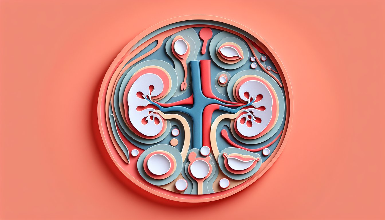
    The genitourinary embryology quiz often starts with the three kidney systems: pronephros, mesonephros, and metanephros, remembered by the mnemonic "Pee, Pee, Pee." These structures form in a cranial-to-caudal sequence, but only the metanephros persists as the permanent adult kidney (Sadler, Langman's Medical Embryology).

  2. Ureteric Bud - Metanephric Mesenchyme Interaction -

    Branching morphogenesis is driven by GDNF/RET signaling between the ureteric bud and metanephric mesenchyme, a core concept in the genitourinary system development quiz. Proper reciprocal induction ensures formation of nephrons and collecting ducts (Moore & Persaud, Clinically Oriented Anatomy).

  3. Ascent of the Kidneys and Vascular Changes -

    During weeks 6 - 9 the metanephric kidneys "ascend" from the pelvic region to their lumbar position, temporarily supplied by sequential branches of the abdominal aorta. Aberrant renal arteries or malrotation can lead to hydronephrosis, a classic topic in the embryology of genitourinary system quiz (Netter's Atlas of Human Anatomy).

  4. Duct Differentiation: Mesonephric vs. Paramesonephric -

    In males, SRY induces Sertoli cells to secrete Müllerian inhibiting substance, causing paramesonephric duct regression, whereas the mesonephric ducts form the epididymis, vas deferens, and seminal vesicles. In females the absence of AMH allows paramesonephric duct development into fallopian tubes and uterus, a must-review point for any embryology anatomy quiz (Gilbert, Developmental Biology).

  5. Common Congenital Anomalies -

    Horseshoe kidney (fusion at lower poles), unilateral renal agenesis, and Müllerian agenesis are key high-yield facts in medical embryology trivia. Understanding their embryonic basis aids in clinical correlation and ultrasound diagnosis (American Urological Association guidelines).

Powered by: Quiz Maker