Unlock hundreds more features
Save your Quiz to the Dashboard
View and Export Results
Use AI to Create Quizzes and Analyse Results

Sign inSign in with Facebook
Sign inSign in with Google

Bones of the Foot Practice Quiz

Test your knowledge with our interactive foot quiz

Difficulty: Moderate
Grade: Grade 10
Study OutcomesCheat Sheet
Paper art illustrating Foot Bone Frenzy, a trivia quiz for high school biology students.

Which bone is known as the heel bone?
Calcaneus
Talus
Navicular
Cuboid
The calcaneus is the largest tarsal bone and is commonly known as the heel bone. It plays a major role in weight-bearing and shock absorption during movement.
How many metatarsal bones are found in the human foot?
5
4
6
7
There are exactly 5 metatarsal bones in the human foot, one corresponding to each toe. They help form the structure of the forefoot and play a role in weight distribution.
Which group of bones forms the forefoot?
Metatarsals and Phalanges
Tarsals and Metatarsals
Tarsals and Phalanges
Navicular and Cuboid
The forefoot is comprised of the metatarsal bones and the phalanges, which make up the toes. This grouping is essential for understanding the structure and movement of the foot.
What is the primary function of the calcaneus?
Provide heel support
Connect the leg to the foot
Protect the foot muscles
Support the arch
The calcaneus serves as the heel bone and provides critical support during locomotion. It also plays an important role in absorbing shock as forces are transmitted through the foot.
Which of the following bones is found in the hindfoot?
Talus
Cuboid
Navicular
Medial Cuneiform
The hindfoot is composed of the calcaneus and the talus, both of which are essential for proper foot mechanics. The talus, in particular, plays a significant role in the ankle joint by articulating with the leg bones.
Which bone in the foot articulates with the tibia and fibula to form the ankle joint?
Talus
Calcaneus
Navicular
Cuboid
The talus is the critical bone that connects with the tibia and fibula to form the ankle joint, also known as the talocrural joint. This joint is essential for movements such as dorsiflexion and plantarflexion.
What are the three main regions of the foot?
Hindfoot, Midfoot, Forefoot
Rearfoot, Midfoot, Frontfoot
Heel, Arch, Toes
Medial, Lateral, Central
The human foot is anatomically divided into the hindfoot, midfoot, and forefoot. Each region contains distinct groups of bones and serves unique functions in support and movement.
Which bone is NOT considered one of the tarsal bones?
Metatarsal
Calcaneus
Talus
Navicular
The tarsal bones include the calcaneus, talus, navicular, cuboid, and three cuneiform bones. Metatarsals are not part of the tarsal group; they are located in the forefoot.
How many phalanges are present in the human foot?
14
10
12
16
There are 14 phalanges in the human foot; this includes two phalanges in the big toe and three in each of the other toes. This anatomical detail is crucial for accurately understanding toe structure.
Which bone is considered the keystone of the medial longitudinal arch?
Navicular
Cuboid
Talus
Calcaneus
The navicular bone is often referred to as the keystone of the medial longitudinal arch due to its central location and supportive role. Its positioning is vital for maintaining the integrity of the arch during movement.
Which of the following bones is most commonly fractured in the foot during high-impact injuries?
Calcaneus
Talus
Cuboid
Navicular
The calcaneus, being the heel bone, is frequently subjected to high-impact forces and is one of the most commonly fractured bones in the foot. Its role in weight-bearing makes it particularly vulnerable during traumatic injuries.
Which bone is located between the talus and the metatarsals in the midfoot region?
Navicular
Cuboid
Calcaneus
Lateral Cuneiform
The navicular bone is positioned in the midfoot between the talus and the cuneiform/metatarsal bones. This strategic location contributes significantly to the stability and mobility of the foot.
How many tarsal bones are there in an adult human foot?
7
6
5
8
An adult human foot contains 7 tarsal bones which include the calcaneus, talus, navicular, cuboid, and three cuneiform bones. This foundational information is key to understanding foot anatomy.
Which of the cuneiform bones is the most medial in the foot?
Medial Cuneiform
Intermediate Cuneiform
Lateral Cuneiform
Cuboid
The medial cuneiform is the most medially positioned among the three cuneiform bones. Its location is essential for forming the medial longitudinal arch and contributes to overall foot stability.
The metatarsophalangeal joints are formed by the articulation of the metatarsal bones with which other bones?
Phalanges
Tarsals
Carpals
Sesamoids
The metatarsophalangeal joints are created by the articulation between the metatarsal bones and the phalanges. These joints are vital for flexion, extension, and overall toe movement during gait.
During foot inversion injuries, which fracture is most commonly associated with the calcaneus and subtalar joint?
Intra-articular calcaneal fracture
Avulsion fracture of the navicular
Comminuted talus fracture
Stress fracture of the metatarsal
In foot inversion injuries, the calcaneus is prone to intra-articular fractures, particularly involving the subtalar joint. This type of fracture can affect the alignment and function of the hindfoot significantly.
Which structural feature of the calcaneus contributes most to shock absorption during gait?
The trabecular structure of the calcaneus
The shape of the talus
The rigidity of the metatarsals
The thinness of the phalanges
The trabecular architecture within the calcaneus allows it to absorb and dissipate impact forces during walking and running. This built-in shock absorption mechanism is essential for reducing stress on the foot and lower limb.
How does the arrangement of the tarsal bones contribute to the foot's overall flexibility and stability?
Their interlocking articulations and robust ligament support
Their uniform shape and size
Their complete immobility
Their reliance on articular cartilage only
The tarsal bones are arranged in an interlocking pattern supported by strong ligaments, which allows for both flexibility and stability. This arrangement permits essential movements while maintaining the structural integrity of the foot.
What is the impact of a disrupted medial longitudinal arch on foot biomechanics?
It can lead to flatfoot and altered weight distribution
It causes the foot to become more rigid
It results in an elevated arch with excessive propulsion
It enhances balance due to tighter ligaments
A disrupted medial longitudinal arch often leads to flatfoot, impacting the way weight is distributed across the foot during movement. Such alterations in biomechanics can result in pain and functional impairments over time.
Why is the navicular bone considered so critical in surgical reconstructions of the foot's arch?
It serves as a central keystone in the medial longitudinal arch
It is the largest bone in the foot
It is the only bone that articulates with the femur
It has no significant articulations
The navicular bone acts as the keystone of the medial longitudinal arch, providing essential support and alignment. Its central location makes it a critical focus in reconstructive surgeries aimed at restoring proper foot biomechanics.
0
{"name":"Which bone is known as the heel bone?", "url":"https://www.quiz-maker.com/QPREVIEW","txt":"Which bone is known as the heel bone?, How many metatarsal bones are found in the human foot?, Which group of bones forms the forefoot?","img":"https://www.quiz-maker.com/3012/images/ogquiz.png"}

Study Outcomes

  1. Identify the major bones of the foot.
  2. Analyze the anatomical structure and spatial relationships of foot bones.
  3. Understand the functions and biomechanical roles of each key bone.
  4. Apply anatomical knowledge to interpret foot bone injuries and deformities.
  5. Evaluate differences in bone morphology to differentiate among foot regions.

Bones of the Foot Cheat Sheet

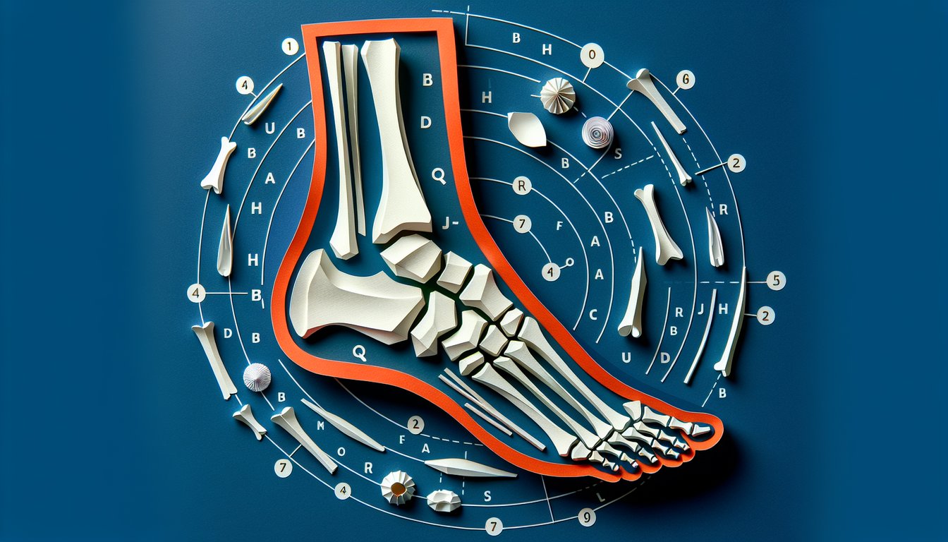
  1. Foot Bone Groups - Your foot is a miniature marvel made of 26 bones split into three squads: 7 tarsals, 5 metatarsals, and 14 phalanges. Mastering this lineup is the key to understanding how your foot bears weight and moves with grace. healthline.com: Bones of the Foot
  2. Tarsal Bone Team - The tarsals are the VIPs in your ankle and heel, including the talus, calcaneus, navicular, cuboid, and three cuneiforms (medial, intermediate, lateral). They squad up to form the base of your foot's structure and stability. teachmeanatomy.info: Tarsals Explained
  3. Mnemonic Magic - Remember "Tiger Cubs Need MILC" to ace your tarsal bone recall: Talus, Calcaneus, Navicular, Medial cuneiform, Intermediate cuneiform, Lateral cuneiform, Cuboid. This fun phrase is your exam-time lifesaver! artofmemory.com: Tarsal Mnemonic
  4. Metatarsal Map - Five metatarsals stretch between your tarsals and toes, numbered I through V from the big toe to the little piggy. They're crucial in distributing your body's weight and keeping you balanced on every step. teachmeanatomy.info: Metatarsals Overview
  5. Phalangeal Flexibility - Your toes pack three phalanges each (proximal, middle, distal), except the big toe, which owns just two. These tiny bones give your toes the dexterity needed for gripping, pushing, and quick footwork. teachmeanatomy.info: Phalanges Breakdown
  6. Talent of the Talus - The talus sits atop the foot, linking up with the tibia and fibula to make the ankle joint. It's the heavyweight champ for transferring body weight down into your foot's framework. teachmeanatomy.info: Talus Insights
  7. Calcareous Calcaneus - Better known as the heel bone, the calcaneus is the biggest tarsal of them all. It forms a sturdy foundation at the back of your foot and gives you the leverage needed for every stride. teachmeanatomy.info: Calcaneus Facts
  8. Navicular Navigator - Shaped like a little boat, the navicular sits on the medial side and connects with the talus and cuneiforms. It's a key player in maintaining your foot's arch and smooth motion. teachmeanatomy.info: Navicular Role
  9. Cuboid Cruiser - The cube-shaped cuboid bone hangs out on the lateral side, linking the calcaneus to the fourth and fifth metatarsals. It's vital for stabilizing your foot during side-to-side movements. teachmeanatomy.info: Cuboid Details
  10. Cuneiform Crew - The medial, intermediate, and lateral cuneiforms are wedge-shaped bones nestled between the navicular and the first three metatarsals. Together, they sculpt the transverse arch, giving your foot both flexibility and strength. teachmeanatomy.info: Cuneiform Collective
Powered by: Quiz Maker