Unlock hundreds more features
Save your Quiz to the Dashboard
View and Export Results
Use AI to Create Quizzes and Analyse Results

Sign inSign in with Facebook
Sign inSign in with Google

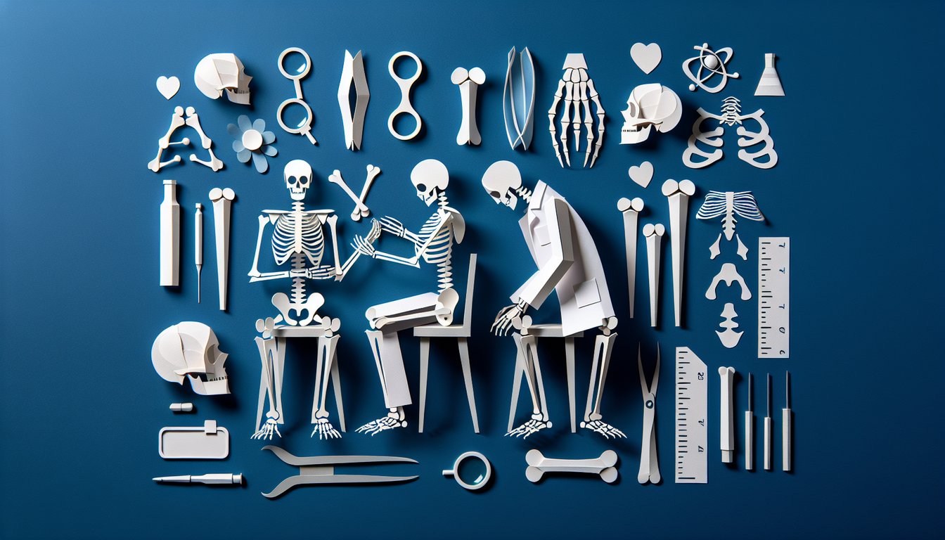
Skeleton Bone Labeling Practice Quiz

Test your anatomy knowledge in a fun quiz

Difficulty: Moderate
Grade: Grade 8
Study OutcomesCheat Sheet
Paper art depicting Bone Labeling Bonanza, a trivia quiz for high school biology students.

Which bone is commonly known as the collarbone?
Clavicle
Scapula
Sternum
Humerus
The clavicle is also known as the collarbone due to its location and shape. The sternum forms the center of the chest and the scapula and humerus serve different roles in the shoulder and arm, respectively.
Which bone forms the upper arm?
Humerus
Radius
Ulna
Carpal
The humerus is the long bone of the upper arm, essential for arm movement. The radius and ulna are found in the forearm, while the carpals make up the wrist.
What is the largest bone in the human body?
Femur
Tibia
Fibula
Patella
The femur, or thigh bone, is the largest bone in the human body. The tibia and fibula form the lower leg, while the patella is the kneecap.
Which bone protects the heart and lungs in the chest?
Sternum
Rib
Vertebrae
Clavicle
The sternum, or breastbone, is centrally located and provides protection to the heart and lungs. While the ribs also contribute to this protection, the sternum is a key structural element in the chest.
Which bone is found in the lower leg and bears most of the weight?
Tibia
Fibula
Femur
Patella
The tibia, commonly called the shinbone, bears most of the body's weight in the lower leg. The fibula is thinner and aids in stability, while the femur and patella are located in the upper leg and knee respectively.
Which two bones form the forearm?
Radius and Ulna
Humerus and Radius
Ulna and Femur
Scapula and Clavicle
The forearm is comprised of the radius and the ulna, allowing for a range of rotational movements. The other options include bones that are not part of the forearm.
What is the primary function of the vertebrae in the human skeleton?
They protect the spinal cord
They support the rib cage
They house the brain
They form the pelvic girdle
The vertebrae form the spinal column, creating a protective canal for the spinal cord. While they also help support the body, their main function is to safeguard the nervous system.
Which bone is a key component of the shoulder girdle and helps connect the arm to the torso?
Scapula
Clavicle
Sternum
Humerus
The scapula, or shoulder blade, is essential in linking the arm to the torso and serves as an attachment point for multiple muscles. Although the clavicle also plays a role, the scapula is the primary bone in the shoulder girdle.
Which bone forms the anterior (front) portion of the pelvis?
Pubis
Ilium
Ischium
Sacrum
The pubis is the bone that makes up the front portion of the pelvic ring. The ilium and ischium contribute to the sides and back of the pelvis, while the sacrum connects the spine to the pelvis.
What is the common function of most bones in the human body?
Support and protect body organs
Produce blood cells exclusively
Store fat exclusively
Regulate body temperature exclusively
Bones provide structural support and shield vital organs from injury. They also play roles in blood cell production and mineral storage, yet their primary function is to support and protect the body.
Which bone does the name 'mandible' refer to?
Jawbone
Cheekbone
Skullcap
Collarbone
The mandible is the jawbone that forms the lower part of the facial skeleton. The other options refer to different bones in the skull that serve other functions.
Which bone is classified as a flat bone?
Scapula
Femur
Ulna
Tibia
The scapula is a flat bone, characterized by its broad and thin structure. In contrast, the femur, ulna, and tibia are categorized as long bones.
Which bone is located in the lower leg and is thinner than its counterpart?
Fibula
Tibia
Femur
Patella
The fibula is the slender bone in the lower leg, running alongside the thicker tibia which bears most of the weight. The femur and patella are not located in the lower leg.
Which bone forms the posterior portion of the skull?
Occipital bone
Temporal bone
Parietal bone
Frontal bone
The occipital bone is situated at the back of the skull and forms its posterior part. The temporal, parietal, and frontal bones are located on the sides or front of the skull.
Which bone in the forearm is primarily responsible for allowing the rotation of the hand?
Radius
Ulna
Humerus
Scapula
The radius rotates around the ulna, enabling the forearm to twist during movements such as pronation and supination. The other bones do not significantly contribute to the rotational movement of the hand.
Which bone exhibits significant sexual dimorphism, aiding in the determination of biological sex?
Pelvis
Skull
Femur
Clavicle
The pelvis shows marked differences between males and females, making it a key indicator for determining biological sex in forensic studies. The skull, femur, and clavicle do not display such pronounced sexual dimorphism.
Which skeletal feature is most commonly analyzed by forensic anthropologists to estimate an adult's age?
Pubic symphysis
Skull suture lines
Rib curvature
Clavicle length
The pubic symphysis undergoes predictable morphological changes with age, making it a reliable feature for age estimation. Although skull suture lines, rib curvature, and clavicle length are observed, they are less precise for this purpose.
Which component of the axial skeleton, if improperly aligned, is most indicative of scoliosis?
Vertebrae
Ribs
Skull
Sternum
Scoliosis is characterized by an abnormal curvature of the spine, making the vertebrae the primary indicators of this condition. The ribs, skull, and sternum are less directly involved in this alignment issue.
Which structure located near the ends of long bones is responsible for longitudinal growth during development?
Epiphyseal plate
Diaphysis
Periosteum
Medullary cavity
The epiphyseal plate is a cartilaginous growth plate responsible for the lengthening of long bones during development. Damage to this structure can lead to abnormalities in growth, unlike the diaphysis, periosteum, or medullary cavity which have different functions.
In the assessment of hip joint stability, which feature of the femur is key for proper articulation with the pelvis?
Femoral head
Femoral neck
Greater trochanter
Lesser trochanter
The femoral head is the rounded, ball-like structure that fits into the acetabulum of the pelvis, ensuring smooth and stable hip joint movement. The femoral neck and trochanters serve as attachment sites for muscles and contribute to the structural integrity but are not the primary articulating surface.
0
{"name":"Which bone is commonly known as the collarbone?", "url":"https://www.quiz-maker.com/QPREVIEW","txt":"Which bone is commonly known as the collarbone?, Which bone forms the upper arm?, What is the largest bone in the human body?","img":"https://www.quiz-maker.com/3012/images/ogquiz.png"}

Study Outcomes

  1. Identify key bones of the human skeleton.
  2. Label anatomical structures with accuracy.
  3. Demonstrate understanding of bone locations and relationships.
  4. Assess personal knowledge of skeletal anatomy through quiz performance.

Labeling The Bones of the Skeleton Cheat Sheet

  1. Familiarize yourself with major bones - Get to know where the skull, spine, ribs, and limbs sit and what roles they play in your body. Mapping these landmarks will anchor your understanding and boost your labeling accuracy. differentmedicalcareers.com
  2. differentmedicalcareers.com
  3. Use mnemonic devices - Turn a long list of carpal bones into a catchy phrase like "Some Lovers Try Positions That They Can't Handle" to make memorization stick. Mnemonics are your brain's secret weapon for turning jargon into joyful jingles! kenhub.com
  4. kenhub.com
  5. Spot the key differences - Remember that the tibia is the stouter, weight-bearing bone and the fibula is its slender, sidekick on the outer leg. Knowing their unique features helps you avoid mix-ups in exams and real life! classroom.synonym.com
  6. classroom.synonym.com
  7. Master the skull bones - Use "Old People From Texas Eat Spiders" to lock in Occipital, Parietal, Frontal, Temporal, Ethmoid, and Sphenoid. Associating faces with fun phrases makes cranium study a breeze! classroom.synonym.com
  8. classroom.synonym.com
  9. Practice with visual aids - Flashcards, diagrams, or bone-shaped stickers help you quiz your inner anatomy geek anytime. Seeing shapes and positions in vivid detail cements your memory faster than plain text alone. sciencing.com
  10. sciencing.com
  11. Master the anatomical position - Always start from the standard "standing straight, palms forward" stance so left and right never get scrambled. It's the golden rule for describing bone locations and directions accurately. differentmedicalcareers.com
  12. differentmedicalcareers.com
  13. Learn upper limb bones - Memorize humerus, radius, ulna, carpals, metacarpals, and phalanges with "How Rare U Cook Mesquite Pork." Turning lists into silly sentences will keep those arm bones locked in your mind! rishacademy.com
  14. rishacademy.com
  15. Study lower limb bones - From femur and patella down through tibia, fibula, tarsals, metatarsals, to phalanges, use "From Pennies To Fives They May Pay" to nail the sequence. Your study sessions will feel more like playtime with catchy chants! rishacademy.com
  16. rishacademy.com
  17. Map the vertebral column - Recall cervical (7), thoracic (12), lumbar (5) by thinking of breakfast at 7, lunch at 12, and dinner at 5. Don't forget the sacrum and coccyx to complete your spine storyline! classroom.synonym.com
  18. classroom.synonym.com
  19. Quiz yourself regularly - Turn review sessions into quick games: name that bone, point to that landmark, or race against the clock. Consistent self-testing cements knowledge and builds exam-day confidence! sciencing.com
  20. sciencing.com
Powered by: Quiz Maker