Unlock hundreds more features
Save your Quiz to the Dashboard
View and Export Results
Use AI to Create Quizzes and Analyse Results

Sign inSign in with Facebook
Sign inSign in with Google

Ultimate Appendicular Skeleton Practice Quiz

Test Your Knowledge on Bones and Muscles

Difficulty: Moderate
Grade: Grade 11
Study OutcomesCheat Sheet
Colorful paper art promoting the Appendicular Anatomy Challenge for high school and college students.

Easy
Which group of bones comprises the appendicular skeleton?
Bones of the limbs and their girdles
Bones of the skull and vertebral column
Ribs and sternum
Base of the skull and facial bones
The appendicular skeleton includes the bones of the limbs along with the pectoral and pelvic girdles. This distinguishes it from the axial skeleton which comprises the skull, vertebral column, and rib cage.
What is the primary structural component of a long bone's shaft?
Diaphysis
Epiphysis
Metaphysis
Periosteum
The diaphysis refers to the shaft of a long bone, providing strength and support. In contrast, the epiphyses are the ends of the bone involved in joint formation.
Which of the following is a typical feature of a long bone?
Central marrow cavity
Multiple thin layers of spongy bone
Large, flat surface for muscle attachment
Irregular shape with no definite structure
Long bones are characterized by a central marrow cavity, a defined shaft (diaphysis), and expanded ends (epiphyses). This structure is essential for supporting weight and facilitating movement.
The humerus is found in which part of the body?
Upper limb
Lower limb
Skull
Torso
The humerus is the bone of the upper arm and forms part of the appendicular skeleton. It plays a key role in connecting the shoulder to the forearm.
Which part of a long bone is primarily responsible for growth in length during development?
Epiphyseal plate
Diaphysis
Periosteum
Articular cartilage
The epiphyseal plate is the region where new bone is added during growth, contributing to the lengthening of the bone. Once growth has ceased, this area ossifies into the epiphyseal line.
Medium
Which of the following best describes the composition of a typical long bone?
A combination of compact bone forming the outer layer and spongy bone within
Primarily spongy bone with minimal compact bone
Evenly distributed cartilage throughout the bone
A hollow bone with no dense tissue
Long bones have a dense outer layer known as compact bone that provides strength, while the inner spongy bone helps reduce weight and houses the marrow. This arrangement is crucial for both structural support and mobility.
How many main bones are found in the forearm?
Two
One
Three
Four
The forearm is composed of two primary bones: the radius and the ulna. These bones work together to allow a range of movements including rotation and flexion.
Which of the following distinguishes the radius from the ulna?
The radius is located laterally while the ulna is medial in anatomical position
The radius is longer than the ulna
The radius has a single head while the ulna has two
The radius is a flat bone, whereas the ulna is a long bone
In the forearm, the radius is positioned on the thumb side (lateral) and the ulna on the side of the little finger (medial). Their distinct positions facilitate the complex movements of the wrist and forearm.
The epiphysis of a long bone primarily serves what function?
Articulation with other bones and weight distribution
Storage of fat only
Site for muscle attachment
Protection of the central marrow
The epiphysis, located at the ends of long bones, is crucial for articulating with adjacent bones and distributing the forces transmitted through joints. Its spongy bone structure also helps to absorb impact.
Which bone, known as the shoulder blade, is a key component of the pectoral girdle?
Scapula
Clavicle
Humerus
Sternum
The scapula, commonly known as the shoulder blade, forms a major part of the pectoral girdle. It helps in connecting the upper limb to the axial skeleton and facilitates a wide range of shoulder movements.
Which of the following bones is NOT found in the upper limb?
Femur
Humerus
Radius
Ulna
The femur is the large thigh bone located in the lower limb, while the humerus, radius, and ulna are all part of the upper limb. This distinction is essential in differentiating between appendicular regions.
The patella is best classified as what type of bone?
Sesamoid bone
Long bone
Short bone
Flat bone
The patella is a sesamoid bone, meaning it is embedded within a tendon and serves to protect the tendon while improving its mechanical leverage. Its unique location and function set it apart from the typical long, short, or flat bones.
What defines the diaphysis of a long bone?
It is the central shaft that provides structural support
It is the end portion that articulates with other bones
It is the growth plate located near the epiphysis
It is the outer fibrous covering of the bone
The diaphysis is the long, central shaft of a bone that provides strength and support. It differs from the epiphysis, which is involved in joint articulation.
Which of the following correctly differentiates the femur from other limb bones?
It is the longest and strongest bone in the body
It is the only bone with a diaphysis
It is a sesamoid bone found in a tendon
It is primarily composed of cartilage
The femur stands out as the longest and strongest bone in the body due to its robust structure. This enables it to efficiently support body weight and withstand high levels of stress.
Articular cartilage on long bone ends primarily functions to:
Reduce friction and absorb shock within joints
Store calcium for bone growth
Produce red blood cells
Anchor muscles to the bone surface
Articular cartilage provides a smooth, lubricated surface for joint movement, reducing friction and absorbing shock. This function is essential for maintaining joint health and minimizing wear during repetitive motion.
Hard
How do the structural differences between the proximal and distal epiphyses of the humerus relate to their functions?
The proximal epiphysis forms a ball-and-socket joint for shoulder mobility, while the distal epiphysis articulates in a hinge-like fashion at the elbow
Both epiphyses are identical in structure, providing similar functions at the shoulder and elbow
The proximal epiphysis is flat, and the distal is rounded, enhancing stability in both joints
The proximal epiphysis is non-articular, whereas the distal epiphysis forms the joint surface
The proximal epiphysis of the humerus is shaped to form a ball-and-socket joint with the glenoid fossa, allowing for a wide range of shoulder movements. In contrast, the distal epiphysis is designed for hinge-like articulation at the elbow, ensuring stability during flexion and extension.
In what way does the arrangement of compact and spongy bone in a long bone optimize its mechanical function?
Compact bone on the exterior provides strength and protection, while spongy bone inside reduces weight and absorbs impact
Spongy bone on the exterior decreases the bone's overall stability
Both types of bone are uniformly distributed, making no contribution to mechanical function
Compact bone is only found near the joints, where it acts as a cushion
The dense outer layer of compact bone provides the necessary strength and rigidity, while the inner spongy bone reduces weight and acts as a shock absorber. This combination allows long bones to be both strong and light, optimizing them for weight bearing and impact resistance.
How does the incorporation of sesamoid bones, like the patella, enhance the function of tendons in limb movement?
They increase the lever arm of the tendon, improving force transmission
They limit the movement of tendons around the joints
They serve solely as sites for muscle attachment
They decrease the overall tension within the tendon
Sesamoid bones like the patella are embedded in tendons where they act to increase the tendon's lever arm. This modification enhances the mechanical advantage during muscle contraction, allowing for more efficient movement.
Which anatomical features of the femur contribute most significantly to its role in bearing body weight?
Its thick diaphysis, broad epiphyses, and strategic trabecular bone arrangement
Its elongated, slender shaft with minimal supporting structures
Its lack of an epiphyseal region to increase flexibility
Its purely cartilaginous composition which absorbs shock
The femur's robust structure, including a thick diaphysis and wide epiphyses, provides the strength necessary to support body weight. Additionally, the arrangement of trabecular bone within the epiphyses helps to distribute and absorb the forces experienced during movement.
How does mechanical stress influence the remodeling and alignment of trabecular bone in the appendicular skeleton?
It stimulates osteoblast activity, leading to a denser and more aligned trabecular network
It causes trabecular bone to dissolve, making the bone more porous
It has no significant effect on bone architecture
It only affects the outer compact bone without influencing trabecular structure
Mechanical stress triggers osteoblastic activity which results in the reinforcement and realignment of trabecular bone. This remodeling process strengthens the bone in areas experiencing higher loads, adapting its architecture to better support stress.
0
{"name":"Which group of bones comprises the appendicular skeleton?", "url":"https://www.quiz-maker.com/QPREVIEW","txt":"Easy, Which group of bones comprises the appendicular skeleton?, What is the primary structural component of a long bone's shaft?","img":"https://www.quiz-maker.com/3012/images/ogquiz.png"}

Study Outcomes

  1. Understand the anatomical terminology related to limb bones.
  2. Identify and differentiate between the bones of the upper and lower extremities.
  3. Analyze the structural features of limb bones for accurate interpretation.
  4. Apply anatomical concepts to assess mastery of the appendicular skeleton.
  5. Evaluate practical examples to prepare for tests and exams in anatomy.

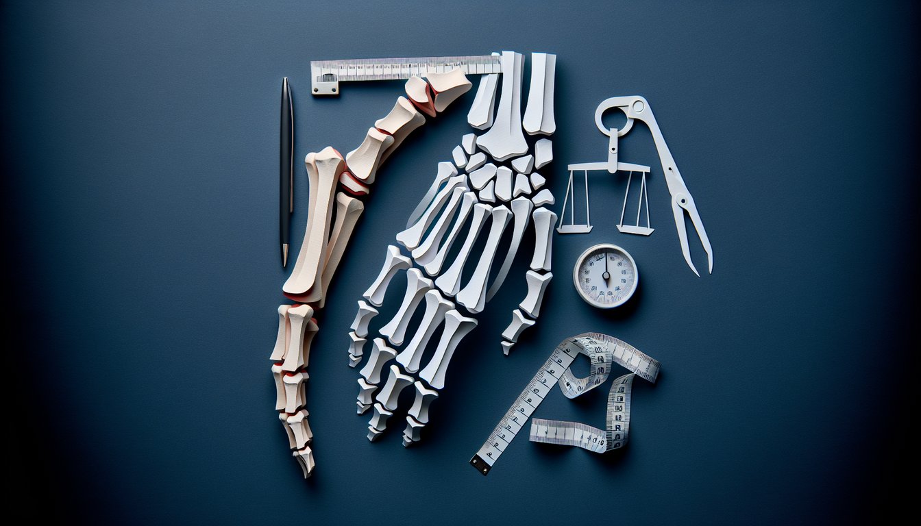
Appendicular Skeleton Quiz: Labeling & Bones Cheat Sheet

  1. The appendicular skeleton - Think of this as your body's dynamic toolkit: 126 bones from your shoulders to your toes that support movement and adventures. It includes the pectoral girdles, upper limbs, pelvic girdle, and lower limbs, so you can high-five, type, and sprint with ease. Ready to dive deeper? training.seer.cancer.gov
  2. The pectoral girdle - This bony frame of clavicles and scapulae attaches your arms to the axial skeleton and acts like a swivel socket for epic shoulder moves. It lets you throw a basketball, lift weights, or strike a superhero pose with maximum flair. Get the 360° view: visiblebody.com
  3. The upper limbs - Your humerus, radius, ulna, carpals, metacarpals, and phalanges team up to perform everything from delicate brush strokes to powerful pull‑ups. These bones form an intricate lever system, giving you the precision to play piano and the strength to carry groceries. Check out the bone lineup: visiblebody.com
  4. The pelvic girdle - Fusion of the ilium, ischium, and pubis creates a sturdy ring that supports your torso and links your legs to the spine. It's your body's weight-bearing champ, balancing stability with flexibility for walking, dancing, and cartwheels. Learn the pelvic power moves: visiblebody.com
  5. The lower limbs - The femur, tibia, fibula, patella, tarsals, metatarsals, and phalanges form the backbone of your mobility, powering every step, leap, and skate. These bones absorb shocks, maintain balance, and propel you from a crawl to a full sprint. Want the full tour? visiblebody.com
  6. The clavicle's unique growth - Unlike most appendicular bones, the clavicle ossifies intramembranously, starting around week five of development and wrapping up at about age 25. This early start makes it the first long bone to begin forming, giving shoulders their sturdy scaffolding. Discover the developmental timeline: openstax.org
  7. Carpal bone mnemonic - Remember "Some Lovers Try Positions That They Can't Handle" to list Scaphoid, Lunate, Triquetrum, Pisiform, Trapezium, Trapezoid, Capitate, and Hamate. This playful phrase turns wrist anatomy into a memory game you can ace on quiz day. Practice the wrist wizardry here: visiblebody.com
  8. The femur - As the longest and strongest bone in your body, the femur connects the hip to the knee, supporting hefty loads and high-impact moves. It's your personal pillar, letting you jump, lunge, and balance with confidence. Explore the powerhouse bone: visiblebody.com
  9. The patella - This sesamoid marvel sits within your quadriceps tendon, shielding the knee joint and boosting the leverage of your thigh muscles. It's the unsung hero behind every squat, kick, and Olympic lift you attempt. Shine the spotlight on your kneecap: visiblebody.com
  10. Foot arches - Your tarsal and metatarsal bones form arches that act like built-in shock absorbers, distributing weight and springing you forward with each step. These curves adapt to terrain, maintain balance, and keep you light on your toes - literally! Step into the details: visiblebody.com
Powered by: Quiz Maker