JPOSNA May 2020: Fracture Quiz

This trauma ‘quiz’ represents an interactive session where POSNA members are able to work through 9 clinical scenarios to challenge how they would address the fracture problems.  Associated with each scenario is an OITE-style question and multiple choice options that seek the most preferred answer from the clinician.  In addition to the answer and follow-up radiographs, we have enlisted the opinions of three POSNA experts in trauma to provide an evidenced-based approach to thinking through each trauma problem. 

Our panel of experts include:
 
Jeff R. Sawyer, MD.  Le Bonheur Children’s Hospital, Campbell Clinic, University of Tennessee Health Science center.  Dr. Sawyer has published >50 trauma-related articles, and has established himself as one of our society’s leaders in pediatric orthopedic trauma. 
 
Tony I. Riccio, MD.  Texas Scottish Rite Hospital for Children, Children’s Health Dallas, UT Southwestern Medical Center.  Dr. Riccio, among many trauma-related endeavors, has spearheaded the effort to promote partnership between POSNA and the OTA, and has taught and published extensively on pediatric orthopedic trauma.
 
Susan A. Scherl, MD.  Children’s Hospital & Medical Center Omaha, University of Nebraska Medical Center.  Dr. Scherl has dedicated her career to the expert care of children with fractures and has published on all aspects of pediatric orthopedic trauma – from NAT to adolescent fractures to supracondylar humerus fractures.
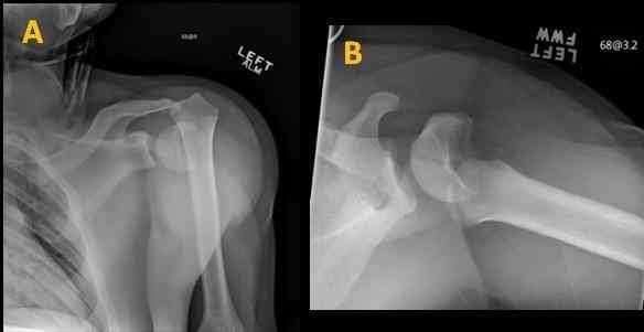
A 14-year-old male defensive lineman was tackling the ball carrier and felt/heard a ‘pop.’ He had immediate pain and inability to use the left arm. Figure 1 A-B demonstrate the injury. What is the next best step in his treatment?
 
Figure 1A & 1B
Closed reduction and sling immobilization in the Emergency Department.
Hanging arm cast and recheck x-rays in 3 days.
Closed reduction and retrograde flexible nail stabilization.
Open reduction via deltoid split approach and screw fixation.
Closed reduction and cannulated screw fixation.
A 12-year-old male injured his left knee 7 days ago at football practice and presents to your clinic with swelling and difficulty moving his knee. Radiographs in Figure 2 A-B demonstrate his injury. What is the preferred method of treatment?
 
Closed reduction, percutaneous retrograde cross-pinning with smooth pins buried beneath skin.
Closed reduction and long leg cylinder cast.
Open reduction and horizontal screw placement in the metaphysis through the Thurston-Holland fragment.
CT scan to better characterize fracture, open reduction and internal fixation.
Closed reduction, percutaneous transphyseal cannulated screw fixation.
4 months ago, a 12-year-old future NBA prospect underwent closed reduction and pinning for a left Salter-Harris II distal femur fracture. Current images are shown in Figure 3A-D. A CT demonstrates 35% of growth plate involvement and his bone age is 11. What is the preferred management?
 
Q3ABCD
Guided growth to prevent expected genu valgum.
Completion epiphysiodesis of left distal femur and utilizing a shoe lift for future limb length discrepancy.
Physeal bar resection with fat graft interposition.
Bilateral distal femur epiphysiodesis.
Observation and eventual magnetic intramedullary nail lengthening.
Figure 4 A-D demonstrate the injury that occurred to a 10-year-old male (55 kg) that tripped and fell walking to school. He had no antecedent left leg pain, but did have immediate pain and inability to walk after his injury. What would be the most appropriate treatment strategy?
 
Q4ABCD
Immediate Spica Cast with an MRI.
Biopsy and titanium elastic nail fixation.
Open biopsy/culture, submuscular plating if benign.
Trochanteric entry antegrade intramedullary nail.
Percutaneous culture, antibiotics and external fixation.
Radiographs in Figure 5 A-B are of an 8-year-old male who fell out of a tree and landed on his right outstretched arm. His skin is intact but he has antecubital puckering and ecchymosis. The right hand is pink but without a palpable pulse. Motor and sensory exam is normal. What is the most appropriate treatment?
 
Closed reduction and splinting prior to elective treatment at surgery center next week.
Emergent open reduction, vascular exploration/repair followed by fracture fixation.
Closed reduction and splinting in the ED overnight, first start trauma room fixation the next day.
Vascular consult, CT angiography, closed reduction and percutaneous pinning.
Emergent closed reduction and percutaneous pinning, confirm pulse returns, observation for neurovascular checks.
7-year-old male (38 kg), was playing baseball and slid into second base. He was safe. The boy felt a ‘pop’ and had immediate pain and inability to walk. Radiographs in the ED are seen in Figures 6 A-B. How would you manage this fracture?
 
Closed reduction and spica casting.
External fixation.
Titanium elastic nails.
Submuscular plating.
Trochanteric-entry, rigid antegrade nail.
8-year-old male with a history of a right proximal femur cyst presents with new onset hip pain, inability to bear weight on the right leg, and with the imaging represented in Figure 7 A-E. What is the preferred method of treatment?
 
Q7AB
Q7CDE
Spica cast until healed.
Injection of cyst with BMP.
CT Chest/Abdomen/Pelvis for staging and open biopsy of lesion.
Implant removal, biopsy, intralesional curettage, fixation with transphyseal proximal femoral locking plate, and injection of cyst with bone substitute material.
Implant removal, biopsy, cephalomedullary nail, proximal screw stopping short of physis, with cement augmentation of proximal femur cyst.
A 14-year-old male was riding his ATV on Friday night when he crashed into a tree. The images in Figure 8 A-C demonstrate his primary injury. He also has a non-displaced clavicle fracture but no other intra-abdominal or intra-cranial injuries. What is the next best step in treatment?
 
Q8A
Q8BC
Immediate open reduction and internal fixation with 3 laterally-based, cannulated screws short of the growth plate to prevent LLD.
Urgent (<24 hours) open reduction and internal fixation with 3 laterally-based, cannulated transphyseal screws.
Skeletal traction and ORIF with a pediatric screw and side plate to be flown in by Monday.
Open reduction on fracture table with dynamic hip screw short of the physis.
Cephalomedullary nail fixation.
A 15-year-old male was playing basketball, went up for a layup and had immediate pain and inability to ambulate. Figure 9 A-B demonstrate the problem. What is the most appropriate treatment plan?
 
Q9AB
Splinting in the ED, return to ambulatory surgery center within 5-7 days for ACL avulsion fixation.
Admission, observation for compartment syndrome, and urgent inpatient ORIF.
Closed reduction and cylinder casting in the ED.
Referral to physical therapy for worsening Osgood-Schlatter Disease.
Closed reduction and percutaneous screw fixation, cylinder casting.
0
{"name":"JPOSNA May 2020: Fracture Quiz", "url":"https://www.quiz-maker.com/Q72HZ9O","txt":"This trauma ‘quiz’ represents an interactive session where POSNA members are able to work through 9 clinical scenarios to challenge how they would address the fracture problems.  Associated with each scenario is an OITE-style question and multiple choice options that seek the most preferred answer from the clinician.  In addition to the answer and follow-up radiographs, we have enlisted the opinions of three POSNA experts in trauma to provide an evidenced-based approach to thinking through each trauma problem.  Our panel of experts include:   Jeff R. Sawyer, MD.  Le Bonheur Children’s Hospital, Campbell Clinic, University of Tennessee Health Science center.  Dr. Sawyer has published >50 trauma-related articles, and has established himself as one of our society’s leaders in pediatric orthopedic trauma.    Tony I. Riccio, MD.  Texas Scottish Rite Hospital for Children, Children’s Health Dallas, UT Southwestern Medical Center.  Dr. Riccio, among many trauma-related endeavors, has spearheaded the effort to promote partnership between POSNA and the OTA, and has taught and published extensively on pediatric orthopedic trauma.   Susan A. Scherl, MD.  Children’s Hospital & Medical Center Omaha, University of Nebraska Medical Center.  Dr. Scherl has dedicated her career to the expert care of children with fractures and has published on all aspects of pediatric orthopedic trauma – from NAT to adolescent fractures to supracondylar humerus fractures., A 14-year-old male defensive lineman was tackling the ball carrier and felt\/heard a ‘pop.’ He had immediate pain and inability to use the left arm. Figure 1 A-B demonstrate the injury. What is the next best step in his treatment?, A 12-year-old male injured his left knee 7 days ago at football practice and presents to your clinic with swelling and difficulty moving his knee. Radiographs in Figure 2 A-B demonstrate his injury. What is the preferred method of treatment?","img":"https://www.quiz-maker.com/3012/images/ogquiz.png"}
Powered by: Quiz Maker